2. 406 P. Gupta et al. / Biochimica et Biophysica Acta 1846 (2014) 405–424
4.3. Reactiveoxygenspecies,akeymechanismofPEITC'santi-cancereffects. . . . . . . . . . . . . . . . . . . . . . . . . . . . . . 415
4.4. Direct protein modificationbyPEITC . . . . . . . . . . . . . . . . . . . . . . . . . . . . . . . . . . . . . . . . . . . . . . . . 419
4.5. Cancer specificbiomarkers . . . . . . . . . . . . . . . . . . . . . . . . . . . . . . . . . . . . . . . . . . . . . . . . . . . . 419
4.6. miRNAs . . . . . . . . . . . . . . . . . . . . . . . . . . . . . . . . . . . . . . . . . . . . . . . . . . . . . . . . . . . . . 419
4.7. Stemcells . . . . . . . . . . . . . . . . . . . . . . . . . . . . . . . . . . . . . . . . . . . . . . . . . . . . . . . . . . . . 419
4.8. Epigenetics . . . . . . . . . . . . . . . . . . . . . . . . . . . . . . . . . . . . . . . . . . . . . . . . . . . . . . . . . . . 419
5. CombinationtherapywithPEITC . . . . . . . . . . . . . . . . . . . . . . . . . . . . . . . . . . . . . . . . . . . . . . . . . . . . . 419
6. PEITCandtobaccosmokeinducedchanges . . . . . . . . . . . . . . . . . . . . . . . . . . . . . . . . . . . . . . . . . . . . . . . . 420
7. Clinicaladvancement . . . . . . . . . . . . . . . . . . . . . . . . . . . . . . . . . . . . . . . . . . . . . . . . . . . . . . . . . . 420
8. Summary. . . . . . . . . . . . . . . . . . . . . . . . . . . . . . . . . . . . . . . . . . . . . . . . . . . . . . . . . . . . . . . . 420
Acknowledgements . . . . . . . . . . . . . . . . . . . . . . . . . . . . . . . . . . . . . . . . . . . . . . . . . . . . . . . . . . . . . 420
References. . . . . . . . . . . . . . . . . . . . . . . . . . . . . . . . . . . . . . . . . . . . . . . . . . . . . . . . . . . . . . . . . . 420
1. Introduction
Since prehistoric times, nature has been a rich source ofmany drugs
and drug leads,which are currently being used to treat various ailments,
including cancer. According to Newman and Cragg, 74.8% of present an-ticancer
agents originated from natural sources, with almost 48.6%
being the actual natural compound or a direct derivative [1]. These in-clude
severalwidely used anticancer agents like taxanes, vinca alkaloids
and camptothecin class of compounds. Interest in natural products as an
alternative to synthetic drugs for cancer treatment is largely due to low
cost, established historical use in traditional medicinal systems, easy
availability and minimal or no toxicity. Also, the potential for targeting
multiple pathways represents an advantage of natural compounds
over synthetic agents, the latter being more target-specific.
Cancer is a complex manifestation of genomic instability in cells
caused by environmental or genetic factors [2]. Genomic instability
leads to disruption of basic biological functions, such as cell division, dif-ferentiation,
angiogenesis andmigration,which promote cancer [2]. The
involvement of multiple mechanisms in cancer development raises an
urgent need for multi-targeted therapy. The current anti-cancer agents
primarily focus on specific targets. The suppression of unique targets
usually leads to activation of compensatory mechanisms resulting in
failure of therapy or development of resistance to drugs. To circumvent
these problems, combination therapy is currently employed by the on-cologists.
Nonetheless, combination approach has also shown marginal
advantage in the overall therapy due to development of resistance or as-sociated
toxicity.We have reached a threshold in terms of clinical ben-efit
and tolerance in patients.
Of all the isothiocyanates (ITCs), phenethylisothiocyanate (PEITC) is
the only one that has reached the clinical stage of testing. This review
provides a critical and comprehensive analysis of the outcomes of stud-ies
showing the anti-cancer effects of PEITC.
2. Epidemiological evidence for chemo-preventive effects of dietary
intake of cruciferous vegetables
Several epidemiological studies from different parts of the world
provide a strong evidence for the reduced risk of cancer with higher in-take
of cruciferous vegetables [3–7]. Although none of these studies pro-vide
a direct correlation between cancer incidence and intake of a
specific ITC, many studies do identify a correlation between the total
ITC intake in the form of cruciferous vegetables and reduced risk of sev-eral
types of cancers in lung, breast, gastric, bladder, colorectal, pancre-atic,
prostate and kidney (Table 1) [5,8–27]. Interestingly, there are also
few studies where the results obtained were opposite to the preventive
effects of cruciferous vegetables. For example, depending on the dura-tion
of intake and age of subjects, one of the studies has shown differen-tial
effects of cruciferous vegetables [20]. In a prospective study,
Giovannucci et al. did not observe any significant correlation between
prostate cancer risk and a short-term intake of cruciferous vegetables
[20]. However, long-term intake of cruciferous diet showed a stronger
inverse correlation with initial stages of prostate cancer and this effect
was stronger in men less than 65 years of age [20]. In contrast, a few
other studies reported no correlation between the intake of cruciferous
vegetables and cancer incidence [27]. The differences in the outcome
showing no correlation with dietary intake of cruciferous vegetables
could be due to several confounding factors such as, differences in the
subject population, duration of intake of cruciferous vegetables in
subject's lifetime, age of subjects, variation in the isothiocyanate content
in cruciferous vegetables based on geographic locations and whether
the consumed vegetables were raw or cooked. Out of 22 studies per-formed,
one study showed increased risk of thyroid cancer with ITC in-take,
while three studies (urinary, prostate and breast cancers) showed
no correlation of ITC intake with cancer incidence. Interestingly all the
studies on stomach, colon and lung cancers showed reduced cancer in-cidence
with ITC intake. A detailed analysis revealed that studies show-ing
no correlation between ITC intake and cancer incidence typically
had wider trial durations or greater age differences for the subjects. In-terestingly,
a study performed in 2001 showed a significant change in
people’s food habits and lifestyle over the last decade [28]. Hence, the
outcomes from studies performed in the last decade might be affected
due to these confounding factors. Furthermore, themajority of the find-ings
were based on questionnaires for the intake of cruciferous vegeta-bles,
which can have lot of variability. In most of the studies,
questionnaires were not specific in asking whether the vegetables
were raw or cooked.
However, based on the majority of the studied outcomes, we can
conclude that an inverse relationship exists between intake of ITCs in
the form of cruciferous vegetables and the overall incidence of cancer.
But no conclusion can be drawn with respect to any specific isothiocya-nate
at this time.
3. PEITC source and pharmacokinetics
3.1. Cruciferous vegetables—source of PEITC
The isothiocyanates (R–N=C=S) (ITC) are known to be the major
bioactive compounds present in cruciferous vegetables and responsible
for anti-cancer activity. ITCs are released from glucosinolates by the ac-tion
of the enzyme myrosinase. The enzyme myrosinase can be activat-ed
by cutting or chewing the vegetables, but heating can destroy its
activity [29]. However, microbial myrosinase from gut can also release
ITCs in the stomach after ingestion of cruciferous vegetables [30,31].
Studies show that myrosinase as well as isothiocyanates are thermola-bile
[32]. Hence ingestion of only raw vegetables can release ITCs and
cooking of the vegetables can reduce ITC content [32]. Although water
cress and broccoli are known to be the richest source, PEITC can also
be obtained fromturnips and radish. PEITC is present as gluconasturtiin
in cruciferous plants. Like other ITCs, PEITC can be released from gluco-nasturtiin
by the action of myrosinase [30,31]. In a study conducted
with human volunteers, approximately 2 to 6 mg of PEITC was found
to be released by the consumption of one ounce of watercress [33,34].
3. Table 1
Epidemiological evidence for the anti-cancer effects of ITCs.
Cancer form Subjects Year
conducted
Sample size Geographical
location
ITC quantitation method Mechanism proposed Outcome Reference
Bladder
cancer
Bladder cancer patients 1980–1998 239 cases Roswell Park
Cancer Institute,
USA
Dietary ITC intake based on pa-tient
questionnaire
– Improved survival [5]
Mixed population with mean ages 63.3
and 62.5 for cases and controls
respectively
1999 697 cases & 708 controls Texas, USA Dietary cruciferous vegetables
intake based on patient
questionnaire
GSTM1 and GSTT1 null Reduced cancer risk [23]
Cases with ages 25–86 years and
controls with ages 21–92 years
1982–1998 275 cases & 825 controls USA Dietary cruciferous vegetables
intake based on patient
questionnaire
– Reduced cancer risk [24]
Breast cancer Chinese women
Age 20–70 years
1996–2005 3035 cases & 3037 controls Shanghai, China Dietary ITC intake based on pa-tient
questionnaire
GSTP1 polymorphism Reduced cancer risk [14]
Breast cancer survivors – 536 intervention & 620 comparison USA Dietary cruciferous vegetables
intake based on patient
questionnaire
– Reduced cancer
recurrence
[25]
US and Chinese breast cancer survivors 1990–2006 11,390 subjects USA and China Dietary cruciferous vegetables
intake based on patient
questionnaire
– No association with
cancer mortality or
recurrence
[27]
Colorectal
cancer
Compiled from published literature – 24 case control studies and 11 prospective
studies
– Dietary intake of cruciferous
vegetables
– Reduced cancer risk [12]
Gastric cancer Middle aged men; mean age ±65 1986–1989 307 cases and 911 control Shanghai, China Urinary ITC GSTM1 and GSTT1
deletion enhanced
protective effect
Reduced cancer
incidence
[8]
Kidney cancer Age 20–79 years 1999–2003 1097 cases & 1555 controls Europe
(multicenter)
Dietary ITC intake based on pa-tient
questionnaire
GSTM1 and GSTT1
polymorphism
Reduced cancer risk [15]
Non-Asians 1986–1994 1204 cases and 1204 controls California, USA Dietary cruciferous vegetables
intake based on patient
questionnaire
– Reduced cancer risk [26]
Lung cancer African Americans & Caucasians; Age
40–84 years
1991–1994 311 cases & 922 control Los Angeles,
California
Dietary ITC intake based on pa-tient
questionnaire
GSTM1 deletion
enhanced protective
effect
Reduced cancer
incidence
[9]
N18 year adults 1990–2005 274 cases & 1089 control Maryland, USA Dietary ITC intake based on pa-tient
questionnaire
_ Reduced cancer
incidence
[11]
Chinese women
Age 40–70 years
1997–2009 74,914 women Shanghai, China Dietary intake of cruciferous
vegetables
– Reduced cancer risk [13]
Men and women with average ages
62.3 and 60.9 years respectively
– 503 cases & 465 controls Texas, USA Dietary ITC intake based on pa-tient
questionnaire
GSTM1 and GSTT1
polymorphism
Reduced cancer risk [16]
Chinese women 1996–1998 233 cases & 187 controls Singapore Dietary ITC intake based on pa-tient
questionnaire
GSTM1 and GSTT1
polymorphism
Reduced cancer risk [17]
Chinese women 1996–1998 303 cases & 765 controls Singapore Dietary ITC intake based on pa-tient
questionnaire
– Reduced cancer risk [18]
Hospital patients 1992–2000 716 cases and 939 controls Massachusetts,
USA
Dietary cruciferous vegetables
intake based on patient
questionnaire
GSTM1 presence Reduced cancer risk [21]
Prostate
cancer
Men
Age 40–75 years
1986–2000 47,365 subjects USA Dietary cruciferous vegetables
intake based on patient
questionnaire
– Non-significant corre-lation
with cancer risk
[20]
Stomach and
colorectal
cancer
Japanese population – 149 cases & 287 controls for stomach cancer
and 115 cases and 230 controls for colorectal
cancer
Japan Dietary cruciferous vegetables
intake based on patient
questionnaire
– Reduced cancer risk [19]
Thyroid
cancer
Melanesian women 1993–1999 293 cases & 354 control New Caledonia Dietary ITC intake based on pa-tient
questionnaire
Low dietary intake of
iodine
Increased risk of
thyroid cancer
[10]
Urinary
carcinoma
Scottish Terriers – 92 cases & 83 controls – Dietary cruciferous vegetables
intake based on owner
questionnaire
– Non-significant corre-lation
with cancer risk
[22]
P. Gupta et al. / Biochimica et Biophysica Acta 1846 (2014) 405–424 407
4. 408 P. Gupta et al. / Biochimica et Biophysica Acta 1846 (2014) 405–424
Similarly, ingestion of 100 g of broccoli andwatercress can release up to
200 μmol of PEITC in humans [29,33]. Interestingly, several pre-clinical
studies have shown that significant anti-cancer effects can be achieved
at micromolar concentrations of PEITC.
3.2. Pharmacokinetics and metabolism
A significant number of studies have shown a positive pharma-cokinetic
profile for orally administered PEITC (Table 2). PEITC is
highly bioavailable after oral administration. A single dose of
10–100 μmol/kg PEITC in rats resulted in bioavailability ranging be-tween
90 and 114% [35]. The high bioavailability was accompanied
by low clearance as well as high protein binding [35]. Increased bio-availability
of PEITC was also observed in another study with repeat-ed
dosing of PEITC [36]. Furthermore, about 928.5 ± 250 nMpeak
plasma concentration of PEITC was achieved in human subjects,
after the consumption of 100 g watercress. The time to reach peak
plasma concentration was observed to be 2.6 h ± 1.1 h with a t1/2
4.9 ± 1.1 h [37]. This indicates that oral administration of PEITC
can result in effective concentrations in blood plasma. However,
long term studies are required to establish the safety profile of
PEITC, since regular intake of PEITC can cause its accumulation
resulting in cumulative effects, which could be toxic.
PEITC metabolism has been reviewed in detail by two groups of in-vestigators
[38,39]. PEITC is primarily removed as mercapturic acid
from the body (Fig. 2). After oral ingestion, PEITC is converted into glu-tathione
conjugates by the action of enzyme glutathione S-transferases
(GST). Several epidemiological as well as pre-clinical studies showed
that the anti-cancer effects of ITC can vary significantly with GST poly-morphism(
Tables 3A and 3B) [15–17,21,40–63]. These glutathione con-jugates
are exported out of the cells by membrane bound transporter
proteins and are cleaved by γ-glutamyl transferase and dipeptidase.
The conjugation of PEITC with intracellular glutathione and the subse-quent
removal of the conjugate result in depletion of glutathione and al-teration
in redox homeostasis leading to oxidative stress, as explained in
detail in Section 4.3 of this review. Cysteine conjugated PEITC is acety-lated
by N-acetyl transferase in liver and eliminated from the body as
N-acetylcysteine (NAC) conjugates or mercapturic acid (Fig. 2) [64]. In-terestingly,
Tang et al. reported that NAC conjugates are also biologically
active and have similar activity to parent compounds, suggesting
prolonged effects of ITCs [64].
PEITC and/or glutathione conjugates were found to be substrates of
important membrane bound transporter proteins like breast cancer resis-tance
protein (BCRP),multidrug resistance-associated proteins 1 (MRP1)
and 2 (MRP2) [65–67]. BCRP plays an important role in absorption and
disposition of many drugs. Hence, PEITC being a substrate of BCRP can po-tentially
affect the pharmacokinetics and pharmacodynamics of other
BCRP substrates. In addition, PEITC can modulate other important trans-porter
and metabolic proteins like P-glycoprotein, [67] and cytochrome
P450 enzymes [68,69]. PEITC-mediated modulation of transporter pro-teins
could be an important factor for the chemopreventive effects of
PEITC. These effects are explained in Section 4.1 of this review. Neverthe-less,
transporter proteins are important determinants of the efficacy and
toxicity of many clinical drugs and by modulating these proteins, PEITC
can affect the efficacy as well as toxicity of some other drugs.
3.3. GST polymorphism and anticancer activity of PEITC
GST is a polymorphic enzymewhich promotes conjugation of gluta-thione
(GSH) with a wide variety of electrophilic xenobiotics including
carcinogens. The conjugation of GSH with carcinogens catalyzed by GST
and subsequent removal of the conjugate results in reduced
Table 2
Pharmacokinetic parameters of PEITC.
Route of
administration
Dose Vehicle Model Number of samples
(n)
Organ AUC ± SD or (SE)
Oral 10 μmol (50 μmol/kg)
(single)
Corn oil Male F344 rats 3/time point Blood 328.1 nmol ml−1 h (106.93)
Liver 1277.57 nmol ml−1 h
(291.76)
Lungs 346.321 nmol ml−1 h (6.51)
Kidneys 987.941 nmol ml−1 h
(114.88)
Nasal
mucosa
378.50 pmol mg−1 h(341.42)
Esophagus 140.19 pmol mg−1 h (111.69)
Stomach 1409.7 pmol mg−1 h (457.62)
Small
intestine
1691.21 pmol mg−1 h
(292.94)
Cecum 102422.29 pmol mg−1 h
(44129.9)
Colon 361.39 pmol mg−1 h (47.32)
Pancreas 176.0 pmol mg−1 h (29.04)
Spleen 57.71 pmol mg−1 h (30.55)
Heart 94.35 pmol mg−1 h (22.99)
Brain 53.71 pmol mg−1 h (5.4)
Oral (Gastric-intubation)
0.5, mg/kg(single) DMSO Male Wistar albino
rats
4 Blood 1033 ± 174378 μg L−1 h
1.0 mg/kg(single) 4 1646 ± 284 378 μg L−1 h
5.0 mg/kg(single) 4 2438 ± 462 μg L−1 h
0.5 mg/kg/day (4 days) 4 1117 ± 282 378 μg L−1 h
1.0 mg/kg/day (4 days) 4 2311 ± 258 μg L−1 h
5.0 mg/kg/day (4 days) 4 5796 ± 1004 μg L−1 h
Intravenous 0.5 mg/kg (single) 15% hydroxypropyl-β-
cyclodextrin
4 1346 ± 378 μg L−1 h
Intravenous 2 μmol/kg 15% hydroxypropyl-β-
cyclodextrin
Male Sprague Dawley
rats
3–4 Blood 2.96 ± 0.78 μM h
10 μmol/kg 17.3 ± 9.3
100 μmol/kg 322.1 ± 149.6
400 μmol/kg 807.5 ± 66.9
Oral 10 μmol/kg – 19.89 ± 3.27
100 μmol/kg 298 ± 139.4
5. P. Gupta et al. / Biochimica et Biophysica Acta 1846 (2014) 405–424 409
carcinogenic effects [70,71]. Increasing evidence from epidemiological
and pre-clinical studies suggest a significant impact of GST polymor-phism
on the outcomes of dietary ITC intake (Table 3A). A majority of
these studies indicates an important effect of GSTT1, GSTM1 and
GSTP1 polymorphism in ITC mediated anti-cancer effects. The presence
of GSTT1 or GSTM1 or both have been strongly correlated with reduced
or no effects of dietary ITCs on cancer risk. Interestingly, pre-clinical
studies have shown ITC-mediated induction of GST enzymes, suggesting
GST-mediated metabolism and inactivation of ITCs (Table 3B). Interest-ingly,
a study indicates concentration dependent effect of specific GST
polymorphism in catalyzing glutathione conjugation with PEITC [56].
The GSTA1-1 enzyme reacts with micromolar concentrations of PEITC,
while GSTM1a-1a exhibits a significantly lower affinity for PEITC. On
the contrary, Meyer et al suggested that GST catalyzes forward as well
as reverse conjugation of PEITC with GSH [56]. Studies also showed
PEITC mediated tissue specific induction of GST isoforms [55,57,60,63,
72,73]. Van et al. showed significant induction of GST by PEITC in esoph-agus
[57]. Furthermore, PEITC mediated induction of GSTA in liver,
GSTM in stomach and liver as well as GSTP in colon was observed.
PEITC treatment also induced glutathione levels by about 1.6 fold in
colon [57]. PEITC-mediated GSTP modulation was observed in few
other studies as well [58,59]. The induction of GST and glutathione ap-pears
to be an important pathway for PEITC-mediated chemopreventive
effects. In a chemical carcinogen-induced multi-organ cancer model,
differential effects of PEITC were observed in pre- and post-tumor
phases. PEITC treatment during pre-initiation period caused reduced
hyperplasia in esophagus and lungs accompanied with reduction of
GSTP in kidney and liver [58]. However, PEITC treatment during post-initiation
phase increased liver GSTP positive foci along with increased
incidence of urinary bladder hyperplasia and tumors [58]. This suggests
preventive effects of PEITC in pre-initiation conditions, while during
post-initiation, PEITC treatment increases tumor incidence. However,
it is important to note that 0.1% PEITC was administered in the diet in
this study,whereas, in most of the studies, a pure and higher concentra-tion
of PEITC was administered.
The role of GST in chemoprevention has been reported to be depen-dent
on the polymorphic status of GST aswell as the source of induction
[74]. This implies that GST polymorphism and its tissue specific differ-ential
effects are important determinants to correlate cancer risk and
ITC intake. Many studies have also reported the outcomes of ITC intake
and cancer risk with no consideration of GST status in the subjects.
Hence, outcomes fromthese studies need to be re-confirmed using bet-ter
designed studies.
4. Anti-cancer activity of PEITC
Several researchers have demonstrated the anticancer properties of
isothiocyanates in various cancer types [75–81]. The anti-cancer activity
of PEITC can be divided into chemopreventive and chemotherapeutic
effects. Although chemopreventive effects were identified through epi-demiological
evidence, chemical carcinogenesis models and transgenic
mouse models, more extensive mechanistic studies were performed to
evaluate its therapeutic potential in various pre-clinical models.
4.1. Chemopreventive effects of PEITC
The diverse epidemiological studies provided the first evidence of
anticancer activity of ITCs (Table 1). The preventive effects of ITCs
were evaluated in multiple chemically-induced cancer models. For ex-ample
Morse et al and others, demonstrated that administration of
ITCs including PEITC prevented the carcinogenic effects of chemical car-cinogens
in rodents [82–86]. In general, carcinogenesis occurs due to
Table 2
Pharmacokinetic parameters of PEITC.
Cmax ± SD
or (SE)
Tmax ± SD
or (SE)
T1/2a ± SD
or (SE)
T1/2e ± SD
or (SE)
Vd ± SD
or (SE)
F% ± SD
or (SE)
Ref
18.77 pmol/μl (2.9) 2.94 h (0.78) 1.34 h (3.15) α = 2.41 h (6.96)
β = 21.7 h (25.77)
– – [182]
39.79 pmol/μl (3.29) 2.5 h (0.56) 0.44 h (0.14) 20.45 h (5.84) – –
9.73 pmol/μl (0.39) 4.53 h (0.4) 0.97 h (0.14) 21.3 h (2.99) – –
46.29 pmol/μl (2.06) 4.31 h (0.39) 1.18 h (0.2) 11.37 h (1.99) – –
20.4 pmol/mg (7.31) 3.52 (0.66) 0.93 h (1.2) 10.10 (12.16) – –
8.2 pmol/bg (2.14) 2.77 h (1.78) 0.67 h (0.69) 9.72 h (9.89) – –
211.19 pmol/mg (26.44) 1.35 h (0.45) 0.37 h (0.24) 3.57 h (1.93) – –
130.58 pmol/mg (7.64) 4.07 h (0.41) 1.69 h (0.64) 5.23 h (2.22) – –
15.07 pmol/mg (1.78) 19.09 h (11.51) 465.87 h (2061.42) 2.52 h (1.07) – –
16.43 pmol/mg (1.22) 8.08 h (1.15) 5.81 h (37.92) 5.4 h (35.71) – –
9.73 pmol/mg (0.39) 4.53 h (0.4) 54.96 h (11.78) 0.97 h (0.14) – –
9.64 pmol/mg (2.0) 1.2 h (0.71) 3.2 h (2.83) 0.33 h (0.37) – –
5.71 pmol/mg (0.51) 4.36 h (0.74) 7.75 h (3.41) 1.48 h (0.61) – –
2.07 pmol/mg (0.1) 6.46 h (0.57) 12.58 h (2.27) 2.07 h (0.41) – –
318 ± 62 μg/L 0.73 ± 0.15 h 0.2 h (Ka = 3.52 ± 1.58) 2.62 ± 1.37 h 1.53 ± 0.81 L/kg 77 ± 13 [37]
387 ± 73 μg/L 0.45 ± 10 h 0.13 h (Ka = 5.19 ± 2.14) 2.78 ± 0.36 h 1.75 ± 0.29 L/kg 61 ± 11
769 ± 107 μg/L 0.78 ± 0.21 h 0.18 h (Ka = 3.88 ± 0.40) 3.19 ± 0.37 h 2.15 ± 0.40 23 ± 5
404 ± 80 μg/L 0.7 ± 0.24 h 0.16 h (Ka = 4.26 ± 2.31) 2.55 ± 0.26 h 1.5 ± 0.14 L/kg 83 ± 21
647 ± 38 μg/L 0.90 ± 0.12 h 0.3 h (Ka = 2.26 ± 0.48) 1.86 ± 0.2 h 1.05 ± 0.10 L/kg 86 ± 10
2139 ± 444 μg/L 0.95 ± 0.10 h 0.22 h (Ka = 3.02 ± 2.03) 1.62 ± 0.44 h 0.88 ± 0.28 L/kg 43 ± 7
– – – 1.36 ± 0.36 h 0.75 ± 0.06 L/kg 100
– – – 3.52 ± 0.35 h 1.94 ± 0.42 L/kg 100 [36]
– – – 6.92 ± 3.73 h 3.27 ± 2.06 100
– – – 9.19 ± 0.83 h 2.66 ± 1.22 100
– – – 13.1 ± 2.0 5.72 ± 1.10 100
9.2 ± 0.6 μM 0.44 ± 0.1 h – – – 115
42 ± 11.4 2 ± 1 – – – 93
6. 410 P. Gupta et al. / Biochimica et Biophysica Acta 1846 (2014) 405–424
STAT3
NK
Cells
the bio-activation of carcinogens by oxidation, reduction or hydrolysis
via phase I drug metabolizing enzymes. Hence, modulation of phase I
enzymes can affect the carcinogen activation process. PEITC shows a
dual activity on phase I enzymes. For example, PEITC on one hand
causes induction of CYP1A1 and CYP1A2; however, it inhibits activity
of certain CytP450 enzymes, such as CYP2E1, CYP3A4 and CYP2A3 [68,
87–91]. Detailed studies are required to elucidate the differential effects
of PEITC on CYP enzymes and their mechanistic implications in cancer
prevention.
Phase II enzymes, which include mainly transferases, play an im-portant
role in detoxifying xenobiotics and carcinogens. As reviewed
by Cheung et al., various ITCs including PEITC have been shown to in-duce
phase II detoxification enzymes, which can explain their che-mopreventive
activity [92]. GSTs play an important role in
chemopreventive effects of PEITC, as discussed earlier in
Section 3.3. PEITC also acts on other phase II enzymes. Saw et al.
demonstrated induction of Nrf2-antioxidant response element by a
combination of ITC and phyto-indoles [93]. Although in general,
ITCs modulate phase II enzymes, human livers demonstrated a
non-identical response to PEITC [94]. Liver enzymes were found to
be most sensitive to PEITC, while lung enzymes were mostly resis-tant
[95]. Hence, based on current evidence, it can be concluded
that the effects of PEITC on drug metabolizing enzymes are individu-al
as well as tissue specific.
Continuous high levels of ROS also promote carcinogenesis by caus-ing
oxidative DNA damage leading to mutations [96]. Interestingly,
PEITC treatment caused a significant increase in the activities of ROS de-toxifying
enzymes such as glutathione peroxidase1, superoxide
BID
BCL2
BAX
p53
Loss of
growth
inhibitory
signaling
dismutase 1 and 2. This was also confirmed in human study where sub-jects
were administered watercress, a major source of PEITC [61].
4.2. Chemotherapeutic effects of PEITC
Hanahan and Weinberg have defined ten basic common hallmarks
underlying cancer progression [2,97]. All types of cancers require
these basic hallmarks for development and advancement. Hence, effec-tive
inhibition of one or more of these hallmarks can potentially en-hance
the outcomes of current anti-cancer therapeutics. Accumulating
evidence suggests that PEITC targets a broad spectrum of proteins to
suppress cancer growth and progression (Fig. 1). Research group led
by Zigang Dong and An-Ng Tony Kong for the first time in 1998 inde-pendently
demonstrated the apoptosis-inducing effects of PEITC in cul-tured
cancer cells [98–100]. The cytotoxic concentrations of PEITC range
from120nMto 14 μMin cancer cells [101,102]. Tseng et al. have report-ed
that working concentrations of PEITC are similar to chemotherapeu-tic
drug daunomycin in human breast cancer cells as well as in
mammary epithelial cells, implying PEITC's anti-cancer potential com-parable
to conventional drugs [103].
Since the discovery of anticancer effects of PEITC, several mecha-nisms
have been proposed for its activity. Two primary mechanisms
that have been identified are cell cycle arrest and induction of apoptosis
[104–106]. PEITC-mediated generation of reactive oxygen species (ROS)
is known to be a general mechanism of action leading to cytotoxic ef-fects,
especially specific to cancer cells [107,108]. Some studies have
also shown significant inhibition of other important cancer promoting
mechanisms like angiogenesis and metastasis [109–113]. PEITC targets
?
?
?
?
Gly
colysis
?
?
?
?
MMP
NFkB
HIF1A
T
Cells
VEGF
Telomerase
Rb
HER2
PEITC
Suppressed
immune
system
Sustained
growth
signaling
Deregulated
metabolism
Genetic
instability
Metastasis
& Invasion
Angio-genesis
Immortality
In-
Evading
apoptosis
EGFR
MAPK
AKT
ERK
B
cells
STAT1
NFkB
TLR3
Macro-phage
Topo
isomer-ase
miRNA
STAT3
Inte
mTOR grins
p21
DR4,5
Tubulin Vi-mentin
MMP
Survivin
BIM
PUMA
p62
Fig. 1. PEITC and hallmarks of cancer.
7. P. Gupta et al. / Biochimica et Biophysica Acta 1846 (2014) 405–424 411
specific regulatory proteins to inhibit cancer-related oncogenes.Mi et al.
have identified more than 30 different biological targets of PEITC [114].
There are about 350 articles available so far on the anti-cancer effects of
PEITC. Table 4 summarizes the targets and major mechanisms of PEITC
that have been studied in different cancer models by various investiga-tors
during recent years (2011–2014).
4.2.1. Anti-proliferative effects
Sustained proliferative signaling is a major characteristic of cancer
cells. Ras activation in cancer by different oncogenes is a common
mechanism to sustain proliferation. Ras further activates various path-ways
like RAF/MEK-MAPK and PI3K/Akt that are crucial for cell prolifer-ation,
and their inhibition is known to suppress cancer growth. It was
observed that Rasmutation did not correlatewith PEITC-mediated inhi-bition
of tumorigenesis, indicating that Ras may not be a target of PEITC
[115]. However, PEITC inhibits Akt, a component of Ras signaling to in-hibit
tumor growth in several cancer types [116–118]. Akt (protein ki-nase
B) is over-activated in many types of cancers and promotes
multiple cell survival mechanisms. PEITC is also known to inhibit EGFR
and HER2, which are important growth factors and regulators of Akt
in different cancer models [102,117,119]. Hence, Akt inhibition can be
due to the suppression of EGFR or HER2. Overall, it is evident from
these examples that even-though PEITC is unable to suppress Ras,
tumor cell growth suppression is achieved through an alternative
mechanism.
4.2.2. Effects on cell division and cell cycle
Cell proliferation is a tightly regulated process, orchestrated by pro-and
anti-proliferative signals. Generally, the pro-proliferative signals are
activatedwhen newcells are required to replace damaged cells. At other
times, the anti-proliferative signals maintain cells in a quiescent state
and also keep cell division under control. However, cancer cells lock in
the pro-proliferative signals, while suppressing tumor suppressor
genes responsible for anti-proliferative effects. Cells have multiple
check points for proliferation,which are regulated by tumor suppressor
genes such as p53, Retinoblastoma protein (Rb) and PTEN. PEITC causes
activation of Rb protein at the cellular level in prostate cancer cells, lead-ing
to attenuation of cell cycle progression [100,120]. Interestingly,met-abolic
conjugates of PEITC with N-acetyl cysteine (NAC) also inhibit the
phosphorylation of Rb leading to cell cycle arrest [67]. PEITC-mediated
activation of another tumor suppressor, p53 was observed in oral squa-mous
cell carcinoma, causing G0/G1 phase arrest in multiple myeloma,
osteogenic sarcoma, breast cancer and prostate cancer, alongwith inhi-bition
of other proteins regulating G2/Mphase [93,121–125]. Itwas also
observed that lung carcinoma cells with wild type p53 were relatively
less sensitive to PEITC as compared to cells with mutated p53 [100,
114,126,127]. Although, a direct effect on another important tumor sup-pressor
PTEN has not been reported, PEITC inhibits Akt, which is
overexpressed when PTEN is mutated.
Cells have a capability to keep track of the number of divisions un-dergone
so that cell death can be initiated after a certain number of di-visions.
This phenomenon uses the shortening of telomere length with
every division. This can be reversed by enzyme telomerase. Often, can-cer
cells exploit telomerase enzyme to alter the length of telomeres ren-dering
them immortal [128]. PEITC has been shown to inhibit
telomerase activity in prostate and cervical cancer cells [129,130]. Fur-thermore,
it was observed that pre-treatment with PEITC induced apo-ptosis
as well as increased the efficacy of adriyamycin and etoposide by
inhibiting protein kinase C and telomerase [130].
4.2.3. Pro-apoptotic effects
Cells usually undergo apoptosis when proliferation cannot be con-trolled
by cell cycle check points. However, cancer cells develop anti-apoptotic
mechanisms to enable survival. Mounting evidence suggests
strong pro-apoptotic activity of PEITC by diverse mechanisms. Most of
these mechanisms are affected by generation of reactive oxygen species
(ROS), which also has been shown to be the basis of selectivity of PEITC
toward cancer cells leaving normal cells undamaged [107]. A detailed
account of PEITC mediated ROS generation is provided in Section 4.3.
ROS generation by PEITC leads to mitochondrial deregulation and mod-ulation
of proteins like Bcl2, BID, BIMand BAX, causing the release of cy-tochrome
c into cytosol leading to apoptosis [102,121,124,131–136].
However, Wu et al. suggested that cytochrome c does not play a role
in PEITC-induced apoptosis [137,138]. Roy et al have shown an interest-ing
tumor regression due to PEITCmediated inhibition of DDB2 through
ROS generation [139]. In addition, PEITC also induces apoptosis by acti-vation
of extrinsic apoptotic pathway in oral and cervical cancer cells
through the induction of death receptors and Fas-mediated apoptosis
[140–143]. Some studies also show PEITC-mediated suppression of
anti-apoptotic proteins like XIAP and survivin, which are up-regulated
in cancer cells [144]. Taken together, a strong cytotoxic potential of
PEITC cannot be denied, although differential roles have been described.
4.2.4. Autophagy
Autophagy is a stress response mechanism, which leads to degrada-tion
of cellular organelles to preserve cell energy. Autophagy is general-ly
considered as cell survival mechanism. However, it is also a unique
form of cell death, occurring under special circumstances [145]. Investi-gations
show that PEITC induces autophagic cell death in cancer cells
[146–148]. Interestingly, Mi et al. showed the formation of aggresomes
by PEITC treatment [149], indicating induction of autophagy, since these
structures are formed due to proteasome failure and are degraded by
autophagic pathway. Bommareddy et al. showed induction of autopha-gy
by PEITC in prostate cancer cells [147]. Treatment with 3-
methyladenine, an autophagy inhibitor, provided evidence that autoph-agy
induced by PEITC was unable to protect the cells from apoptosis. It
was observed that PEITC-induced autophagy was mediated by Atg5
[147]. A partial role of the inhibition of mTOR/Akt signalingwas also ob-served
in PEITC-induced autophagy. Later PEITC-mediated induction of
Fig. 2. General metabolic pathway for PEITC.
8. Table 3A
Epidemiologic evidence for the effects of GST polymorphism on ITC uptake/metabolism and cancer risk.
Cancer Design Population Location Method of analysis ITC intake/
levels
GST
studied
Status Outcomes Statistics Reference
Adenoma Case control (459 cases; 507 control) Mixed USA Dietary intake High GSTM1 Null Reduced risk P = 0.001 for trend and 0.1 for
interaction
[42]
Breast Case control (740 cases; 810 control) Caucasian women USA Dietary intake High GSTM1/
T1
Null/positive Reduced risk OR = 0.6 (0.4–1.01)
Premenopausal women
OR = 1.0 (0.7–1.4)
Postmenopausal women
[44]
Case control (1052 cases and 1098 control) Women (b65 years
age)
USA Dietary intake – GSTT1 Null Increased risk OR = 1.86 (1.12–3.08)
–
[46]
– GSTM1 Null Increased risk
– GSTP Ile/Ile Increased risk
High/Low GSTT1/
M1/P
Null/Positive No correlation
Colon Case control (213 cases; 1194 control) Chinese men and
women
Singapore Dietary intake High GSTM1/
T1
Null Reduced risk OR = 0.43 (0.2–0.96) [43]
Colorectal Case control (173 cases and 313 controls) Mixed USA Urinary ITC Detectable GSTT1 Null/Positive No correlation – [48]
Detectable GSTM1 Null/Positive No correlation –
Detectable GSTP1 AG or GG Marginally reduced
risk
P = 0.09
Case control (322 cases and 1251 controls) Women 40–70 years China Urinary ITC High GSTT1 Null Reduced risk P = 0.04 [50]
High GSTM1 Null Reduced risk P = 0.07
High GSTT1/
M1
Null Reduced risk OR = 0.51 (0.27–0.95)
Kidney Case control (1097 cases and 1555 control) Mixed Europe Dietary intake Low GSTT1 Null Increased risk OR = 1.86 (1.07–3.23) [15]
Low GSTM1/
T1
Null Increased risk OR = 2.49 (1.08–5.77)
Lung Case control (503 cases; 465 control) Mixed subjects from
Houston
USA Dietary intake Low GSTM1 Null/Current
smoker
Increased risk in OR = 2.22 (1.2–4.1) [16]
Low GSTT1 Null/Current
smoker
Increased risk OR = 3.19 (1.54–6.62)
Low GSTM1
and T1
Null/Current
smoker
Increased risk OR = 5.45 (1.72–17.22)
Low GSTM1 Null/Former
smoker
No correlation OR = 1.14 (0.66–1.96)
Low GSTT1 Null/Former
smoker
Increased risk OR = 1.79 (0.95–3.37)
412 P. Gupta et al. / Biochimica et Biophysica Acta 1846 (2014) 405–424
9. Case control (233 cases; 187 control) Chinese women – Dietary intake High GSTM1/
T1
Null Reduced risk OR = 0.54 (0.3–0.95) [17]
High GSTM1 Positive No effect 1.07 (0.5–2.29)
Case control (232 cases; 710 control) Chinese men China Dietary intake and urinary
ITC analysis
Detectable GSTM1 Null Reduced
risk
Relative
risk = 0.36
(0.2–.63)
[41]
Detectable GSTM1/T1 Null Reduced risk Relative
risk = 0.28
(0.13–0.57)
Case control (716 cases; 939 control) Caucasian women
and men
USA Dietary intake High GSTM1 Positive Reduced risk OR = 0.61 (0.39–0.95) [21]
High GSTM1 Null No correlation OR = 1.15 (0.78–1.68)
High GSTM1 Null/positive No correlation –
High GSTM1/
T1
Null/positive No correlation –
Literature
review
30 studies – Dietary
intake
High GSTT1/M1 Null Reduced risk OR = 0.41 (0.26–
0.65)
[47]
– Cohort Chinese (45–
74 years age)
111 men, 135
women
Singapore Dietary intake and urinary
ITC analysis
– GSTM1 Null No correlation P = 0.61 [40]
– GSTM1 Positive No correlation
– GSTT1 Null Low excretion P = 0.006
– GSTT1 Positive High excretion
– GSTP1 a/a No correlation P = 0.77
– GSTP1 a/b No correlation
– GSTP1 b/b No correlation
Cohort (114 subjects) 18–50 years Mixed USA Urinary ITC and
metabolites
Single
intake
GSTM1 Null High excretion in 62%
subjects
ITC (mol/24 h) = 9.9 ± 1.4 [45]
GSTM1 Positive High excretion in 39%
subjects
ITC (mol/24 h) = 14.0 ± 1.3
(P = 0.48)
GSTT1 Null Marginally
significant
ITC (mol/24 h) = 9.9 ± 2.7
GSTM1 Positive Marginally
significant
ITC (mol/24 h) = 11.6 ± 0.9
(P = 0.05)
GSTP1 A/A Non significant ITC (mol/24 h) = 9.9 ± 1.5
GSTP1 G/A Non significant ITC (mol/24 h) = 14.8 ± 1.4
Cross-over intervention study 20 healthy subjects – Plasma ITC – GSTM1 Null/Positive No correlation – [49]
– GSTT1 Null/Positive No correlation –
48 subjects, 28 GSTT1 and M1 positive and
20 null genotypes
Healthy volunteers – Urinary ITC Watercress
juice
GSTT1/
M1
Null/Positive No correlation – [51]
P. Gupta et al. / Biochimica et Biophysica Acta 1846 (2014) 405–424 413
10. 414 P. Gupta et al. / Biochimica et Biophysica Acta 1846 (2014) 405–424
Table 3B
Pre-clinical evidence for effect of GST polymorphism on isothiocyanate efficacy.
Model Agent used GST polymorph Outcome Reference
Male Wistar rats BITC Subunits 3 & 2 Increased [52]
Rat T9 glioma cells AITC, BITC GSTP Increased marginally [53]
autophagy was shown in a transgenic mice model of prostate cancer
[148]. Taken together, autophagy can be one important anti-cancer
mechanisms of PEITC. Nevertheless, further study is required to deter-mine
whether or not PEITC induces autophagy in other in vivo cancer
models.
4.2.5. Anti-angiogenic effects
Rapidly proliferating cancer cells have increased demand for nutri-ents
and oxygen. Hence, these cells enhance the growth of new blood
vessels (angiogenesis) to meet the increasing nutritional demand.
Targeting vascular endothelial growth factor (VEGF), a major promoter
of angiogenesis, has been an important mechanismof cancer treatment
for the past fewdecades. As reviewed by Loureiro and D'Amore, VEGF is
regulated by two major mechanisms: hypoxic regulation and non-hypoxic
regulation [150]. Importantly, cancer cells are constantly
under hypoxia due to limited oxygen supply. During hypoxia,
hypoxia-inducible factor (HIF1α) accumulation stimulates secretion of
VEGF leading to angiogenesis [150]. A study by Xiao and Singh provided
evidence on the anti-angiogenic effects of PEITC through VEGF suppres-sion,
but the exact mechanism of this inhibition was not clear [113]. It
was shown later that PEITC directly or indirectly suppresses HIF1α
[151–154]. Besides hypoxic regulation, VEGF is also regulated by growth
factors released by cancer cells (heregulin, EGF and TGF), hormones (es-trogen,
testosterone and insulin) as well as oncogenes Wnt and Ras
[150]. In addition, VEGF is negatively regulated by tumor suppressor
genes such as p53 and p73. Based on the effects of PEITC on proliferation
and cell cycle described in Sections 4.2.1 and 4.2.2, it is possible that
PEITC can block angiogenesis by non-hypoxic mechanisms also.
4.2.6. Anti-metastatic effects
In malignant cancer, tumor cells tend to invade blood circulation to
reach other organs of the body leading to spread of cancer. Under
normal conditions, non-cancerous cells undergo apoptosis under
anchorage-independent conditions due to the absence of extracellular
matrix,which provides survival signals. In contrast, cancer cells develop
survival mechanisms that allow them to survive during circulation
under anchorage-independent conditions and spread to distant organs.
This process, referred to as “invasion and metastasis”, is a major reason
for the poor prognosis in majority of cancers. It leads to relapse of can-cer,
which is mostly resistant to conventional chemotherapy. There
are few studies which suggest anti-invasive and anti-metastatic effects
of PEITC. Various studies with PEITC have shown suppression of inva-sion
through inhibition of matrix metalloproteinases along with anti-metastatic
effects caused by suppression of ERK kinase activity and tran-scriptional
activity of NFkB [109,111]. PEITC was also known to inhibit
processes, such as epithelial to mesenchymal transition (EMT), cell in-vasion
and migration, which are essential pre-requisites for metastasis
[155–157]. A recent study by Gupta et al. demonstrated anti-metastatic
potential of PEITC in a novel in vivo model of breast cancer
metastasis [119]. In this model,whenMDA-MB-231 brain seeking lucif-erase
breast cancer cellswere injected into the left ventricle of the heart
of female athymic nude mice, a small percentage of tumor cells lodge in
the brain via blood circulation and growthere as metastatic tumors. Ob-servations
from a pretreatment model suggested that oral administra-tion
of 10 μmol PEITC significantly prevented the migration of breast
cancer cell to the brain in vivo. In another experiment, PEITC administra-tion
after tumor cell implantation not only suppressed the growth of
metastasized tumors in brain but also prolonged the survival of tumor
bearingmice [119]. Although these studies reveal the anti-metastatic ef-ficacy
of PEITC in a breast cancermodel,more evidence is required to es-tablish
similar anti-metastatic effects in other cancer models.
4.2.7. Anti-inflammatory effects
The tumor microenvironment is similar to inflammatory lesions
and plays a very important role in carcinogenesis. Inflammation pro-motes
cancer cells to grow in a bimodal way. Firstly, pre-existing in-flammation
promotes carcinogenesis by stimulating cells through
several chemokines, cytokines and growth factors. Secondly, the same
factors can be secreted by established cancer cells to re-enforce tumor
growth and development. Hence, cancer growth can be inhibited by
controlling the inflammatory process. Several studies have demonstrat-ed
direct modulation of inflammatory process by PEITC. It has been
shown that chemically-induced inflammatory responses in mice were
suppressed by PEITC [141,158]. The mechanistic studies have shown
suppression of inflammatory mediators like nitric oxide, TNF-α, and
IL-10 in LPS-stimulated macrophages, as well as impairment of
AITC, BITC + dibutyryl
cAMP
GSTP Increased
Male ACI/N rats BITC, BTC GSTP Reduced [54]
Kinetic study ITC GSTP1-1 and M1-1 Most efficient catalysis [55]
GATM4-4 Least efficient catalysis
Kinetic study PEITC, BITC GSTA1, A2, P1 Catalyze forward and reverse reactions in a concentration dependent manner [56]
Male Wistar rats PEITC GSTA Increased in liver [57]
GSTM Increased in stomach and liver
GSTP Increased on colon
Male F344 rats PEITC GSTP Reduced in liver (Pre-initiation period of tumors) with reduced hyperplasia in
esophagus and kidney
[58]
Enhanced in liver (Post-initiation period of tumors) with increased urinary bladder
tumors
RL34 cells (rat liver
epithelial)
BITC, PEITC GSTP1 Increased [59]
RL34 cells ITC GSTA1/2 Nrf2 dependent increase [60]
GSTA3
GSTM1
PBMC cells Watercress/PEITC GSTM1-0 Increased GPX and SOD [61]
GSTM1-1 Not increased GPX and SOD
GSTT1 No impact
HL60 cells ITC GSTA
(Overexpression)
Reduced efficacy [62]
Male Wistar rats ITC GSTM Increased in liver [63]
GSTA Increased in Liver
GSTT Increased in kidney
11. P. Gupta et al. / Biochimica et Biophysica Acta 1846 (2014) 405–424 415
macrophagemigration inhibitory factor (MIF) through covalentmodifi-cation
by PEITC [159,160]. A fewreports also connect the immunomod-ulatory
actionwith anti-cancer activity of PEITC. For example, PEITC has
been shown to protect mouse liver and lung from changes caused by
cigarette smoke [153]. A bio-informatic analysis of PEITC treated animal
tumors showed modulation of genes involved in inflammation and ex-tracellular
matrix pathways [161]. Taken together, these findings sug-gest
anti-inflammatory effect as one of the anticancer effects of PEITC.
However, further comprehensive studies between cancer and inflam-mation
are still required to elucidate the anti-cancer mechanisms of
PEITC.
4.2.8. Immunomodulatory effects
The human body's integral defense mechanisms serve as a major
host for future therapeutic opportunities and can help in the fight
against cancer. The immune system is equipped with several check
points to scan and selectively eliminate malignant cells. Interestingly,
cancer cells develop sophisticated mechanisms to evade the immune
system. There is a need for extensive and rigorous studies to discover
agents that can prevent cancer cells from evading the immune system.
Tsou et al. demonstrated the immunomodulatory activities of PEITC
[162]. PEITC treatment caused activation of macrophages and NK cells
in a mouse leukemia model. PEITC treatment also promoted the differ-entiation
of B cells but the proliferation remained unaffected. Converse-ly,
itwas observed that PEITC increased the proliferation of T cellswhile
inhibiting the differentiation of the precursor cells of T cells and macro-phages.
PEITC treatment caused reduction in weight of the spleen and
liver of leukemic mice, although no reduction in the overall weight of
mice was observed [162]. The reduction of the size of the spleen and
liver is more likely a result of killing of the leukemia cells in those or-gans,
with a concomitant reduction in weight of those organs, rather
than a detrimental effect on the immune system. Another study report-ed
in vitro and ex vivo effects of PEITC on toll-like receptor 3 (TLR3).
TLR's are considered to be an important component of innate immune
system. Studies showed that PEITC treatment inhibited TLR3 signaling
[163]. PEITC treatment also inhibited the dimerization of TLR3 receptors
leading to inhibition of IRF3 signaling. These effects were accompanied
by inhibition of anchorage-independent growth and colony formation
of cancer cells. Of note, PEITC also inhibits lipopolysaccharide induced
TLR/IRF3 signaling in leukemia cells [164]. Thus, while theoretically
the inhibition of TLR3 might be considered a negative effect on the im-mune
system, the resulting inhibition of IRF3 signaling inhibits at least
two mechanisms of metastatic tumor development. Further detailed
studies are required to elucidate the complete effects of PEITC on im-mune
system.
4.2.9. Effect of PEITC on cancer cell energetics and metabolism
Cancer cells have a high demand of ATP, which is fulfilled by higher
glycolysis rates, a phenomenon known asWarburg's effect. The discov-ery
of Warburg's effect in cancer cells has given new insight into cancer
cell-specific biological pathways along with the possibility of specific
novel targets. Agents that can inhibit or reverse this metabolic switch
can be pivotal in the advancement of cancer therapeutics with reduced
side effects and improved efficacy. A recent study showed reduced rates
of glycolysis in PEITC-treated cells and depletion of ATP lead to death in
prostate cancer cells [165]. The study showed increased glycolysis and
lactic acid production in cancer cells, as measured through estimation
of oxygen consumption rate and extracellular acidification rate, respec-tively.
PEITC (5 μM) treatment suppressed glycolysis in the cancer cells,
but no changeswere observed in normal cells. In addition, reduced con-centration
of ATP was also observed in cancer cells. Although this study
provides an in vitro evidence of PEITC mediated inhibition of glycolysis,
it is important to confirmthese observations in animal models aswell as
in other cancer types.
4.3. Reactive oxygen species, a key mechanism of PEITC's
anti-cancer effects
Reactive oxygen species (ROS) have a dual role inside cells. In nor-mal
cells, ROS production causes DNA damage that drives cells toward
apoptosis. Conversely, in cancer cells, ROS promotes cell survival by in-ducing
several survival pathways responsible for cell proliferation, apo-ptosis
suppression, cell migration and invasion, aswell as suppression of
the immune system. Cancer cells generate higher ROS levels due to in-creased
oxidative metabolism and shortage of nutrient supply. Howev-er,
increasingly high ROS levels can be toxic to cancer cells and can
induce cell death. The levels and duration of ROS determine the final
outcome (survival or death) of ROS present in cancer cells. In other
words, ROS has to cross a threshold to induce apoptosis. The threshold
is usually low in cancer cells as compared to normal cells, probably
due to consistently high ROS levels. This difference provides an opportu-nity
to target cancer cells by increasing ROS generation to a level that
becomes toxic to cancer cells.
Interestingly, isothiocyanates were originally identified as natural
antioxidants that can reduce ROS levels to serve as a chemo-preventive
component of the diet [166–168]. The antioxidant effect is
achieved at very low ITC levels in normal cells as shown in various ani-mal
models. At higher concentrations, ITCsmay generate ROS by deplet-ing
antioxidant levels. PEITC is known to cause ROS generation, which is
the major mechanism of toxicity in cancer cells [107,119,138]. Several
mechanisms have been explained for PEITC-induced ROS. There is a
continuous leakage of electrons from the electron transport chain
(ETC.), which is major source of ROS production. PEITC causes genera-tion
of endogenous ROS by disrupting mitochondrial respiratory chain
and PEITC-mediated degradation of NADH dehydrogenase Fe–S subunit
3 inhibits complex I functioning [116,169]. In addition, PEITC also in-hibits
mitochondrial complex III activity and reduces the oxygen con-sumption
rate in prostate cancer cells [165]. Another study showed
that PEITC treatment induced the growth factor adaptor protein,
p66Shc, as a mechanism of ROS generation in cancer cells [170]. These
outcomes were observed in the in vitro study using prostate cancer
cells. However, no studies have yet been performed to confirm these ef-fects
in vivo.
Another established mechanism to induce ROS toxicity is by the in-hibition
of ROS detoxification of the cell. Each cell has well-developed
mechanismsto protect against ROS-induced damage. Primary ROS scav-enging
mechanisms include dismutation of superoxide anion to oxygen
and H2O2. Further, hydrogen peroxide is converted intowater by gluta-thione
peroxidase (GPX), or to oxygen and water by the enzymatic ac-tion
of catalase. Glutathione (GSH) is a substrate for GPX. Levels of
GSSG (oxidized form) and GSH (reduced form) reflect the redox status
of cells. PEITC binds to GSH and causes its depletion in cancer cells lead-ing
to ROS-induced cell damage [107,165,169]. Interestingly, the sensi-tivity
towards PEITC correlates with constitutive GSH levels present in
the cells [171,172]. The cells with higher levels of GSH were relatively
more sensitive to PEITC, which may also explain the selectivity toward
cancer cells compared with normal cells, since cancer cells have higher
levels of GSH in normal cells.
Based on the available data, it is well-established that PEITC acts
as a pro-oxidant to initiate ROS generation, leading to apoptosis in
cell culture models. However, according to a recently published arti-cle,
resultant modulation of ROS from an in vitro study can vary sig-nificantly
from an in vivo study with the same agent [173]. Thus,
outcomes based on the in vitro studies may be debatable. Hence, it
is crucial to confirm ROS generation by PEITC and its manifestations
in an in vivo model, along with the mechanisms involved. In another
study, capsaicin treatment was shown to increase ROS levels in im-planted
pancreatic tumors, suggesting the role of ROS in tumor sup-pression.
This action, along with a decrease in SOD activity and an
increase in GSSG/GSH ratio in the tumors, correlated with the overall
tumor growth suppression [174].
14. Table 4 (continued)
Cancer type Mechanism Molecular Targets Outcome Concentration and
duration
Model used Reference
Autophagy p62, LC3 Apoptosis; Tumor growth
suppression
3 μmol PEITC/g diet
(19 weeks)
Mouse model [148]
DNA
Fragmentation
XIAP, Survivin Proliferation inhibition
and apoptosis
0–5 μM (6, 12, 24 h) PC-3; LNCaP [144]
miRNA and
transcriptional
activity
Androgen receptor, miR-17, PCAF Proliferation inhibition 10–20 μM (24–48 h) LNCaP; PC-3; DU145; C4–2B PCa; ALVA31 [183]
Mitchondrial
structure,
autophagy
Mitochondria, β-tubulin Apoptosis 8 μM (4–18 h) LNCaP [146]
Gene expression Insulin-like growth factor binding protein 3, fibronectin,
thyroxine degradation enzyme, and integrin β6
Tumor growth
suppression
3 μmol/g diet (7 weeks) LNCaP [161]
Prostate, head and
neck
Protein
suppression
TLR3, IRF3, Inhibition of anchorage
independent growth and
colony formation; tumor
growth inhibition
2.5, 5, 10 μM; 3 μmol/g
diet (19 weeks)
HEK293; HEK293 derived TLR3 expressing stable cells
(Wt11); RL24 cells; HT1080 cells; RAW264.7 cells; SV40 T
antigen expressing FVB MEF; PCI15A; LNCaP; C57/BL6 mice
[163]
Sarcoma ROS, DNA damage Cyclin A, Cyclin B1, Chk1, p53, catalase, iNOS, Mn-SOD, AIF,
cytochrome c, caspase 9 and 3
Apoptosis; G2/M cell cycle
arrest
5, 7.5, 10, 15 μM(12, 18,
24 h)
U-2 OS [125]
418 P. Gupta et al. / Biochimica et Biophysica Acta 1846 (2014) 405–424
15. 4.4. Direct protein modification by PEITC
P. Gupta et al. / Biochimica et Biophysica Acta 1846 (2014) 405–424 419
Isothiocyanates are chemically characterized by N = C = S, which
imparts electrophilic properties to these compounds. As reviewed by
Mi et al., the electrophilic property of PEITC leads to covalent interaction
with nucleophilic entities like DNA, RNA, or critical amino acids of pro-teins
and peptides [114]. Although, DNA and RNA were not the direct
targets, PEITC was found to bind directly to proteins [120,175]. PEITC
modifies the functionality of several proteins by covalently binding
with their nucleophilic amino acids. Some of themost important target
proteins/peptides of PEITC include glutathione via sulfhydryl group,
Cyp450 and tubulin via cysteine and macrophage migration inhibitory
factor via proline [120,176–178]. The modulation of proteins undoubt-edly
affects several cellular processes leading to cell growth
suppression.
4.5. Cancer specific biomarkers
Cancer occurs due to genetic abnormalities. Several identified
cancer-specific genetic changes are individual-specific. These genetic
signatures demand individualized treatment strategies for patients,
resulting in better therapeutic outcomes. For example, mutations in
BRCA1 and/or BRCA2 account for 5–10% of breast cancer and 10–15%
of ovarian cancer. Another major gene mutation known to enhance
risk of any cancer significantly is p53. Interestingly, PEITC is beneficial
in these cancers and has been shown to increase the expression of
BRCA2 along with induction of tumor suppressors, such as p53 and
p57 [123]. In addition, aberrant expression of several other genes is
known to be responsible for poor prognosis in cancer. An example of
this phenomenon is HER2 and EGFR overexpression, which leads to
poor prognosis in lung, breast and ovarian cancers. PEITC works excep-tionallywell
in suppressing EGFR, HER2 and their oncogenic manifesta-tions
[102,117,119,141]. PEITC has been shown to induce apoptosis in
diverse cancer cell lines with varying levels of HER2. Integrins have re-cently
been identified as important therapeutic targets in various cancer
types. PEITC was found to inhibit major integrins, such as ITGB1, ITGA2
and ITGA6 in prostate cancer cells [179]. Based on the available data on
PEITC on cancer specific biomarkers, its application can be streamlined
for personalized treatment options.
4.6. miRNAs
miRNAs are small single-stranded RNA molecules that can regulate
the function and expression of various genes. miRNAs have recently
been identified to be of significant importance due to their role in tu-morigenesis.
miRNAs commonly act as tumor suppressors, but a few
miRNAs that are overexpressed and cause tumorigenesis also have
been identified. An overall loss of miRNAs has been detected in
human cancers, suggesting mostly tumor suppressor effects. To date,
very few studies have been performed to test miRNA modulation by
PEITC. Izzotti et al demonstrated the chemopreventive effects of PEITC,
using environmental cigarette smoke (ECS)-induced miRNA modula-tions
in rat lungs and liver [153,180,181]. Interestingly, it was observed
thatmiRNAs were more susceptible to changes induced by ECS as com-pared
to proteins. In another study, 484 miRNAs were analyzed in rat
lungs exposed with ECS. About 25 miRNAs were modulated significant-ly
by ECS. However, combination treatment with PEITC and indole 3-
carbinol reversed the effects of ECS on most of these miRNAs [181].
PEITC alone or in combination with indole 3-carbinol reversed the ef-fects
of ECS in rat lungs, but the combination had amuch stronger effect.
The combination reversed the majority of miRNAs down-regulated by
ECS, which were involved in multiple processes related to cancer pro-gression,
such as angiogenesis, cell proliferation and stress response. A
similar study was performed on mouse lungs and liver, where 576
miRNAs were analyzed after exposure to ECS [180]. The effect of PEITC
on miRNAs modulated by ECS was more pronounced in liver as
compared to lungs. A plausible reason for this phenomenon could be
the first pass effect of liver, as suggested by the authors. Conaway
et al. showed significantly higher concentrations of PEITC achieved in
liver as compared to lungs, also suggesting enhanced effect in liver
than lungs [182]. Furthermore, another group observed that PEITC
caused miR-17 mediated suppression of p300/CBP-associated factor
(PCAF), a co-regulator for androgen receptors (AR) leading to inhibition
AR in prostate cancer cells [183]. Jutooru et al. showed the modulation
of miR-27a, miR-20a and miR-17-5p by PEITC in pancreatic cancer
cells, leading to apoptosis [184]. One result of reduction in some
tumor suppressormiRNAs is reduction in ROS (Croce, personal commu-nication).
Taken together these observations confirm that a number of
miRNAs are modulated by PEITC. However, additional animal studies
are required to delineate the implications of these changes in chemo-prevention
or chemotherapeutics.
4.7. Stem cells
Cancer stem cells (CSC) are considered to be the central element in
the process of tumorigenesis. Furthermore, stem cells are resistant to
many agents used to kill cancer cells. Although intensive research in
this field is ongoing, in-depth knowledge on CSCs is still limited. Based
on increasing evidence, the role of CSCs as therapeutic targets cannot
be ignored. There is still lack of evidence for direct activity of PEITC on
cancer stem cells [109,111]. This probably can be due to lack of precise
techniques for isolation and characterization of cancer stem cells. Indi-rect
studies indicate the effects of PEITC on stemcells.Wuet al. demon-strated
reversal of resistance by PEITC in platinumresistant cells,which
are known to have stem cell properties [155]. Furthermore, PEITC in-hibits
developmental genes in the embryonic stem cells, suggesting its
effects on stem cells development. However, this also raises a concern
of potential toxicity during embryonic development [185]. The clear
mechanistic evidence for effects of PEITC on the growth and prolifera-tion
of CSCs is still lacking.
4.8. Epigenetics
Epigenetic modulations have recently gained a significant impor-tance
in cancer therapy. Many new agents are being identified that
can alter gene expression without causing changes in gene sequence.
Although gene array data showed that PEITC caused modulation of
gene expression, it is now well known for epigenetic modulations in
cancer cells [123,186–188]. As discussed in Section 4.4, PEITC has the ca-pability
to bind directly with proteins to alter their function. Moreover,
PEITC also affects a number of miRNAs which can play a critical role in
cancer progression (Section 4.6). As reviewed by Wang et al., PEITC
acts as an inhibitor of CpGmethylation and histone deacetylation toma-nipulate
gene expression [187]. Liu et al. demonstrated that epigenetic
changes like histone tail modifications occur at low non-cytotoxic
concentrations of PEITC,which can play an important role in chemopre-vention
[189]. Furthermore, two other studies showsynergism of PEITC
with paclitaxel through epigenetic mechanisms [190,191]. The
hyperacetylation of α-tubulin induced by the combination of PEITC
and paclitaxel was identified as the major mechanism of synergism in
cancer cells. PEITC also modulates proteins like heat shock proteins
(HSP) and proteases, which play a pivotal role in protein maturation
and folding. For example, inhibition of HSP90, 70, 60, GRP78, ADAM
17 or topoisomerase IIα by PEITC results in epigenetic modifications
[114,121,122,124,175,192–194]. In line with this, PEITC-induced post-translational
modification was observed in p53 mutant lung cancer
cells, but the precise mechanism was not clear [195].
5. Combination therapy with PEITC
Drug resistance is a major obstacle in successful cancer therapeutics
leading to poor clinical outcome. The therapy failure can occur due to
16. 420 P. Gupta et al. / Biochimica et Biophysica Acta 1846 (2014) 405–424
different mechanisms, including reduced drug uptake, increased efflux
of drug, induction of anti-apoptotic mechanism, inactivation of the
drug through metabolic enzymes or reversal of DNA damage by induc-tion
of DNA repair pathways. As discussed earlier in this article, PEITC
has been observed to modify several of these mechanisms. The out-comes
of the studies where PEITC was used to potentiate the effects of
conventional anti-cancer drugs or new anti-cancer agents have been
detailed in our previous review [196]. The proposed mechanisms by
which PEITC enhances the effects of other agents or drugs are: reduction
in the availability of GSH for conjugation leading to efflux and inactiva-tion,
inhibition of drug transporter proteins like MRP and PgP, hence
improving drug availability to the cells and induction of pro-apoptotic
pathways to counteract anti-apoptotic mechanisms. Using pre-clinical
studies, improved outcomes were observed when the conventional
agents, such as docetaxel, metformin, vinblastine, doxorubicin and
HDAC inhibitors were combined with PEITC [102,165,197,198]
(Table 4). PEITC treatment sensitized glioma cells to TRAIL via ROS gen-eration
[199]. Interestingly, cisplatin also showed synergismwith PEITC
in cervical and breast cancer cells, which was mediated by modulation
of MAPkinases, NFkB and death receptors [200]. PEITC treatment also
reversed resistance to cisplatin in gastric cancer cells by inhibition of
transporter proteins PgP and MRP1 [201]. Considering the above facts,
it can be concluded that PEITC can potentiate the effects of classical che-motherapeutic
agents and therefore could be beneficial for drug-resistant
cancers.
6. PEITC and tobacco smoke induced changes
Most of the initial studies used chemicals present in tobacco smoke
induced tumor models to demonstrate efficacy of PEITC against lung tu-mors.
Studies have shown that dietary administration of PEITC signifi-cantly
reduces tumor incidence induced by 4-(methylnitrosamino)-1-
(3-pyridyl)-1-butanone (NNK) and benzoypyrene (BaP) in mouse
models [202–204]. Importantly, these agents are the major carcinogens
of the tobacco smoke. Interestingly, studies showed PEITC mediated in-hibition
of the metabolic activation of these carcinogens leading to re-duced
tumorigenesis [205]. Furthermore, studies also showed that
PEITC inhibits the formation of 4-hydroxy-1-(3-pyridyl)-1-butanone
(HPB) releasing DNA adducts of NNK and BaP [206–209]. The inhibition
of adduct formation was observed in whole lung tissue and in lung cells
except macrophages [209]. Recently, various studies also showed stron-ger
chemopreventive effects of PEITC in combination with myoinositol
or aspirin, in NNK and BaP-induced lung tumor models [210–212]. In-terestingly,
PEITC suppressed cell proliferation in cigarette smoke treat-ed
human bronchial epithelial cells (HBEC) as well as lung cancer A549
cells but not in DMSO treated HBEC cells [210]. Furthermore, adminis-tration
of dietary PEITC also reduced the DNA damage and molecular
changes caused by cigarette smoke, along with reduction in tumor inci-dence
in lung tumor model [213–215]. Taken together, these observa-tions
demonstrate potentially strong anti-cancer effects of PEITC for
smokers and tobacco consumers.
7. Clinical advancement
Copious evidence from preclinical studies indicates efficacy of PEITC
against cancer and supports its further development for clinical applica-tions.
Interestingly, four clinical studies for human testing of PEITCwere
initiated, out of which one study (NCT00968461) was withdrawn for
unexplained reasons. Out of three, one study (NCT00005883) has
been completedwith a goal ofmulti-dose testing of PEITC in lung cancer
patients. Although this phase I study is completed, the results have not
been published yet. Another Phase I clinical study (NCT01790204) is
planned at Georgetown University, with the goal of testing the benefi-cial
effects of PEITC in oral carcinoma with mutant p53. A Phase I
study (NCI CN-55120) has confirmed that after oral intake of 200 mg
PEITC by human volunteers, 10 μM PEITC can be achieved in plasma,
as mentioned by Mi et al. [216]. Interestingly, many studies have
shown the anti-cancer efficacy of PEITC at concentrations less than
10 μM. TheMasonic Cancer Center at the University ofMinnesota, in col-laboration
with National Cancer Institute, is currently conducting a
phase II study with PEITC in lung cancer patients. The results of these
studies will provide a clear idea of the efficacy and toxicity of PEITC in
humans andwill be instrumental in designing plans for further develop-ment
of PEITC.
8. Summary
PEITC is a multifaceted agent that targets multiple processes re-quired
for cancer growth and development. It modulates many proteins
to suppress survival and proliferation of cancer cells. Identification of
specific targets paved ways to develop individualized patient treatment
strategies. In addition, modification of proteins through covalent bind-ingwith
PEITC opens avenues to develop this compound based on its af-finity
or selectivity towards specific cellular proteins. Based on the
mechanisms of action of PEITC, such as ROS generation and inhibition
of cell growth with mutant p53, there is significant evidence to demon-strate
selectivity of PEITC towards cancer cells. Organ selective effects of
PEITC are also evident by studies showing enhanced modulation of en-zymes,
protein targets and miRNAs observed in liver as compared to
lungs, probably explained by the fact that PEITC levels achieved in
liver were significantly higher than in lungs. PEITC exhibits strong
anti-migratory and anti-invasive effects in different cancer forms, sug-gesting
its anti-metastatic potential. Current evidence on these aspects
relies mainly on in vitro studies. Due to the lack of ideal animal models,
in vivo evidence is limited to breast cancer. Combination therapeutics is
a common mode of intervention to circumvent problems due to drug
resistance. Although, some evidence exists for increased efficacy of con-ventional
drugs when used in combination with PEITC, major advance-ment
is still awaited in this area. Moreover, being a natural compound,
PEITC is not expected to present any severe toxicity, unlikemany tradi-tional
anti-cancer agents. However, due to the likelihood of drug inter-actions
as a result of its effects on drug metabolizing and detoxifying
enzymes, more evidence is required for its practical clinical application
in patients. Nonetheless, phase I and phase II clinical trials are in process.
At the same time, missing information in the areas mentioned above is
needed to fill in the gaps in our knowledge. Nonetheless, broad spec-trum
of PEITC justifies the rationale for its clinical development.
Acknowledgements
The work was supported in part by R01 grants CA106953 and
CA129038 (to S.K.S.) awarded by the National Cancer Institute, NIH
andMRC grant 2007-0054931 (to S.H.K.). Dr. Sanjay K. Srivastava is cur-rently
an International Scholar at Kyung Hee University, Seoul, South
Korea.
References
[1] D.J. Newman, G.M. Cragg, Natural products as sources of new drugs over the
30 years from 1981 to 2010, J. Nat. Prod. 75 (2012) 311–335.
[2] D. Hanahan, R.A. Weinberg, The hallmarks of cancer, Cell 100 (2000) 57–70.
[3] J.V. Higdon, B. Delage, D.E. Williams, R.H. Dashwood, Cruciferous vegetables and
human cancer risk: epidemiologic evidence and mechanistic basis, Pharmacol.
Res. 55 (2007) 224–236.
[4] D.A. Boggs, J.R. Palmer, L.A. Wise, D. Spiegelman, M.J. Stampfer, L.L. Adams-
Campbell, L. Rosenberg, Fruit and vegetable intake in relation to risk of breast can-cer
in the Black Women's Health Study, Am. J. Epidemiol. 172 (2010) 1268–1279.
[5] L. Tang, G.R. Zirpoli, K. Guru, K.B. Moysich, Y. Zhang, C.B. Ambrosone, S.E. McCann,
Intake of cruciferous vegetables modifies bladder cancer survival, Cancer
Epidemiol. Biomark. Prev. 19 (2010) 1806–1811.
[6] S. Palmer, Diet, nutrition, and cancer, Progr. Food Nutr. Sci. 9 (1985) 283–341.
[7] D.T. Verhoeven, R.A. Goldbohm, G. van Poppel, H. Verhagen, P.A. van den Brandt,
Epidemiological studies on brassica vegetables and cancer risk, Cancer Epidemiol.
Biomark. Prev. 5 (1996) 733–748.
[8] K.A. Moy, J.M. Yuan, F.L. Chung, X.L.Wang, D. Van Den Berg, R.Wang, Y.T. Gao,M.C.
Yu, Isothiocyanates, glutathione S-transferase M1 and T1 polymorphisms and
17. P. Gupta et al. / Biochimica et Biophysica Acta 1846 (2014) 405–424 421
gastric cancer risk: a prospective study ofmen in Shanghai, China, Int. J. Cancer 125
(2009) 2652–2659.
[9] C.L. Carpenter, M.C. Yu, S.J. London, Dietary isothiocyanates, glutathione S-transferase
M1 (GSTM1), and lung cancer risk in African Americans and Cauca-sians
from Los Angeles County, California, Nutr. Cancer 61 (2009) 492–499.
[10] T. Truong, D. Baron-Dubourdieu, Y. Rougier, P. Guenel, Role of dietary iodine and
cruciferous vegetables in thyroid cancer: a countrywide case–control study in
New Caledonia, Cancer Causes Control 21 (2010) 1183–1192.
[11] T.K. Lam, I. Ruczinski, K.J. Helzlsouer, Y.Y. Shugart, L.E. Caulfield, A.J. Alberg, Crucif-erous
vegetable intake and lung cancer risk: a nested case–control study matched
on cigarette smoking, Cancer Epidemiol. Biomark. Prev. 19 (2010) 2534–2540.
[12] Q.J.Wu, Y. Yang, E. Vogtmann, J.Wang, L.H. Han, H.L. Li, Y.B. Xiang, Cruciferous veg-etables
intake and the risk of colorectal cancer: a meta-analysis of observational
studies, Ann. Oncol. 24 (2013) 1079–1087.
[13] Q.J.Wu, L. Xie,W. Zheng, E. Vogtmann, H.L. Li, G. Yang, B.T. Ji, Y.T. Gao, X.O. Shu, Y.B.
Xiang, Cruciferous vegetables consumption and the risk of female lung cancer: a
prospective study and a meta-analysis, Ann. Oncol. 24 (2013) 1918–1924.
[14] S.A. Lee, J.H. Fowke, W. Lu, C. Ye, Y. Zheng, Q. Cai, K. Gu, Y.T. Gao, X.O. Shu, W.
Zheng, Cruciferous vegetables, the GSTP1 Ile105Val genetic polymorphism, and
breast cancer risk, Am. J. Clin. Nutr. 87 (2008) 753–760.
[15] L.E. Moore, P. Brennan, S. Karami, R.J. Hung, C. Hsu, P. Boffetta, J. Toro, D. Zaridze, V.
Janout, V. Bencko, M. Navratilova, N. Szeszenia-Dabrowska, D. Mates, A. Mukeria, I.
Holcatova, R. Welch, S. Chanock, N. Rothman, W.H. Chow, Glutathione S-transferase
polymorphisms, cruciferous vegetable intake and cancer risk in the
Central and Eastern European Kidney Cancer Study, Carcinogenesis 28 (2007)
1960–1964.
[16] M.R. Spitz, C.M. Duphorne, M.A. Detry, P.C. Pillow, C.I. Amos, L. Lei, M. de Andrade,
X. Gu, W.K. Hong, X. Wu, Dietary intake of isothiocyanates: evidence of a joint ef-fect
with glutathione S-transferase polymorphisms in lung cancer risk, Cancer
Epidemiol. Biomark. Prev. 9 (2000) 1017–1020.
[17] B. Zhao, A. Seow, E.J. Lee,W.T. Poh, M. Teh, P. Eng, Y.T. Wang,W.C. Tan, M.C. Yu, H.P.
Lee, Dietary isothiocyanates, glutathione S-transferase -M1, −T1 polymorphisms
and lung cancer risk among Chinese women in Singapore, Cancer Epidemiol.
Biomark. Prev. 10 (2001) 1063–1067.
[18] A. Seow,W.T. Poh, M. Teh, P. Eng, Y.T.Wang,W.C. Tan, K.S. Chia, M.C. Yu, H.P. Lee,
Diet, reproductive factors and lung cancer risk among Chinese women in
Singapore: evidence for a protective effect of soy in nonsmokers, Int. J. Cancer 97
(2002) 365–371.
[19] M. Hara, T. Hanaoka, M. Kobayashi, T. Otani, H.Y. Adachi, A. Montani, S. Natsukawa,
K. Shaura, Y. Koizumi, Y. Kasuga, T. Matsuzawa, T. Ikekawa, S. Sasaki, S. Tsugane,
Cruciferous vegetables, mushrooms, and gastrointestinal cancer risks in a multi-center,
hospital-based case–control study in Japan, Nutr. Cancer 46 (2003)
138–147.
[20] E. Giovannucci, E.B. Rimm, Y. Liu,M.J. Stampfer,W.C.Willett, A prospective study of
cruciferous vegetables and prostate cancer, Cancer Epidemiol. Biomark. Prev. 12
(2003) 1403–1409.
[21] L.I.Wang, E.L. Giovannucci, D. Hunter, D. Neuberg, L. Su, D.C. Christiani, Dietary in-take
of Cruciferous vegetables, Glutathione S-transferase (GST) polymorphisms
and lung cancer risk in a Caucasian population, Cancer Causes Control 15 (2004)
977–985.
[22] M. Raghavan, D.W. Knapp, P.L. Bonney, M.H. Dawson, L.T. Glickman, Evaluation of
the effect of dietary vegetable consumption on reducing risk of transitional cell car-cinoma
of the urinary bladder in Scottish Terriers, J. Am. Vet. Med. Assoc. 227
(2005) 94–100.
[23] H. Zhao, J. Lin, H.B. Grossman, L.M. Hernandez, C.P. Dinney, X.Wu, Dietary isothio-cyanates,
GSTM1, GSTT1, NAT2 polymorphisms and bladder cancer risk, Int. J. Can-cer
120 (2007) 2208–2213.
[24] L. Tang, G.R. Zirpoli, K. Guru, K.B. Moysich, Y. Zhang, C.B. Ambrosone, S.E. McCann,
Consumption of raw cruciferous vegetables is inversely associated with bladder
cancer risk, Cancer Epidemiol. Biomark. Prev. 17 (2008) 938–944.
[25] C.A. Thomson, C.L. Rock, B.J. Caan, S.W. Flatt, W.A. Al-Delaimy, V.A. Newman, R.A.
Hajek, J.A. Chilton, J.P. Pierce, Increase in cruciferous vegetable intake in women
previously treated for breast cancer participating in a dietary intervention trial,
Nutr. Cancer 57 (2007) 11–19.
[26] J.M. Yuan, M. Gago-Dominguez, J.E. Castelao, J.H. Hankin, R.K. Ross, M.C. Yu, Crucif-erous
vegetables in relation to renal cell carcinoma, Int. J. Cancer 77 (1998)
211–216.
[27] S. Nechuta, B.J. Caan,W.Y. Chen, M.L. Kwan,W. Lu, H. Cai, E.M. Poole, S.W. Flatt,W.
Zheng, J.P. Pierce, X.O. Shu, Postdiagnosis cruciferous vegetable consumption and
breast cancer outcomes: a report fromthe After Breast Cancer Pooling Project, Can-cer
Epidemiol. Biomark. Prev. 22 (2013) 1451–1456.
[28] Health and Behavior: The Interplay of Biological, Behavioral, and Societal Influ-ences,
2001. (Washington (DC)).
[29] T.A. Shapiro, J.W. Fahey, K.L. Wade, K.K. Stephenson, P. Talalay, Chemoprotective
glucosinolates and isothiocyanates of broccoli sprouts: metabolism and excretion
in humans, Cancer Epidemiol. Biomark. Prev. 10 (2001) 501–508.
[30] T.A. Shapiro, J.W. Fahey, K.L.Wade, K.K. Stephenson, P. Talalay, Humanmetabolism
and excretion of cancer chemoprotective glucosinolates and isothiocyanates of
cruciferous vegetables, Cancer Epidemiol. Biomark. Prev. 7 (1998) 1091–1100.
[31] S.M. Getahun, F.L. Chung, Conversion of glucosinolates to isothiocyanates in
humans after ingestion of cooked watercress, Cancer Epidemiol. Biomark. Prev. 8
(1999) 447–451.
[32] D. Van Eylen, I. Oey, M. Hendrickx, A. Van Loey, Kinetics of the stability of broc-coli
(Brassica oleracea Cv. Italica) myrosinase and isothiocyanates in broccoli
juice during pressure/temperature treatments, J. Agric. Food Chem. 55 (2007)
2163–2170.
[33] F.L. Chung, M.A. Morse, K.I. Eklind, J. Lewis, Quantitation of human uptake of the
anticarcinogen phenethyl isothiocyanate after a watercress meal, Cancer
Epidemiol. Biomark. Prev. 1 (1992) 383–388.
[34] S.S. Hecht, F.L. Chung, J.P. Richie Jr., S.A. Akerkar, A. Borukhova, L. Skowronski, S.G.
Carmella, Effects of watercress consumption on metabolism of a tobacco-specific
lung carcinogen in smokers, Cancer Epidemiol. Biomark. Prev. 4 (1995) 877–884.
[35] Y. Ji, Y. Kuo, M.E. Morris, Pharmacokinetics of dietary phenethyl isothiocyanate in
rats, Pharm. Res. 22 (2005) 1658–1666.
[36] N. Konsue, J. Kirkpatrick, N. Kuhnert, L.J. King, C. Ioannides, Repeated oral adminis-tration
modulates the pharmacokinetic behavior of the chemopreventive agent
phenethyl isothiocyanate in rats, Mol. Nutr. Food Res. 54 (2010) 426–432.
[37] Y. Ji, M.E.Morris, Determination of phenethyl isothiocyanate in human plasma and
urine by ammonia derivatization and liquid chromatography-tandem mass spec-trometry,
Anal. Biochem. 323 (2003) 39–47.
[38] X. Wu, Q.H. Zhou, K. Xu, Are isothiocyanates potential anti-cancer drugs? Acta
Pharmacol. Sin. 30 (2009) 501–512.
[39] M.E. Morris, R.A. Dave, Pharmacokinetics and pharmacodynamics of phenethyl iso-thiocyanate:
implications in breast cancer prevention, AAPS J. 16 (4) (2014) 705–713.
[40] A. Seow, C.Y. Shi, F.L. Chung, D. Jiao, J.H. Hankin, H.P. Lee, G.A. Coetzee,M.C. Yu, Uri-nary
total isothiocyanate (ITC) in a population-based sample of middle-aged and
older Chinese in Singapore: relationship with dietary total ITC and glutathione S-transferase
M1/T1/P1 genotypes, Cancer Epidemiol. Biomark. Prev. 7 (1998)
775–781.
[41] S.J. London, J.M. Yuan, F.L. Chung, Y.T. Gao, G.A. Coetzee, R.K. Ross, M.C. Yu, Isothio-cyanates,
glutathione S-transferase M1 and T1 polymorphisms, and lung-cancer
risk: a prospective study of men in Shanghai, China, Lancet 356 (2000) 724–729.
[42] H.J. Lin, N.M. Probst-Hensch, A.D. Louie, I.H. Kau, J.S. Witte, S.A. Ingles, H.D. Frankl,
E.R. Lee, R.W. Haile, Glutathione transferase null genotype, broccoli, and lower
prevalence of colorectal adenomas, Cancer Epidemiol. Biomark. Prev. 7 (1998)
647–652.
[43] A. Seow, J.M. Yuan, C.L. Sun, D. Van Den Berg, H.P. Lee, M.C. Yu, Dietary isothiocy-anates,
glutathione S-transferase polymorphisms and colorectal cancer risk in the
Singapore Chinese Health Study, Carcinogenesis 23 (2002) 2055–2061.
[44] C.B. Ambrosone, S.E. McCann, J.L. Freudenheim, J.R. Marshall, Y. Zhang, P.G. Shields,
Breast cancer risk in premenopausal women is inversely associated with consump-tion
of broccoli, a source of isothiocyanates, but is not modified by GST genotype, J.
Nutr. 134 (2004) 1134–1138.
[45] S.E. Steck, M.D. Gammon, J.R. Hebert, D.E. Wall, S.H. Zeisel, GSTM1, GSTT1, GSTP1,
and GSTA1 polymorphisms and urinary isothiocyanatemetabolites following broc-coli
consumption in humans, J. Nutr. 137 (2007) 904–909.
[46] S.E. Steck, M.M. Gaudet, J.A. Britton, S.L. Teitelbaum, M.B. Terry, A.I. Neugut, R.M.
Santella, M.D. Gammon, Interactions among GSTM1, GSTT1 and GSTP1 polymor-phisms,
cruciferous vegetable intake and breast cancer risk, Carcinogenesis 28
(2007) 1954–1959.
[47] T.K. Lam, L. Gallicchio, K. Lindsley, M. Shiels, E. Hammond, X.G. Tao, L. Chen, K.A.
Robinson, L.E. Caulfield, J.G. Herman, E. Guallar, A.J. Alberg, Cruciferous vegetable
consumption and lung cancer risk: a systematic review, Cancer Epidemiol.
Biomark. Prev. 18 (2009) 184–195.
[48] M. Epplein, L.R. Wilkens, M. Tiirikainen, M. Dyba, F.L. Chung, M.T. Goodman, S.P.
Murphy, B.E. Henderson, L.N. Kolonel, L. Le Marchand, Urinary isothiocyanates;
glutathione S-transferase M1, T1, and P1 polymorphisms; and risk of colorectal
cancer: the Multiethnic Cohort Study, Cancer Epidemiol. Biomark. Prev. 18
(2009) 314–320.
[49] P. Riso, A. Brusamolino, M. Moro, M. Porrini, Absorption of bioactive compounds
from steamed broccoli and their effect on plasma glutathione S-transferase activi-ty,
Int. J. Food Sci. Nutr. 60 (Suppl. 1) (2009) 56–71.
[50] G. Yang, Y.T. Gao, X.O. Shu, Q. Cai, G.L. Li, H.L. Li, B.T. Ji, N. Rothman, M. Dyba, Y.B.
Xiang, F.L. Chung, W.H. Chow, W. Zheng, Isothiocyanate exposure, glutathione S-transferase
polymorphisms, and colorectal cancer risk, Am. J. Clin. Nutr. 91
(2010) 704–711.
[51] M. Dyba, A. Wang, A.M. Noone, D. Goerlitz, P. Shields, Y.L. Zheng, R. Rivlin, F.L.
Chung, Metabolism of isothiocyanates in individuals with positive and null
GSTT1 and M1 genotypes after drinking watercress juice, Clin. Nutr. 29 (2010)
813–818.
[52] R.M. Vos, M.C. Snoek, W.J. van Berkel, F. Muller, P.J. van Bladeren, Differential in-duction
of rat hepatic glutathione S-transferase isoenzymes by hexachlorobenzene
and benzyl isothiocyanate. Comparison with induction by phenobarbital and 3-
methylcholanthrene, Biochem. Pharmacol. 37 (1988) 1077–1082.
[53] A. Hara, N. Sakai, H. Yamada, T. Tanaka, K. Kato, H. Mori, K. Sato, Induction of glu-tathione
S-transferase, placental type in T9 glioma cells by dibutyryladenosine
3′,5′-cyclic monophosphate and modification of its expression by naturally occur-ring
isothiocyanates, Acta Neuropathol. 79 (1989) 144–148.
[54] S. Sugie, A. Okumura, T. Tanaka, H.Mori, Inhibitory effects of benzyl isothiocyanate
and benzyl thiocyanate on diethylnitrosamine-induced hepatocarcinogenesis in
rats, Jpn. J. Cancer Res. 84 (1993) 865–870.
[55] R.H. Kolm, U.H. Danielson, Y. Zhang, P. Talalay, B. Mannervik, Isothiocyanates as
substrates for human glutathione transferases: structure–activity studies,
Biochem. J. 311 (Pt 2) (1995) 453–459.
[56] D.J. Meyer, D.J. Crease, B. Ketterer, Forward and reverse catalysis and
product sequestration by human glutathione S-transferases in the reaction
of GSH with dietary aralkyl isothiocyanates, Biochem. J. 306 (Pt 2) (1995)
565–569.
[57] E.M. van Lieshout, W.H. Peters, J.B. Jansen, Effect of oltipraz, alpha-tocopherol,
beta-carotene and phenethylisothiocyanate on rat oesophageal, gastric, colonic
and hepatic glutathione, glutathione S-transferase and peroxidase, Carcinogenesis
17 (1996) 1439–1445.

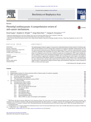
![406 P. Gupta et al. / Biochimica et Biophysica Acta 1846 (2014) 405–424
4.3. Reactiveoxygenspecies,akeymechanismofPEITC'santi-cancereffects. . . . . . . . . . . . . . . . . . . . . . . . . . . . . . 415
4.4. Direct protein modificationbyPEITC . . . . . . . . . . . . . . . . . . . . . . . . . . . . . . . . . . . . . . . . . . . . . . . . 419
4.5. Cancer specificbiomarkers . . . . . . . . . . . . . . . . . . . . . . . . . . . . . . . . . . . . . . . . . . . . . . . . . . . . 419
4.6. miRNAs . . . . . . . . . . . . . . . . . . . . . . . . . . . . . . . . . . . . . . . . . . . . . . . . . . . . . . . . . . . . . 419
4.7. Stemcells . . . . . . . . . . . . . . . . . . . . . . . . . . . . . . . . . . . . . . . . . . . . . . . . . . . . . . . . . . . . 419
4.8. Epigenetics . . . . . . . . . . . . . . . . . . . . . . . . . . . . . . . . . . . . . . . . . . . . . . . . . . . . . . . . . . . 419
5. CombinationtherapywithPEITC . . . . . . . . . . . . . . . . . . . . . . . . . . . . . . . . . . . . . . . . . . . . . . . . . . . . . 419
6. PEITCandtobaccosmokeinducedchanges . . . . . . . . . . . . . . . . . . . . . . . . . . . . . . . . . . . . . . . . . . . . . . . . 420
7. Clinicaladvancement . . . . . . . . . . . . . . . . . . . . . . . . . . . . . . . . . . . . . . . . . . . . . . . . . . . . . . . . . . 420
8. Summary. . . . . . . . . . . . . . . . . . . . . . . . . . . . . . . . . . . . . . . . . . . . . . . . . . . . . . . . . . . . . . . . 420
Acknowledgements . . . . . . . . . . . . . . . . . . . . . . . . . . . . . . . . . . . . . . . . . . . . . . . . . . . . . . . . . . . . . 420
References. . . . . . . . . . . . . . . . . . . . . . . . . . . . . . . . . . . . . . . . . . . . . . . . . . . . . . . . . . . . . . . . . . 420
1. Introduction
Since prehistoric times, nature has been a rich source ofmany drugs
and drug leads,which are currently being used to treat various ailments,
including cancer. According to Newman and Cragg, 74.8% of present an-ticancer
agents originated from natural sources, with almost 48.6%
being the actual natural compound or a direct derivative [1]. These in-clude
severalwidely used anticancer agents like taxanes, vinca alkaloids
and camptothecin class of compounds. Interest in natural products as an
alternative to synthetic drugs for cancer treatment is largely due to low
cost, established historical use in traditional medicinal systems, easy
availability and minimal or no toxicity. Also, the potential for targeting
multiple pathways represents an advantage of natural compounds
over synthetic agents, the latter being more target-specific.
Cancer is a complex manifestation of genomic instability in cells
caused by environmental or genetic factors [2]. Genomic instability
leads to disruption of basic biological functions, such as cell division, dif-ferentiation,
angiogenesis andmigration,which promote cancer [2]. The
involvement of multiple mechanisms in cancer development raises an
urgent need for multi-targeted therapy. The current anti-cancer agents
primarily focus on specific targets. The suppression of unique targets
usually leads to activation of compensatory mechanisms resulting in
failure of therapy or development of resistance to drugs. To circumvent
these problems, combination therapy is currently employed by the on-cologists.
Nonetheless, combination approach has also shown marginal
advantage in the overall therapy due to development of resistance or as-sociated
toxicity.We have reached a threshold in terms of clinical ben-efit
and tolerance in patients.
Of all the isothiocyanates (ITCs), phenethylisothiocyanate (PEITC) is
the only one that has reached the clinical stage of testing. This review
provides a critical and comprehensive analysis of the outcomes of stud-ies
showing the anti-cancer effects of PEITC.
2. Epidemiological evidence for chemo-preventive effects of dietary
intake of cruciferous vegetables
Several epidemiological studies from different parts of the world
provide a strong evidence for the reduced risk of cancer with higher in-take
of cruciferous vegetables [3–7]. Although none of these studies pro-vide
a direct correlation between cancer incidence and intake of a
specific ITC, many studies do identify a correlation between the total
ITC intake in the form of cruciferous vegetables and reduced risk of sev-eral
types of cancers in lung, breast, gastric, bladder, colorectal, pancre-atic,
prostate and kidney (Table 1) [5,8–27]. Interestingly, there are also
few studies where the results obtained were opposite to the preventive
effects of cruciferous vegetables. For example, depending on the dura-tion
of intake and age of subjects, one of the studies has shown differen-tial
effects of cruciferous vegetables [20]. In a prospective study,
Giovannucci et al. did not observe any significant correlation between
prostate cancer risk and a short-term intake of cruciferous vegetables
[20]. However, long-term intake of cruciferous diet showed a stronger
inverse correlation with initial stages of prostate cancer and this effect
was stronger in men less than 65 years of age [20]. In contrast, a few
other studies reported no correlation between the intake of cruciferous
vegetables and cancer incidence [27]. The differences in the outcome
showing no correlation with dietary intake of cruciferous vegetables
could be due to several confounding factors such as, differences in the
subject population, duration of intake of cruciferous vegetables in
subject's lifetime, age of subjects, variation in the isothiocyanate content
in cruciferous vegetables based on geographic locations and whether
the consumed vegetables were raw or cooked. Out of 22 studies per-formed,
one study showed increased risk of thyroid cancer with ITC in-take,
while three studies (urinary, prostate and breast cancers) showed
no correlation of ITC intake with cancer incidence. Interestingly all the
studies on stomach, colon and lung cancers showed reduced cancer in-cidence
with ITC intake. A detailed analysis revealed that studies show-ing
no correlation between ITC intake and cancer incidence typically
had wider trial durations or greater age differences for the subjects. In-terestingly,
a study performed in 2001 showed a significant change in
people’s food habits and lifestyle over the last decade [28]. Hence, the
outcomes from studies performed in the last decade might be affected
due to these confounding factors. Furthermore, themajority of the find-ings
were based on questionnaires for the intake of cruciferous vegeta-bles,
which can have lot of variability. In most of the studies,
questionnaires were not specific in asking whether the vegetables
were raw or cooked.
However, based on the majority of the studied outcomes, we can
conclude that an inverse relationship exists between intake of ITCs in
the form of cruciferous vegetables and the overall incidence of cancer.
But no conclusion can be drawn with respect to any specific isothiocya-nate
at this time.
3. PEITC source and pharmacokinetics
3.1. Cruciferous vegetables—source of PEITC
The isothiocyanates (R–N=C=S) (ITC) are known to be the major
bioactive compounds present in cruciferous vegetables and responsible
for anti-cancer activity. ITCs are released from glucosinolates by the ac-tion
of the enzyme myrosinase. The enzyme myrosinase can be activat-ed
by cutting or chewing the vegetables, but heating can destroy its
activity [29]. However, microbial myrosinase from gut can also release
ITCs in the stomach after ingestion of cruciferous vegetables [30,31].
Studies show that myrosinase as well as isothiocyanates are thermola-bile
[32]. Hence ingestion of only raw vegetables can release ITCs and
cooking of the vegetables can reduce ITC content [32]. Although water
cress and broccoli are known to be the richest source, PEITC can also
be obtained fromturnips and radish. PEITC is present as gluconasturtiin
in cruciferous plants. Like other ITCs, PEITC can be released from gluco-nasturtiin
by the action of myrosinase [30,31]. In a study conducted
with human volunteers, approximately 2 to 6 mg of PEITC was found
to be released by the consumption of one ounce of watercress [33,34].](data:image/gif;base64,R0lGODlhAQABAIAAAAAAAP///yH5BAEAAAAALAAAAAABAAEAAAIBRAA7)