2. 3160 J. Med. Plant. Res.
ancient times (Rahman, 2007). Medicinal actions of garlic
include lipid-lowering, antibacterial, antiviral, antifungal,
antihypertensive, blood glucose lowering, antithrombotic,
antioxidant and antiplatelet actions (Borrelli et al., 2007;
Reinhart et al., 2009; El-Khayat et al., 2010). Although
garlic is one of the popular food or herbal supplement in
Korea and USA (Kim and Kwon, 2009; Blumenthal et al.,
2006), some people express their discomfort due to
strong odor and flavor of garlic. Thermal processes are
commonly used in food manufacturing. One of the
important objectives of thermal processes is to raise the
sensory quality of foods, their palatability and to extend
the range of colors, tastes, aromas and textures in food
(Capuano and Fogliano, 2010).
Beside to these effects, heating processes leads to the
formation of biological compounds that are not originally
present in food. Recently, aged black garlic (ABG) is one
of garlic-processed product available on the market in
Korea (Choi et al., 2008; Lee et al., 2009). ABG is
produced by ageing raw garlic through thermal process at
high temperature and high humidity. During these
processing, unstable and odorous compounds of raw
garlic are converted to stable and odorless compounds S-
allylcysteine (SAC) or decomposed to organosulfur
compounds such as diallyl sulphide (DAS), diallyl
disulphide (DADS), diallyl trisulphide (DATS), dithiins and
ajoene (Amagase, 2006; Corzo-Martinez et al., 2007). It
is also reported that ABG contained antioxidant activity
and content of total polyphenol higher than fresh garlic
(Choi et al., 2008; Shin et al., 2008). Yet, to date, only
limited studies for beneficial effect of ABG is available.
Several studies have investigated that ABG exerted
hypoglycemic and hypocholsterolemic effects in animal
model of type 2 diabetes mellitus or in vitro antioxidant
effect on human low density lipoprotein (LDL) (Yang,
2007; Lee et al., 2009; Seo et al., 2009). Therefore, little
is known about the effect of ABG on regulation of body
weight, level of lipid parameters and antioxidant activity in
vivo animal model of obesity. For this reason, we investi-
gated whether the effects of ABG on the accumulation of
body fat, and the lipid levels and antioxidant activity in
serum and liver of rats fed a high-fat diet.
MATERIALS AND METHODS
Chemicals and reagents
Simvastatin (SIM) was purchased from Sigma Chemical Company
(St. Louis, USA). All other chemicals and reagent, unless noted
otherwise, were obtained from Invitrogen (USA) or Sigma (USA). All
the chemicals used were of the highest grade available.
Preparation of samples
Raw garlic was purchased from Uisung agricultural association
(Gyeongsangbuk-do, Republic of Korea) in 2009. The unpeeled
garlic packed in airtight containers, and was aged at 75°C and 50%
humidity level for 14 days in constant temperature and humidity
chamber. Aged black garlic were peeled off, mixed with 10 volumes
of water, and blended. The samples were extracted with water for 3
h at 100°C and concentrated in a rotary evaporation at 50°C. The
extracts were lyophilized to be used for the study.
3T3-L1 preadipocyte culture and differentiation
3T3-L1 preadipocytes were purchased from the Korean Cell Line
Bank (KCLB). 3T3-L1 cells were cultured in Dulbecco’s modified
Eagle’s medium (DMEM) containing 10% calf serum (CS) in 100
U/ml of penicillin and 100 mg/ml of streptomycin. Two days post
confluence was reached (D0), differentiation was induced by
changing the medium to DMEM supplemented with 10% fetal
bovine serum (FBS) plus 0.5 mM/L isobutylmethylxanthine (IBMX),
1 mg/ml insulin and 1 mM/L dexamethazone (DEX). After two days
(D2), the medium was changed to DMEM containing 10% FBS plus
1 mg/ml insulin. On day 4 (D4), the medium was replaced with
DMEM containing 10% FBS and repeated with changes of medium
every two days until day 8 (D8). The ABG extract was added to the
media on D0, which is the day of the initiation of differentiation. All of
cultures were incubated in humidified atmosphere of 5% CO2 at
37°C.
Oil red O staining
Oil red O Staining and quantification of lipid droplet in 3T3-L1
adipocytes were carried out using adipogeneis assay kit (Cayman,
USA). In brief, the cells were removed medium from the wells and
then washed with PBS. The cells were added fixative to each well
and incubated for 15 min at room temperature. The fixed cells were
rinsed with wash solution twice for 5 min each and then dried
completely. The well was added oil red O working solution and
incubated for 20 min. The stained cells were washed with dH2O 4
times and then added dye extraction solution to each well gently
mixing for 30 min. The absorbance was measured at 510 nm by
using microplate reader.
Experimental animals and design
Male Sprague-Dawley rats (5 weeks old) weighing 130 to 150 g
were obtained from Shizuoka Laboratory Center Inc. (Hamamatsu,
Japan) and acclimated for 1 week prior to experimental use. Sixty
rats were randomly divided into 2 groups: normal (n = 10) and high-
fat groups (n = 50). Normal group was fed the AIN-93G purified
rodent diet while high-fat groups were fed hyperlipidemic diet
containing 45% fat. The AIN-93 G purified diet contained 16% fat
(soybean oil), 20% protein and 64% carbohydrates, as energy
content. The high-fat diet contained 45% fat (soybean oil and lard),
20% protein and 35% carbohydrates, as energy content. The
animals fed high-fat diets were then randomly assigned to five
groups (n = 10/group): control, ABG extract-treated groups and
SIM-administered group. The treated group was orally given the
ABG extract with a multiple dose of 100, 250 and 500 mg/kg body
weight daily and SIM with a dose of 1 mg/kg body weight daily for 5
weeks. The normal and control group received the vehicle at the
same volume during the same period. For oral feeding of ABG
extract or vehicles, all groups were consumed ad libitum water and
AIN-93M diet. The animals were housed at 2 to 3 per plastic cage.
They were kept in a temperature-controlled environment animal
facility at 22 ± 2°C with a 12 h light/dark cycle. This experiment was
approved by the Animal Experiment Committee of National
Academy of Agricultural Science (NAAS) in Rural Development
Administration (Republic of Korea). All animals were maintained and
managed in accordance with NAAS animal use and care protocols.
3. Blood and tissue collection
After treatment period, animals were deprived of diets for at least 8
h. The rats were anesthetized by overexposure of CO2 gas and
exsanguinated through cardiac puncture. Blood samples were
rapidly centrifuged at 3,000 rpm for 15 min at 4°C. The isolated
serum was stored at -20°C before analysis. Hepatic and adipose
tissue were quickly removed, weighed, placed in ice-cold saline and
stored at -20°C for further studies.
Serum and hepatic lipids
Serum total cholesterol (TC) and triglyceride (TG) concentrations
were determined using commercial kits (Asan, Korea) according to
the instructions of the manufacturer. Total lipids from the liver were
extracted using the procedure developed by Folch et al. (1957). The
extracted samples were dried under nitrogen gas, resolved in 2-
propanol containing 10% Triton X-100 (w:w). Hepatic TC and TG
concentrations were analyzed with the same enzymatic kit used
serum analyses.
Measurement of GSH:GSSG ratio
Reduced glutathione (GSH):oxidized gluthathione (GSSG) ratio in
serum and liver were assayed using commercial kit (Calbiochem,
USA). The analysis was done according to the manufacturer’s
instructions.
Assay of lipid peroxidation (LPO) level
Thiobarnituric acid-reactive substance (TBARS) concentrations in
serum and liver were quantified spectrophotometrically. The levels
of TBARS were estimated using commercial kit (Cayman Chemical,
USA) according to the protocol of the manufacturer. Briefly, the 100
µl of serum was mixed with the same volume of sodium dodecyl
sulfate (SDS) solution and the mixture was swirled to mix. The
sample was mixed with thiobarbituric acid (TBA) reagent. The
mixture was boiled at 95°C for 60 min and then immediately cooled
in the ice bath. After 10 min, the sample was centrifuged at 4,000
rpm for 10 min at 4°C. The absorbance of the supernatants was
measured using a microplate reader (Molecular Devices, USA) at
535 nm. In liver, TBARS were assayed using 25 mg of hepatic
tissue homogenized in 250 µl of RIPA buffer with protease inhibitor
cocktail. The homogenates were centrifuged at 3,000 rpm for 10
min at 4°C. The measurement of TBARS level in hepatic tissue was
evaluated using the same method of that in serum, described above.
The TBARS levels were expressed as mM/mg protein.
Total protein determination
The protein content of the supernatant was determined using a
commercial protein assay kit (Bio-Rad Protein Assay Kit, Bio-Rad
Laboratories, Inc., CA, USA). Crystalline bovine serum albumin
(BSA) was used for the standard curve. The concentration was
measured using a spectrophotometer at 595 nm.
Statistical analysis
Data were expressed as means ± S.E. The differences between 2
groups were assessed by independent t-test. The results were also
analyzed for statistically significant experimental differences using
one-way analysis of variance (ANOVA) and post-hoc Duncan’s
multiple range tests. Differences were considered statistically
Kim et al. 3161
significant at p-value < 0.05. All statistical analyses were performed
using SPSS (version 12.0).
RESULTS
Inhibitive effect on ABG extract on 3T3-L1 adipocyte
differentiation
Firstly, we investigated the effect of the ABG extracts to
inhibit mature adipocyte differentiation in vitro. To assess
the degree of differentiation, Oil red O staining was
performed on day 8 (D8) of differentiation. The 3T3-L1
cells treated with AGE (Figure 1B, C, D) showed less
color intensity as compared to the non-treated cells
(Figure 1A). The Oil red O quantification showed the ABG
extracts treatment significantly inhibited differentiation and
decreased lipid cells (Figure 1E). At 10 µg/ml
concentration, lipid accumulation was significant
decreased to approximately 15% compared to the ABG
extracts non-treated group.
Effect of ABG extract on weight of body and white
adipose tissues
We verified whether the animals fed high-fat 45% diet
were definitely induced obesity and hypercholesterolemia
compared with rats fed normal diet for 5 weeks (Figure 2).
The body weight, serum total cholesterol and adipose
tissue weight of rats fed high-fat 45% diet for 5 weeks
were increased significantly higher than those of rats fed
the normal diet. After 5 weeks of administering vehicle,
ABG extract or SIM, the body weights were significantly
lower in the normal, ABG extract and SIM-treated groups
than in the control group (Table 1). The paratesticular
white adipose tissue (WAT) weights were significantly
lower in the SIM-treated group than in the control group.
The abdominal and perirenal WAT weights were shown
significant decreases in the normal and ABG extract-
treated groups compared to the control group. Therefore,
total WAT weights were significantly shown decreases in
the ABG extract and SIM-treated groups near to the level
of the normal group. However, the food intakes were not
significantly different among the groups.
Effect of ABG extract on lipid parameters of serum
and hepatic tissue
The concentrations of serum and hepatic lipids are shown
in Table 2. The serum TC concentration was not
significantly different among the groups. A significant
increase of serum TG concentration in the control group
was observed relative to the normal group. However, AB
G extract showed significant serum TG lowering effect
compared to the control and SIM-treated group. The
hepatic TC concentration in the control group was
4. 3162 J. Med. Plant. Res.
Figure 1. ABG extract inhibited preadipocyte differentiation in a dose-dependent manner. Fully
differentiated 3T3-L1 cells both non-treated (A) and treated (B, 1 µg/ml; C, 5 µg/ml; D, 10 µg/ml)
with ABG extract were stained with Oil red O staining solution. The absorbance at 510 nm from
3T3-L1 cells treated with different concentrations of ABG extract (E).
significantly increased higher than that in the normal
group while ABG extract or SIM-treated groups were
shown significant decreases near to the normal group.
The hepatic TG concentration in the control group was
also significantly higher than those of the normal group
while the administration of ABG extract or SIM resulted in
significant lower hepatic TG levels compared to that of the
control group.
Effect of ABG extract on GSH:GSSG ratio in serum
and hepatic tissue
The GSH:GSSG ratio in serum was significantly in the
control group lower than the normal group (normal group,
124.85 ± 1.42; control group, 107.32 ± 3.15) while that of
the ABG extract or SIM-treated groups (AGE100, 122.29
± 2.64; AGE 250, 128.68 ± 1.49; AGE 500, 124.29 ± 3.47;
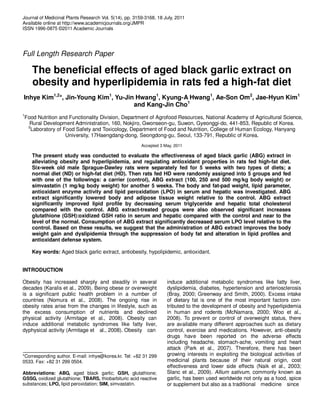
5. Kim et al. 3163
Figure 2. High-fat diet induced obesity and hypercholesterolemia in animals. The rats in
each group were fed normal or high-fat 45% diet for 5 weeks (A). Serum total cholesterol
level of normal and obese groups at 5-week were compared (B). Adipose tissue weight
was measured in visceral and epididymal fats (C). Data are represented as mean ± S.E.
for 10 rats per group. Normal, normal diet group; Obese, high-fat 45% diet group.
*Significant at P < 0.05 compared to the Normal group.
SIM, 125.81 ± 3.28) were shown significant increases
near to the normal group (Figure 3A). In liver, the
GSH:GSSG ratio was significantly in the control group
lower than the normal group (normal group, 221.52 ±
31.4; control group, 86.52 ± 3.43; Figure 3B). The
supplementations of ABG extract or SIM significantly
increased hepatic GSH:GSSG ratio compared to the
control group. Especially, 250 and 500 mg/kg of ABG
extract-administered groups were shown no significant
differences compared to the normal group (AGE 250,
6. 3164 J. Med. Plant. Res.
Table 1. Effects of ABG extract on body weight and white adipose tissue (WAT) weight.
Groups Body weight (g)
WAT weight (g/100 bw)
Food intake(g/d)
Paratesticular Abdorminal Perirenal Total WAT
Normal 515.8 ± 8.0
b
2.33 ± 0.10
ab
1.98 ± 0.09
b
3.65 ± 0.07
b
7.74 ± 0.13
b
30.1 ± 1.7
ns
Control 571.6 ±13.2a
2.84 ± 0.17a
2.52 ± 0.11a
4.02 ± 0.08a
9.25 ± 0.34a
31.9 ± 1.0
AGE 100 532.6 ± 9.1
b
2.31 ± 0.08
ab
2.08 ± 0.17
b
3.48 ± 0.07
b
7.74 ± 0.38
b
32.0 ± 1.2
AGE 250 527.7 ± 15.6
b
2.39 ± 0.08
ab
2.07 ± 0.10
b
3.39 ± 0.76
b
7.79 ± 0.23
b
31.0 ± 0.5
AGE 500 524.5 ± 12.2
b
2.42 ± 0.10
ab
2.11 ± 0.11
b
3.36 ± 0.11
b
7.95 ± 0.25
b
28.8 ± 0.9
SIM 530.0 ± 11.4
b
2.24 ± 0.30
b
2.28 ± 0.15
ab
4.11 ± 0.22
a
7.96 ± 0.61
b
30.6 ± 0.2
Values are represented as mean ± S.E. for 10 rats per group.
a,b
Values with different letters are significantly different among columns at
P-values < 0.05 by Duncan’s multiple range test.
ns
Not significant.
Table 2. Effects of supplementation of ABG extract on concentrations of serum and hepatic lipid
parameters.
Groups
Serum (mg/dl) Hepatic (mg/100 mg)
TC TG TC TG
Normal 128.5 ± 5.7
ns
150.0 ± 8.9
d
78.1 ± 3.8
b
148.4 ± 5.8
b
Control 119.3 ± 3.7 305.6 ± 9.4
a
118.9 ± 9.6
a
176.4 ± 9.4
a
AGE100 118.3 ± 3.3 186.4 ± 7.9
c
70.3 ± 5.4
b
154.0 ± 9.8
b
AGE250 128.5 ± 5.5 185.0 ± 8.5c
63.5 ± 3.7b
158.9 ± 9.0b
AGE500 125.5 ± 6.7 147.1 ± 7.3
d
75.3 ± 3.3
b
158.7 ± 5.2
b
SIM 130.9 ± 8.3 244.3 ± 9.1b
80.0 ± 7.4b
140.3 ± 9.1b
Values are represented as mean ± S.E. for 10 rats per group.
a,b,c,d
Values with different letters are
significantly different among columns at P-values < 0.05 by Duncan’s multiple range test.
ns
Not significant.
269.75 ± 22.47; AGE, 247.75 ± 20.62).
Effect of ABG extract on LPO in serum and hepatic
tissue
The serum TBARS level of the control group (45.33 ±
1.37 mM/mg protein) was significantly higher than those
of the normal group (21.08 ± 1.30 mM/mg protein, Figure
4A). However, the administration of ABG extract or SIM
(AGE 100, 27.22 ± 5.29 mM/mg protein; AGE 250, 22.98
± 2.38 mM/mg protein; AGE 500, 19.54 ± 1.12 mM/mg
protein; SIM, 27.44 ± 1.52 mM/mg protein) significantly
decreased serum TBARS levels compared with the
control group. There were no significant differences in
hepatic TBARS levels among the groups (Figure 4B).
DISCUSSION
Obesity has become a major public health problem
worldwide. The concerns associated with obesity are
related to numerous symptoms of metabolic syndrome,
including hypertension, dyslipidemia, insulin resistance
and glucose intolerance. A variety of approaches could be
used to prevent and control obesity in human. Since
perfect cure or prevention for obesity are yet to be found
and most of anti-obesity medications could have side
effects, there has been growing interests in investigating
the discovery of new naturally occurring materials
(Monteiro et al., 2008). Among these, extensive
researches carried out on garlic have reported the
biological effects, which include hypolipidemic (Ejaz et al.,
2003), hypocholesteroliemic (Asdaq et al., 2009) and anti-
obesity effect (Elkayam et al., 2003). Garlic contains a
high content of organosulfur components and flavonoids.
The major compounds in garlic that are known to contri-
bute to the potent biological activity are sulfur-containing
compounds. However, it has been known that allicin, the
major biologically active component of garlic, is absent
from garlic and garlic preparations because allicin is an
unstable and transient compound with oxidant activity
(Lawson et al., 1992; Lawson and Gardner, 2005). There-
fore, allicin is nearly undetectable in blood circulation after
garlic consumption due to decomposition into other
organosulfur compounds. The ABG is a type of garlic-
processed foods in the Korea, Thailand and Japan (Lee
el al., 2011). It is manufactured by ageing whole bulbs of
garlic at high temperature and high humidity.
During the ageing process, γ-glutamyl cysteine in intact
garlic bulbs is converted to water-soluble SAC, the major
unique organosulfur compound in ABG, which contributes
7. Kim et al. 3165
Figure 3. ABG extract recovered from reduction of the GSH:GSSG ratio by inducing high fat
diet for 5 weeks. The GSH:GSSG ratio was measured in serum (A) and liver (B). Data are
represented as mean ± S.E. for 10 rats per group. Normal, normal diet + orally vehicles; Control,
high-fat 45% diet group + orally vehicles; AGE100, high-fat 45% diet + orally 100 mg/kg bw of
ABG extract; AGE250, high-fat 45% diet + orally 250 mg/kg bw of ABG extract; AGE500, high-
fat 45% diet + orally 500 mg/kg bw of ABG extract; SIM, high-fat 45% diet + orally 1 mg/kg bw of
SIM. *Significant at P < 0.05 compared with among the groups. abc
Values with different letters
are significantly different among the groups at P-values<0.05 by Duncan’s multiple range tests.
to the health benefits (Imai et al., 1994; Amagase, 2006).
The phenolic compounds in ABG are also reported higher
than fresh garlic. It was demonstrated that the total
polyphenol content of ABG was approximately 10.0 mg/g
while raw garlic contained 3.67 mg/g (Jang et al., 2008).
In the present study, we tried to determine the possibility
the effect of ABG extract for 5 weeks on antiobesity,
control of lipid parameters and antioxidant activity in high-
fat diet induced obese rats. We measured the inhibitive
effect of ABG extract on mature adipocyte differentiation
of 3T3-L1 preadipocyte. The results of the present study
showed ABG extract inhibited 3T3-L1 preadipocyte
differentiation and fat accumulation compared with the
untreated cells. This is in agreement with Ambati et al.
(2009) who reported that ajoene, a sulfur-containing com-
pound of garlic, significantly decreased lipid accumulation
in maturing 3T3-L1 preadipocyte. A significant decrease
in final body weight was observed in the ABG extract and
SIM-administered groups compared with the control
group. In association with lower body weight, reduced
WAT weights in the abdominal and perirenal were
observed in the ABG extract-administered groups to the
level of the normal group although the food intake was
approximately the same for all groups. Previous studies
reported that ABG consumption at the level of 5% of the
diet did not influence body weight in db/db mice, type 2
diabetes mellitus animal model, during 7 weeks (Lee et
al., 2009; Seo et al., 2009). This finding is shown the
possibility that the body weight-lowering effect of ABG
extract on high-fat diet induced obese male rats. We
assume that these differences between the previous
studies and ours are caused by dose of ABG, routes of
8. 3166 J. Med. Plant. Res.
Figure 4. Effect of ABG extract on LPOs of rats. The LPO levels of the groups
were compared in serum (A) and liver (B). Data are represented as mean ± S.E.
for 10 rats per group. Normal, normal diet + orally vehicles; Control, high-fat 45%
diet group + orally vehicles; AGE100, high-fat 45% diet + orally 100 mg/kg bw of
ABG extract; AGE250, high-fat 45% diet + orally 250 mg/kg bw of ABG extract;
AGE500, high-fat 45% diet + orally 500 mg/kg bw of ABG extract; SIM, high-fat
45% diet + orally 1 mg/kg bw of SIM.*Significant at P < 0.05 compared with among
the groups.
administering ABG and animal model. We measured the
effect of ABG extract on serum and hepatic lipid profile. In
this study, consumption of ABG extract significantly
decreased serum TG, hepatic TC and TG compared with
the control group. However, Seo et al. (2009) reported
that ABG significantly decreased not only serum TC
concentration, but also serum TG concentration.
Pourkabir et al. (2010) also reported that garlic powder
supplementation at the level of 2.5 and 5% of the diet
improved serum TG, TC, VLDL-C and LDL-C levels in
hypercholesterolemic guinea pigs. Gorinstein et al. (2006)
reported that the garlic samples of raw and boiled at
100°C for 20 min significantly hindered the cholesterol-
induced increases in plasma TC and LDL-C but not
plasma TG. In agreement with our research results, Lee
et al. (2011) demonstrated that, by means of red garlic
extract (light color than black garlic) supply, the TC level
of serum did not show significant differences between
obese group (control) and the groups fed red garlic
extract while a significant decrease of TG level was
observed in the groups fed red garlic extract compared to
the control groups. These inconsistent results of re-
searches could be attributable, in part, to methodological
limitations of non-standardized extract used previous
studies. Further study is needed to characterize the
composition and standardize the ABG extract.
The superoxide anions including reactive oxygen
species (ROS) and reactive nitrogen species (RNS) are
generated in cells and tissues under increased oxidative
stress induced by obesity and hyperlipidemia (Furukawa
et al., 2004). The GSH is a central role to the cellular
antioxidant defense system and acts as an essential
cofactor for antioxidant enzymes such as glutathione
peroxidase (GPx). GSH is consumed by the glutathione
reductase (GR) to detoxify peroxides produced due to
increased lipid peroxidation (Kumar et al., 2011).
9. Therefore, GSH:GSSG ratio is a good indicator of
oxidative stress in cells and tissues. In this study, our
results were shown that a significant decrease in the
GSH:GSSG ratio was observed in the serum and hepatic
tissue of the control group compared to the normal group,
whereas the GSH:GSSG ratios in serum and hepatic
tissue of the ABG extract-administered groups were
comparable to the normal group and higher than the
control group. This suggested that the balance between
GSH and GSSG was maintained in the serum and liver
when ABG extract was administered under oxidative
stress status induced by high fat-diet. This is in agree-
ment with earlier reports showing that ABG could be more
effective in removing superoxide anions (Lee et al., 2009).
The antioxidant action of ABG could be responsible for
the higher content of the polyphenolic compounds and
SAC. It was reported that the total polyphenol content of
ABG was 3 times higher than that of raw garlic (Jang et
al., 2008). The measurement of TBARS concentration is
commonly used as an indicator of LPO process and
indirectly of oxidative stress (Beltowski et al., 2000). We
have observed a significant increase in the serum TBARS
level of the control group, and a decrease that of the ABG
extract-administrated groups to the normal group. Lee et
al. (2009) investigated the effect on TBARS level in db/db
mice, an animal model of type 2 diabetes mellitus, fed
ABG-contained diet. They observed that ABG
supplementation was effective in lowering hepatic TBARS
compared to the control and garlic groups. These findings
support our results that ABG triggers the decrease of LPO
under oxidative stress status such as obesity,
hyperlipidemia and diabetes mellitus.
In conclusion, our study showed evidences that the
administration of ABG extract is capable of ameliorating
weight control and lipid parameters, and restoring anti-
oxidant balance in high-fat diet induced obesity. Based on
the results obtained from present study, ABG extract may
play a role in reducing body fat, lowering hyperlipidemia
and protecting against oxidative stress in obese status.
However, further studies are needed to elucidate the
underlying mechanisms.
ACKNOWLEDGMENTS
This work was carried out with the support of
“Cooperative Research Program and Fundamental
Research Program for Agriculture Science and
Technology Development (Project No.PJ006706 and
PJ007714)” from the National Academy of Agricultural
Science and Rural Development Administration, Republic
of Korea.
REFERENCES
Ambati S, Yang JY, Rayalam S, Park HJ, Della FMA, Baile CA (2009).
Ajoene exerts potent effects in 3T3-L1 adipocytes by inhibiting
adipogenesis and inducing apoptosis. Phytother. Res., 23(4): 513-518.
Kim et al. 3167
Amagase H (2006). Clarifying the real bioactive constituents of garlic. J.
Nutr., 136: 716S-725S.
Armitage JA, Poston L, Taylor PD (2008). Developmental origins of
obesity and the metabolic syndrome: the role of maternal obesity.
Front. Horm. Res., 36: 73-84.
Asdaq SM, Inamdar MN, Asad M (2009). Effect of conventional
antihypertensive drugs on hypolipidemic action of garlic in rats. Indian
J. Exp. Biol., 47(3): 176-181.
Blumenthal M, Ferrier GKL, Cavaliere C (2006). Total sales of herbal
supplements in United States show steady growth. Herbalgram, 71:
64-66.
Borrelli F, Capasso R, Izzo AA (2007). Garlic (Allium sativum L.):
adverse effects and drug interactions in humans. Mol. Nutr. Food
Res., 51: 1386-1397.
Bray GA (2000). A concise review on the therapeutics of obesity.
Nutrition, 16: 953-960.
Beltowski J, Wójcicka G, Górny D, Marciniak A (2000). The effect of
dietary-induced obesity on lipid peroxidation, antioxidant enzymes
and total plasma antioxidant capacity. J. Physiol. Pharmacol., 51(4):
883-896.
Capuano E, Fogliano V (2010). Acrylamide and 5-hydroxymethylfurfu
ral (HMF): a review on metabolism, toxicity, occurrence in food and
mitigation strategies.
Choi DJ, Lee SJ, Kang MJ, Cho HS, Sung NJ, Shin JH (2008).
Physicochemical characteristics of black garlic (Allium sativum L.). J.
Korean. Soc. Food Sci. Nutr., 37(4): 465-471.
Corzo MM, Corzo N, Villamiel M (2007). Biological properties of onions
and garlic. Trends Food Sci. Technol., 18: 609-625.
Ejaz S, Woong LC, Ejaz A (2003). Extract of garlic (Allium sativum) in
cancer chemopreventions. Exp. Oncol., 24: 93-97.
Elkayam A, Mirelman D, Peleg E, Wilchek M, Miron T, Rabinkov A, Oron
HM, Rosenthal T (2003). The effects of allcin on weight in fructose-
induced hyperinsulinemic, hyperlipidemic, hypertensive rats. Am. J.
Hypertens., 16(12): 1053-1056.
EL KZ, Rasheed W, Ramzy T, Hussein J, Agaiby M, Morsy S, Morsy F,
Shaffie N (2010). Protective effect of garlic oil against liver injury in
experimental animals. J. Med. Plants Res., 4(22): 2359-2369.
Folch J, Lees M, Sloan SGH (1957). A simple method for isolation and
purification of total lipids from animal tissues. J. Biol. Chem., 226:
497-509.
Greenway FL, Smith SR (2000). The future of obesity research. Nutrition,
16: 976-982.
Furukawa S, Fujita T, Shimabukuro M, Iwaki M, Yamada Y, Nakajima Y,
Nakayama O, Makishima M, Matsuda M, Shimomura I (2004).
Increased oxidative stress in obesity and its impact on metabolic
syndrome. J. Clin. Invest., 114(12): 1752-1761.
Gorinstein S, Leontowicz M, Leontowicz H, Najman K, Namiesnik J,
Park YS, Jung ST, Kang SG, Trakhtenberg S (2006). Supplementation
of garlic lowers lipids and increases antioxidant capacity in plasma of
rats. Nutr. Res., 26: 362-368.
Imai J, Ide N, Nagae S, Moriguchi T, Matsuura H, Itakura Y (1994).
Antioxidants and free radical scavenging effects of aged garlic extract
and its constituents. Planta. Med., 60: 417-420.
Jang EK, Seo JH, Lee SP (2008). Physiological activity and
antioxidative effects of aged black garlic (Allium sativum L.) extract.
Korean. J. Food Sci. Technol., 40: 443-448.
Karalis KP, Giannogonas P, Kodela E, Koutmani Y, Zoumakis M, Teli T
(2009). Mechanisms of obesity and related pathology: linking immune
responses to metabolic stress. FEBS. J., 276: 5747-5754.
Kim JY, Kwon O (2009). Garlic intake and cancer risk: an analysis using
the food and drug administration’s evidence-based review system for
the scientific evaluation of health claims. Am. J. Clin. Nutr., 89: 257-
264.
Kumar S, Srivastava N, Gomes J (2011). The effect of lovastatin on
oxidative stress and antioxidant enzymes in hydrogen peroxide
intoxicated rat. Food Chem. Toxicol., doi:10.1016/j.fct.2010.12.014.
Lawson LD, Ransom DK, Hujgs BG (1992). Inhibition of whole blood
platelet aggregation by compounds in garlic clove extracts and
commercial garlic products. Thromb. Res., 65: 141-156.
Lawson LD, Gardner CD (2005). Composition, stability, and
bioavailability of garlic products being used in a clinical trial. J. Agric.
Food Chem., 53(16): 6254-6261.
10. 3168 J. Med. Plant. Res.
Lee YM, Gweon OC, Seo YJ, Im J, Kang MJ, Kim MJ, Kim JI (2009).
Antioxidant effect of garlic and aged black garlic in animal model of
type 2 diabetes mellitus. Nutr. Res. Pract., 3(2): 156-161.
McNamara DJ (2000). Dietary cholesterol and atherosclerosis. Biochim.
Biophys. Acta, 1529: 310-320.
Monteiro R, Assunção M, Andrade JP, Neves D, Calhau C, Azevedo I
(2008). Chronic green tea comsumption decreases body mass,
induces aromatase expression, and changes proliferation and
apoptosis in adult male rat adipose tissue. J. Nutr., 138: 2156-2163.
Naik GH, Priyadarsini KI, Satav JG, Banavalikar MM, Sohani DP, Biyani
MK, Mohan H (2003). Comparative antioxidant activity of individual
herbal components used in ayurvedic medicine. Phytochemistry, 63:
97-104.
Lee EN, Choi YW, Kim HK, Park JK, Kim HJ, Kim MJ, Lee HW, Kim KH,
Bae SS, Kim BS, Yoon S (2011). Chloroform extract of aged black
garlic attenuates TNF-α-induced ROS generation, VCAM-1
expression, NF-κB activation and adhesiveness for monocytes in
human umbilical vein endothelial cells. Phytother. Res., 25: 92-100.
Lee SJ, Kim RJ, Ryu JH, Shin JH, Kang MJ, Kim IS, Sung NJ (2011).
Effects of the red garlic extract for anti-obesity and hypolipidemic in
obese rats induced high fat diet. J. Life Sci., 21(2): 211-220.
Nomura S, Ichinose T, Jinde M, Kawashima Y, Tachiyashiki K,
Imaizumi K (2008). Tea catechins enhance the mRNA expression of
uncoupling protein 1 in rat brown adipose tissue. J. Nutr. Biochem.,
19: 840-847.
Park YS, Yoon Y, Ahn HS (2007). Platycodon grandiflorum extract
represses up-regulated adipocyte fatty acid binding protein triggered
by a high fat feeding Iobese rats. World Gastroenterol., 13(25): 3493-
3499.
Pourkabir M, Shomali T, Asadi F (2010). Alterations in serum lipid,
lipoprotein and visceral abdominal fat pad parameters of
hypercholeterolemic quinea pigs in response to short term garlic
comsumption. Afr. J. Biotechnol., 9(46): 7930-7933.
Rahman MS (2007). Allicin and other functional active components in
garlic: health benefits and bioavailability. Int. J. Food Prop., 10: 245-
268.
Reinhart KM, Talati R, White CM, Coleman CI (2009). The impact of
garlic on lipid parameters: a systematic review and meta-analysis.
Nutr. Res. Rev., 22: 39-48.
Seo YJ, Gweon OC, Im J, Lee YM, Kang MJ, Kim JI (2009). Effect of
garlic and aged blacl garlic on hyperglycemia and dyslipidemia in
animal model of type 2 diabetes mellitus. J. Food Sci. Nutr., 14: 1-7.
Shin JH, Choi DJ, Lee SJ, Cha JY, Sung NJ (2008). Antioxidant activity
of black garlic (Allium sativum L.). J. Korean Soc. Food Sci. Nutr.,
37(8): 965-971.
Slanc P, Doljak B, Kreft S, Lunder M, Janes D, Strukelj B (2009).
Screening of selected food and medicinal plant extracts for pancreatic
lipase inhibition. Phytother. Res., 23(6): 874-877.
Woo MN, Bok SH, Lee MK, Kim HJ, Jeon SM, Do GM, Shin SK, Ha TY,
Choi MS (2008). Anti-obesity and hypolipidemic effects of a
proprietary herb and fiber combination (S&S PWH) in rats fed high-fat
diets. J. Med. Food, 11(1): 169-178.
Yang ST (2007). Antioxidant activity of extracts of aged black garlic on
oxidation of human low density lipoprotein. J. Life Sci., 17(10): 1330-
1335.