1. Histological study of
Thyroid gland
- Asst Prof Chetana
.S.Kanekar
M.Sc Zoology( SET Life sciences)
2. Introduction
Thyroid consist of two
lateral lobes and the
connecting part called
isthmus.
Largest gland in body.
Endodermal in origin.
Gland secretes
thyroid hormones
called thyroxine.
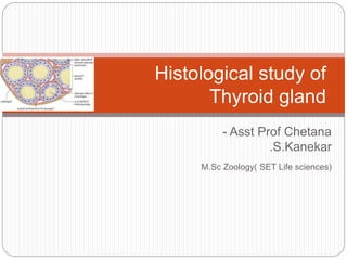
4. Microscopic structure
Structural unit is follicle
or acinus.
Follicle consist of layer
of simple epithelium,
enclosing cavity called
the follicular cavity.
The cavity is usually
filled with gel-like
viscous iodine-rich
material called colloid.
Interfollicular spaces
are filled by reticular
connnective tissue,
adipose tissue and
blood vessels.
5. Follicular cells
These are cuboidal epithelial cells with their basal
ends resting on basement membrane.
These cells show the changes in shape
depending on state of gland.
When gland is inactive, cells exhibit squamous
structure and columnar when hyperactive.
The follicular cells show central or basal round
nucleus with one or more excentric nuclei.
The apical tips of cells extend microvilli in the
cavity.
6. Parafollicular cells
In Interfollicular spaces spaces there are some
special parafollicular cells.
They found in singly or in groups.
They secrete hormone thyrocalcitonin, which
lowers the calcium level.
7. Colloid
Cavity of thyroid follicle is filled with semi-fluid or
gel like substance, called thyroid colloid.
It is the endocrine secretion of epithelial cells and
composed of nucleoproteins, thyroglubolin and
proteolytic enzymes.
Among the endocrine glands, thyroid is unique
because it utilizes an inorganic element iodine for
the synthesis of its hormones.
9. Distinguishing features of thyroid
Follicle is structural unit.
Follicle contains follicular epithelial cells and
parafollicular cells.
Follicle shows follicular cavity in which contains
gel-like colloid.
Inter-follicular spaces are filled by reticular
connective tissue and blood vessels.