SlideShare ist ein Scribd-Unternehmen logo
1 von 110
Common sports-related
shoulder injuries
S
houlder pain is commonly treated in general practice; its causes
are often
multi-factorial. The focus of this article is on sports-related
shoulder injuries
likely to be seen in the community. This article aims to
overview the presen-
tation, assessment and management of these conditions in
general practice.
The GP curriculum and common sports-related shoulder injuries
Clinical module 3.20: Care of people with musculoskeletal
problems lists the learning objectives required
for a GP to manage common sports-related shoulder injuries in
the community or refer for specialist management. In
particular, GPs are expected to be able to:
. Communicate health information effectively to promote better
outcomes
. Explore the perceptions, ideas or beliefs the patient has about
the condition and whether these may be acting as
barriers to recovery
. Use simple techniques and consistent advice to promote
activity in the presence of pain and stiffness
. Agree treatment goals and facilitate supported self-
management, particularly around pain, function and physical
activity
. Assess the importance and meaning of the following
presenting features:
. pain: nature, location, severity, history of trauma
. variation of symptoms over time
. loss of function – weakness, restricted movement, deformity
and disability, ability to perform usual work or
occupation
. Understand that reducing pain and disability rather than
achieving a complete cure could be the goal of
treatment
. Understand indications and limitations of plain radiography,
ultrasound, and magnetic resonance scans
. Diagnose common, regional soft-tissue problems that can be
managed in primary care
. Understand the challenge that many musculoskeletal
conditions might be better and more confidently managed
by other healthcare personnel rather than GPs, because most
GPs do not gain the necessary treatment skills
during their training
. Refer those conditions which may benefit from early referral
to an orthopaedic surgeon
The four most common categories of shoulder pain
seen in primary care are (Mitchell, Adebajo, Hay, &
Carr, 2005):
. Rotator cuff disorders (85% tendinopathy)
. Glenohumeral disorders
. Acromioclavicular joint disease, and
. Referred neck pain.
There are many different types of sports that can cause
acute or chronic shoulder injuries. In professional English
Rugby Union, for example, the most common match
injury is of the acromioclavicular joint (32% overall) and
the most severe injury requiring the longest time off
(mean of 81 days) is shoulder dislocation (Headey,
Brooks, & Kemp, 2007).
Shoulder injuries can also occur in non-contact sports,
such as golf, tennis, swimming and weightlifting.
Although shoulder injuries may be more common in con-
tact sports, the injury may have a larger impact on the
performance of individuals playing non-contact sports.
For example, golfers require very precise manoeuvres
of their dominant shoulder to swing a golf club with
accuracy...
..
..
..
..
..
..
..
..
..
..
..
..
..
..
..
..
..
..
..
..
..
..
..
..
! The Author(s) 2016. Reprints and permissions:
sagepub.co.uk/journalsPermissions.nav
30
...................
InnovAiT, 10(1), 30–38 DOI: 10.1177/1755738016678436
...............................................................................................
...............................................................................................
............
http://crossmark.crossref.org/dialog/?doi=10.1177%2F17557380
16678436&domain=pdf&date_stamp=2016-11-22
Anatomy
...........................................................
The shoulder complex (Fig. 1) consists of the
glenohumeral joint, the acromioclavicular joint and the
sternoclavicular joint. These work collectively with the
scapulothoracic joint to achieve normal shoulder girdle
movements.
The glenohumeral joint is a ball-and-socket joint that con-
sists of the glenoid cavity, glenoid labrum, glenohumeral
ligaments and the joint capsule. These components all
articulate with the head of the humerus and the scapula.
Four muscles form the rotator cuff that controls move-
ments of the glenohumeral joint. The deltoid muscle and
the muscles that control the scapula also provide move-
ment and stability of the shoulder. These muscles and
their primary actions are summarised in Table 1.
History
...........................................................
In general practice, a history of the shoulder problem is
probably your most helpful tool when trying to formulate
a diagnosis. Establish the nature, location, duration, and
the exact mechanisms of injury. When asking about the
shoulder pain, enquire about locking, clicking, catching,
popping, stiffness, swelling, redness, warmth, giving
way, night pain, weakness, and paraesthesia. A training
history to include frequency, intensity and any changes
to training routine may also be relevant.
Ask about any referred pain, and do not forget possible
gastrointestinal or cardiac causes of shoulder pain. Look
out for other more sinister features, such as night sweats,
fevers, weight loss and history of cancer.
Common shoulder
sports injuries
...........................................................
Rotator cuff disorders
Rotator cuff tendinopathy is an overuse condition
whereby the rotator cuff tendons at a microscopic level
demonstrate hypercellularity, neovascularisation and col-
lagen matrix disruption. The tendons are typically swol-
len, but have minimal or absent inflammation (Rees,
Maffulli, & Cook, 2009). They mainly occur in sports
that involve overhead movements of the shoulder and
patients may report a history of overuse, such as an
increase in training intensity for a competition.
Figure 1. The acromioclavicular joint and associated
structures.
Reproduced from Brukner P., Khan, K. (2012). Brukner &
Khan’s Clinical Sports Medicine (4th ed.), with permission from
McGraw-Hill Education. ..
..
..
..
..
..
..
..
..
..
..
..
..
..
..
..
..
..
..
..
..
..
..
..
..
..
..
..
..
..
..
..
..
..
..
..
..
..
..
..
..
..
..
..
..
..
..
..
..
..
..
..
..
..
..
..
..
..
..
..
..
..
..
..
..
..
..
..
..
..
..
..
..
..
..
..
..
..
..
..
..
..
..
..
..
..
..
..
..
..
..
..
..
..
..
..
..
..
..
..
..
..
..
..
..
..
..
..
..
..
..
..
..
..
..
..
..
..
..
..
..
.
Table 1. The primary action of the muscles
around the scapula and shoulder.
Shoulder muscles Primary actions
Rotator cuff muscles
Supraspinatus Abduction of humerus
Infraspinatus External rotation of humerus
Teres minor External rotation of humerus
Subscapularis Internal rotation of humerus
Other muscles
Deltoid Abduction, flexion and
extension of humerus
Trapezius Retracts, elevates, rotates and
stabilises scapula
Serratus anterior Protracts and laterally rotates
scapula
Rhomboids Retracts and medially rotates
scapula
Levator scapulae Elevates scapula
Pectoralis minor Protracts, medially rotates
scapula
31
....................
InnovAiT
..................................................................................... ..........
...............................................................................................
............
Patients typically present with a gradual onset dull shoul-
der pain exacerbated by overhead activities or reaching
above the shoulder level. They may get symptoms from
reaching behind the back and with lifting movements.
Night pain can also be a feature, and weakness can
develop in late stages (Factor & Dale, 2014).
The rotator cuff muscles or tendons can also tear, espe-
cially in older patients. Tears can be partial or full thick-
ness, and may present acutely or develop gradually.
Other symptoms include night pain, stiffness, weakness
and clicking sensations.
Glenoid labral injuries
The glenoid labrum is a ring of fibrocartilaginous tissue
attached to the rim of the glenoid cavity, it allows the
cavity size to be expanded and therefore provide more
stability to the joint (Brukner & Khan, 2012). The long
head of the biceps brachii tendon attaches to the labrum.
Patients with labral injuries present with pain usually in
the posterior aspect of the shoulder exacerbated by
overhead activities with associated features of shoulder
weakness, popping, catching, and grinding sensations.
Mechanisms of labral injuries include a single traumatic
event, such as a direct blow or fall onto the shoulder, a
forceful or excessive traction on the labrum, such as drop-
ping then catching a heavy object, or repeated micro-
trauma to the labrum, such as from cocking the shoulder
in throwing sports (Wilk et al., 2005). It is always important
to consider a diagnosis of labral injury, as early referral to a
shoulder specialist is indicated for best results.
Shoulder instability and dislocation
Shoulder instability refers to symptomatic laxity within
the glenohumeral joint that can be caused by over-
stretched or torn shoulder ligaments, muscles, tendons
or labrum. Dislocation occurs when the humeral head no
longer sits within the glenoid cavity (i.e. a complete dis-
ruption to the joint). A partial disruption to the joint is
termed subluxation.
The causes of subluxation or dislocation can be traumatic
or atraumatic. Traumatic dislocation is common in ath-
letes, and approximately 95% of first time shoulder dis-
locations result from a forceful collision, a fall onto an
outstretched arm or a sudden twisting movement: 98%
of traumatic dislocations occur in an anterior direction.
Approximately 5% of dislocations overall are atraumatic in
nature, and these individuals may have altered muscle
control of the shoulder, capsular laxity, or both (Hayes,
Callanan, Walton, Paxinos, & Murrell, 2002).
Patients present with anterior or posterior shoulder pain,
visible deformity, popping or catching sensations, weak-
ness, unstable feeling and night pains. They may have a
history of recurrent dislocations and be involved in
specific high-risk activities, for example, throwing a jav-
elin, being a pitcher in baseball or swimming.
Clavicle fracture
Clavicle fractures, particularly the mid-third of the clav-
icle, are the most common acute shoulder injuries and
account for one in twenty adult fractures. Fractures
located more laterally can disrupt the acromioclavicular
joint. Over 80% of clavicle fractures can be managed
conservatively (Quillen, Wuchner, & Hatch, 2004).
These injuries usually occur from a fall onto the clavicle
or, less frequently, a direct blow to the clavicle.
Patients may be involved with contact sports or other at-
risk sports such as horse-riding and cycling. They present
with acute localised pain with swelling and sometimes
visible deformity. Acute injuries are more likely to present
to the hospital Accident & Emergency Department than
primary care.
Acromioclavicular joint injury
Acute acromioclavicular joint (ACJ) injuries can occur due
to a direct force to the acromion typically with the shoul-
der adducted, or from an indirect force elsewhere in the
body, for example, a fall onto an outstretched arm
(Simovitch, Sanders, Ozbaydar, Lavery, & Warner,
2009). Patients present with acute localised pain, swelling
and sometimes redness. Injuries can range from a simple
acromioclavicular ligament sprain that can be managed
conservatively, to ligament tears with ACJ displacement
that often require surgery.
Chronic ACJ pain can occur following acute ACJ injuries
or from repeated irritation to the joint that can develop
into osteolysis or osteoarthritis. These chronic changes
can be caused by sports that involve throwing or lifting
weights. The symptoms will be similar to acute ACJ, but
the pain develops insidiously.
Biceps tendinopathy
The biceps brachii muscle has both a short head and a
long head. The short head originates from the coracoid
process of the scapula, whereas the long head originates
from the supraglenoid tubercle of the scapula where it
attaches to the superior aspect of the glenoid labrum.
The long head of the biceps (LHB) tendon exits the
glenohumeral joint and travels through the bicipital
groove between the greater and lesser tuberosities.
Biceps tendinopathy usually refers to an overuse injury of
the LHB tendon. It has a close relationship with the glenoid
labrum and the insertion sites of the rotator cuff tendons.
An accurate diagnosis of LHB tendinopathy can therefore
be difficult; and other shoulder injuries, such as rotator
cuff disease, labral lesions, and shoulder instability, may
co-exist (Krupp, Kevern, Gaines, Kotara, & Singleton,
2009). The typical features are gradual onset dull anterior..
..
..
..
..
..
..
..
..
..
..
..
..
..
..
..
..
..
..
..
..
..
..
..
..
..
..
..
..
..
..
..
..
..
..
..
..
..
..
..
..
..
..
..
..
..
..
..
..
..
..
..
..
..
..
..
..
..
..
..
..
..
..
..
..
..
..
..
..
..
..
..
..
..
..
..
..
..
..
..
..
..
..
..
..
..
..
..
..
..
..
..
..
..
..
..
..
..
..
..
..
..
..
..
..
..
..
..
..
..
..
..
..
..
..
..
..
..
..
..
..
..
32
...................
...............................................................................................
...............................................................................................
................................
shoulder pain in someone with risk factors for biceps over-
use such as weightlifters and bench pressers.
Examination
...........................................................
Have a system for examining joints. Several exist; how-
ever, the ‘Look, Feel, Move’ system is perhaps the sim-
plest. Start by inspecting the shoulder from the front,
side and back, looking for any swelling, asymmetry,
deformity, skin changes and scarring.
Palpate from the sternoclavicular joint, move laterally to the
ACJ, then across the spine of the scapula and along the
border of the scapula. It is also important to feel the corac-
oid process, humeral head, bicipital groove and surround-
ing shoulder musculature for any swelling or tenderness.
It is important to test the range of both passive and active
movements of the shoulder in all directions. Table 2 sum-
marises ‘normal’ range of shoulder movement (ROM), how-
ever, in reality, normal range varies between individuals and
it is therefore better to compare ROM with the unaffected
side for patients with unilateral shoulder injuries.
Special tests
...........................................................
BLISS is a helpful mnemonic to remember which condi-
tions to examine for using special tests. It stands for:
. B – Biceps tendon
. L – Labral
. I – ‘Impingement’
. S – ‘Scarf’ (referring to the ACJ), and
. S – Stability tests
A systematic review of 45 studies evaluating specific
shoulder tests, demonstrated that the diagnostic accuracy
for many of these tests is limited (Hegedus et al., 2008).
Therefore, special tests should only be used to provide
supporting evidence for or against a shoulder injury sus-
pected on the basis of history and general examination.
Biceps tendon tests
The Yergason’s test and Speed’s test are the two com-
monly used tests for biceps tendon pathology. They may
also be positive in labral lesions, as the LHB tendon is
attached to the labrum. They have low sensitivities (43%
and 32%, respectively), but are moderately specific (79%
and 75%, respectively). The positive predictive value
(PPV) of Yergason’s test and that of Speed’s test is 60%
and 50%, respectively, compared with shoulder arthros-
copy (Holtby & Razmjou, 2004).
Yergason’s test is performed with the patient’s elbow
flexed to the side at 90� with forearm pronated. The
patient is then asked to actively supinate the forearm
against the examiner’s resistance. A positive test will
elicit pain in the bicipital groove.
Speed’s test is performed with the patient’s elbow
extended and forearm supinated. The patient is then
asked to elevate (forward flex) their humerus against
the examiner’s resistance. A positive test will elicit pain
in the bicipital groove.
Labral pathology tests
There are a variety of special tests for labral lesions, but a
test called ‘Biceps Load 2’ (Fig. 2) has the highest sensi-
tivity (89.7%) and specificity (96.9%) with a PPV of 92.1%
(Kim, Ha, Ahn, & Choi, 2001). The test is performed
with the patient supine, arm abducted to 120� and max-
imally externally rotated with elbow flexed at 90� and
forearm supinated. The patient is then asked to actively
flex his or her elbow against the examiner’s resistance. In
a positive test, this manoeuvre should increase or repro-
duce the shoulder pain.
Impingement tests
Impingement itself is actually a syndrome or clinical sign
rather than a specific diagnosis. It is usually due to narrow-
ing of the subacromial space, and causes include rotator
cuff tendinopathy, rotator cuff calcification, bony spurs
under the acromion, and subacromial bursal pathology.
With impingement syndrome, a ‘painful arc’ can be
detected when assessing ROM for abduction whereby
pain occurs between 60� and 120� degrees of abduction
and eases above 120�. The Hawkins–Kennedy test and
Neer’s sign are two other tests for impingement that
have moderate sensitivities of 79%, and lower specifici-
ties of 59% and 53%, respectively (Hegedus et al., 2008).
Table 2. Types of shoulder movement and
their ’normal’ range of movement
Types of shoulder
movement
‘Normal’ range
of movement
(degrees)
Forward flexion 180
Extension 50
Abduction 180
Adduction 30
Internal rotation with elbows
at the side flexed at 90�
70
External rotation with elbows
at the side flexed at 90�
90
..
..
..
..
..
..
..
..
..
..
..
..
..
..
..
..
..
..
..
..
..
..
..
..
..
..
..
..
..
..
..
..
..
..
..
..
..
..
..
..
..
..
..
..
..
..
..
..
..
..
..
..
..
..
..
..
..
..
..
..
..
..
..
..
..
..
..
..
..
..
..
..
..
..
..
..
..
..
..
..
..
..
..
..
..
..
..
..
..
..
..
..
..
..
..
..
..
..
..
..
..
..
..
..
..
..
..
..
..
..
..
..
..
..
..
..
..
..
..
..
..
.
33
....................
InnovAiT
...............................................................................................
...............................................................................................
............
The Hawkins–Kennedy test is performed by the exam-
iner taking the patient’s arm into 90� of forward flexion
then flexing the patient’s elbow to 90� followed by pas-
sive internal rotation of the humerus. A positive test
occurs when symptoms are reproduced on passive inter-
nal rotation.
Neer’s sign is performed with the examiner stabilising the
patient’s scapula, internally rotating the patient’s arm,
and passively forward flexing the patient’s arm. A posi-
tive test occurs when symptoms are reproduced on pas-
sive forward flexion of the arm.
ACJ tests
The ‘Scarf’ test, also known as the cross-body adduction
test, has a sensitivity of 77%, but only a 20% PPV, for
chronic ACJ pathology (Chronopoulos, Kim, Park,
Ashenbrenner, & McFarland, 2004). The test is per-
formed with the patient’s arm and elbow flexed and
then forcibly adducted by the examiner across the
chest. A positive test will reproduce pain in the ACJ.
There is, however, a far simpler ACJ test. ACJ tenderness
on palpation has a sensitivity of 96% and a 52% PPV for
chronic ACJ pathology (Walton et al., 2004).
Stability tests
The ‘apprehension test’ for anterior shoulder instability
has shown sensitivities between 53 and 72% and specifi-
cities between 96 and 99% (Biederwolf, 2013). The test is
best performed with the patient supine with arm pos-
itioned in 90� abduction with external rotation. The
examiner gradually applies more external rotation while
observing the patient for apprehension (not just pain
alone). Care must be taken when performing this test,
as there is a risk of acutely dislocating an unstable
shoulder.
A test of postero-inferior instability can be performed with
the ‘Jerk’s test’ that has a sensitivity of 73% and specificity
of 98% (Biederwolf, 2013). The test is performed with the
patient’s arm abducted to 90� with internal rotation while
the examiner stabilises the patient’s scapula with one hand
and grasps the elbow with the other. The examiner then
proceeds to apply an axial load to the humerus while
horizontally adducting the patient’s arm across the body
(Fig. 3). A positive test is indicated by reproduction of
pain, or reproduction of a click or clunk.
Clinical examination
findings
...........................................................
The clinical signs for each of the common causes of
sports-related shoulder pain are summarised in Table 3.
It is important to understand that clinical signs may over-
lap in different conditions, as the structures within the
shoulder are so closely associated with each other. There
may also be more than one pathological process occur-
ring in the shoulder simultaneously...
..
..
..
..
..
..
..
..
..
..
..
..
..
..
..
..
..
..
..
..
..
..
..
..
..
..
..
..
..
..
..
..
..
..
..
..
..
..
..
..
..
..
..
..
..
..
..
..
..
..
..
..
..
..
..
..
..
..
..
..
..
..
..
Figure 2. Biceps load 2 test with patient lying supine.
34
...................
...............................................................................................
...............................................................................................
................................
Investigations
...........................................................
There are potentially three radiological investigations that
can be requested from general practice, although direct
access does vary from area to area in the UK. These are
X-rays, ultrasound scans, and magnetic resonance ima-
ging (MRI).
X-rays
In the context of sporting injuries, X-rays are primarily
indicated for detecting any bony fractures or disloca-
tions. If there is any suspicion of these injuries then the
patient needs to be referred for immediate X-ray or, if
this is not available, to the local emergency or minor
injuries unit.
For rotator cuff disorders, an X-ray is not indicated, but it
may reveal calcified tendons. X-ray can be helpful for
chronic ACJ injuries as it may show osteoarthritic changes
and osteolytic lesions suggestive of a stress fracture.
Ultrasound
A meta-analysis of five studies (311 shoulders) that used
an ultrasound scan (USS) to diagnose rotator cuff dis-
orders found a sensitivity level of 79% and a specificity
of 94% (Roy et al., 2015). Ultrasound scanning is, there-
fore, a very valuable investigation for rotator cuff
disorders.
An USS can also identify pathologies in and around the
subacromial space, which is useful if patients have
impingement signs. However, before requesting a shoul-
der USS, the age of the patient needs to be taken into
consideration, as the incidence of asymptomatic rotator
cuff tears increases after the age of 40 (Oschman, Janse
van Rensburg, Maritz, Boraine, & Owen, 2007).
MRI
MRI can be used to evaluate both soft tissue and bony
injuries in detail. It is unclear whether MRI is more accur-
ate at detecting rotator cuff tendinopathy compared with
USS, but both are equally effective in detecting partial or
full thickness tears (Gazzola & Bleakney, 2011). Labral
injuries can be detected with MRI, but a meta-analysis
revealed that magnetic resonance arthrography is super-
ior to MRI (Smith, Hilton, Toms, Donell, & Hing, 2011).
The cost implications of referring patients for a
shoulder MRI need to be carefully considered in general
practice, and onward referral to a specialist rather than
requesting a MRI maybe a better management option in
some cases.
Management
...........................................................
Rotator cuff disorder
In rotator cuff tendinopathy, it is important to counsel
patients about relative rest from the sport or the specific
activities that trigger the pain. Non-steroidal anti-inflam-
matory drugs (NSAIDs) and application of ice can pro-
vide symptomatic relief. Subacromial corticosteroid
injections do result in a reduction in pain symptoms,
but multiple injections over a short period of time and
injection into the tendon itself increases the risk of
tendon rupture (Gomoll, Katz, Warner, & Millett, 2004).
Referral to physiotherapy for rotator cuff muscle
strengthening and scapulohumeral work is recom-
mended for all patients. The treatment of a tear in a
rotator cuff is similar to rotator cuff tendinopathy, with
the exception that young sports-people with a full thick-
ness rotator cuff tear normally require referral for surgical
repair (Brukner & Khan, 2012).
Figure 3. Jerk’s test viewed from the side.
..
..
..
..
..
..
..
..
..
..
..
..
..
..
..
..
..
..
..
..
..
..
..
..
..
..
..
..
..
..
..
..
..
..
..
..
..
..
..
..
..
..
..
..
..
..
..
..
..
..
..
..
..
..
..
..
..
..
..
..
..
..
..
..
..
..
..
..
..
..
..
..
..
..
..
35
....................
InnovAiT
...............................................................................................
...............................................................................................
............
Glenoid labral injuries
Conservative management of glenoid labral injuries is usu-
ally unsuccessful (Brukner & Khan, 2012). These injuries
need to be referred to an orthopaedic shoulder specialist
for an arthroscopic repair (reattachment) of the labrum, or,
if the labral injury is stable, arthroscopic debridement. Post-
operative physiotherapy input is needed to gradually
rehabilitate and restore normal shoulder function.
Shoulder instability and dislocation
In non-acute atraumatic shoulder instability, physiother-
apy referral is required to provide progressive scapular
stabilisation exercises, strengthen the rotator cuff mus-
cles, and control the glenohumeral translation (Hayes
et al., 2002). If physiotherapy is unsuccessful, then cap-
sular surgery can be considered.
In acute or traumatic shoulder dislocation, the patient
should be referred to the local Accident & Emergency
Department for diagnostic X-rays, reduction of the shoul-
der with appropriate analgesia, and post-reduction X-rays.
Clavicle fractures
Patients with a clavicle fracture need analgesia and
prompt referral to the Accident & Emergency
Department for X-rays. If there is low risk of clavicle
foreshortening then a broad arm sling (or figure-of-
eight bandage) is usually applied, and the fracture grad-
ually heals over a month or so. Surgical intervention is
indicated if there is a risk of clavicle foreshortening,
delayed union, or non-union.
ACJ injuries
In acute ACJ injuries, patients should be given analgesia
and a sling for immobilisation if available. If an ACJ sprain
is suspected, then it may be reasonable to treat with ice,
analgesia, and immobilisation followed by a review 48
hours after the acute episode. If the ACJ appears
deformed, the pain is difficult to control, or there is uncer-
tainty about the degree of ACJ injury, then referral to the
Accident & Emergency Department or to the on-call
Table 3. Sports-related shoulder conditions
and their possible clinical signs.
Condition Possible clinical signs
Rotator cuff
tendinopathy
. Asymmetry and muscle wasting
. Palpation tenderness at the
greater tubercle (insertion sites of
three rotator cuff muscles)
. Reduced active ROM
. Passive ROM intact
. Reduced power on resisted
movements
. Impingement signs
Rotator cuff
tears
. Shoulder shrug appearance
. Partial or no active ROM
. Passive ROM often intact
. Reduced power on resisted
movements
. Drop-arm sign in complete tears if
arm cannot be actively maintained
at 90� abduction
. Impingement signs
Glenoid
labral injury
. Palpation tenderness in the
anterior shoulder structures
. Swelling if acute
. Reduced external rotation and or
abduction ROM
. Reduced power on resisted
movements
. Biceps load 2 test positive if
superior labrum anterior posterior
(SLAP) tear
. Speed’s test positive if SLAP
. Yergason’s test positive if SLAP
. Jerk’s test positive if postero-
inferior labral injury
Shoulder
instability
and
dislocation
. Prominent humeral head if anter-
ior dislocation
. Swelling if acute
. Reduced active and passive ROM
if dislocated
. Increased active or passive ROM
if instability with laxity
. Apprehension test positive if
anterior dislocation
. Jerk’s test positive if postero-
inferior dislocation
. Upper arm axillary nerve sensa-
tion can be reduced in anterior
dislocations
Clavicle
fracture
. Localised swelling with deformity
. Clavicle shortened or angulated
. Localised bony tenderness
(continued)
Table 3. Continued.
Condition Possible clinical signs
ACJ injury . Step deformity and swelling if
acute
. Localised ACJ tenderness
. Scarf test positive
. Impingement signs if chronic
Biceps
tendinopathy
. Bicipital groove tenderness
. Speed’s test positive
. Yergason’s test positive
..
..
..
..
..
..
..
..
..
..
..
..
..
..
..
..
..
..
..
..
..
..
..
..
..
..
..
..
..
..
..
..
..
..
..
..
..
..
..
..
..
..
..
..
..
..
..
..
..
..
..
..
..
..
..
..
..
..
..
..
..
..
..
..
..
..
..
..
..
..
..
..
..
..
..
..
..
..
..
..
..
..
..
..
..
..
..
..
..
..
..
..
..
..
..
..
..
..
..
..
..
..
..
..
..
..
..
..
..
..
..
..
..
..
..
..
..
..
..
..
..
..
36
...................
...............................................................................................
...............................................................................................
................................
orthopaedic team is appropriate. Surgery is indicated for
tears in both the acromioclavicular and coracoclavicular
ligaments with displacement of the clavicle (Tauber, 2013).
The management of chronic ACJ injuries requires relative
rest and activity modification, such as reducing the
weight load when bench pressing. NSAIDs and cortico-
steroid injections into the ACJ can have therapeutic
benefits, particularly in younger patients (Hossain,
Ayekoloye, Odumala, & Jacobs, 2003). Referral to
physiotherapy for range of motion and strength training
can aid recovery. In chronic persistent ACJ pain, ortho-
paedic referral for consideration of surgical excision of
the distal clavicle may be necessary.
Biceps tendinopathy
Initial treatment of biceps tendinopathy involves a period
of relative rest, withdrawal from exacerbating activities,
ice and NSAIDs. The next step is referral for physiother-
apy rehabilitation that involves restoration of passive
biceps ROM, followed by active biceps ROM exercises,
and then finally rotator cuff strengthening. If symptoms
fail to respond after 6–8 weeks of conservative measures
then corticosteroid injections into the subacromial space
can be beneficial, and in resistant cases, injections into
the biceps tendon sheath can be tried (Krupp et al.,
2009). There are a variety of surgical options for biceps
tendinopathy, such as decompression, debridement, ten-
otomy and tendon transfer, but choice of procedure is a
specialist decision.
Specialist referral
...........................................................
Patients presenting acutely with sports-related injuries
who have severe pain, and/or symptoms in keeping
with acute bony injury or joint dislocation should be
referred for immediate Accident & Emergency
Department review.
Most other sports-related shoulder injuries can be man-
aged in primary care with rest, ice, analgesia, physiother-
apy and possible local steroid injection. Specialist sports
physiotherapy input can be helpful if available and may
be particularly useful in helping patients to avoid repeat
injury when they return to their sport.
Consider specialist orthopaedic referral if:
. No clear cause of the patient’s pain can be found –
this referral should be urgent if the patient has a past
history of cancer, the pain is worsening, or the pain is
associated with any constitutional symptoms such as
night sweats or raised erythrocyte sedimentation rate
. Pain is not resolving despite conservative measures in
primary care
. The patient is young (under 40 years) and is found to
have a full thickness rotator cuff tear on imaging
. The patient has a glenoid labral tear
Key points
. Sports-related shoulder injuries are common and
can cause both acute and chronic symptoms
. A concise history and a targeted examination is
essential for initial diagnosis of sports-related
shoulder injuries in primary care
. Special tests in the examination of sports-related
shoulder injuries are useful only to provide sup-
porting evidence for a specific suspected injury
. X-ray, USS and MRI may all be useful in further
evaluation of patients with sports-related shoulder
pain depending on local availability
. The majority of patients with sports-related shoul-
der injuries can be managed in primary care; some
acute injuries will require prompt referral to the
Accident and Emergency Department for further
imaging and immediate management
. Refer to an orthopaedic shoulder specialist if the
diagnosis is unclear, the symptoms fail to settle
with primary care management, if the patient is
young and has a full-thickness rotator cuff tear,
or if the patient has a glenoid labral tear
References and further information
. Biederwolf, N. E. (2013). A proposed evidence-
based shoulder special testing examination algo-
rithm: Clinical utility based on a systematic
review of the literature. International Journal of
Sports Physical Therapy, 8(4), 427–440.
Retrieved from www.ncbi.nlm.nih.gov/pmc/arti
cles/PMC3812837/
. Brukner, P., & Khan, K. (2012). Brukner & Khan’s
clinical sports medicine, fourth edition. New York:
McGraw-Hill Medical
. Chronopoulos, E., Kim, T. K., Park, H. B.,
Ashenbrenner, D., & McFarland, E. G. (2004).
Diagnostic value of physical tests for isolated
chronic acromioclavicular lesions. American
Journal of Sports Medicine, 32(3), 655–661. doi:
10.1177/0363546503261723
. Factor, D., & Dale, B. (2014). Current concepts of
rotator cuff tendinopathy. International Journal of
Sports Physical Therapy, 9(2), 274–288. Retrieved
from www.ncbi.nlm.nih.gov/pmc/articles/
PMC4004132/
. Gazzola, S., & Bleakney, R. R. (2011). Current ima-
ging of the rotator cuff. Sports Medicine and
Arthroscopy Review, 19(3), 300–309. doi:
10.1097/JSA.0b013e3182189468
. Gladstone, J. N., & Rosen, A. L. (1999). Disorders
of the acromioclavicular joint. Current Opinion in
Orthopaedics, 10(4), 316–321. doi: 10.1097/
00001433-199908000-00011
. Gomoll, A. H., Katz, J. N., Warner, J. J., & Millett, P.
J. (2004). Rotator cuff disorders: Recognition and
management among patients with shoulder pain.
Arthritis and Rheumatism, 50(12), 3751–3761. doi:
10.1002/art.20668..
..
..
..
..
..
..
..
..
..
..
..
..
..
..
..
..
..
..
..
..
..
..
..
..
..
..
..
..
..
..
..
..
..
..
..
..
..
..
..
..
..
..
..
..
..
..
..
..
..
..
..
..
..
..
..
..
..
..
..
..
..
..
..
..
..
..
..
..
..
..
..
..
..
..
..
..
..
..
..
..
..
..
..
..
..
..
..
..
..
..
..
..
..
..
..
..
..
..
..
..
..
..
..
..
..
..
..
..
..
..
..
..
..
..
..
..
..
..
..
..
.
37
....................
InnovAiT
...............................................................................................
...............................................................................................
............
www.ncbi.nlm.nih.gov/pmc/articles/PMC3812837/
www.ncbi.nlm.nih.gov/pmc/articles/PMC4004132/
. Hayes, K., Callanan, M., Walton, J., Paxinos, A., &
Murrell, G. A. (2002). Shoulder instability:
Management and rehabilitation. Journal of
Orthopaedic Sports Physical Therapy, 32(10),
497–509. doi: 10.2519/jospt.2002.32.10.497
. Headey, J., Brooks, J. H., & Kemp, S. P. (2007).
The epidemiology of shoulder injuries in English
professional rugby union. American Journal of
Sports Medicine, 35(9), 1537–1543. doi:
10.1177/0363546507300691
. Hegedus, E. J., Goode, A., Campbell, S., Morin,
A., Tamaddoni, M., Moorman, C. T., & Cook, C.
(2008). Physical examination tests of the shoulder:
A systematic review with meta-analysis of individ-
ual tests. British Journal of Sports Medicine, 42(2),
80–92. discussion p. 92. doi: 10.1136/bjsm.2007.
038406
. Holtby, R., & Razmjou, H. (2004). Accuracy of the
Speed’s and Yergason’s tests in detecting biceps
pathology and SLAP lesions: Comparison with
arthroscopic findings. Arthroscopy, 20(3),
231–236. doi: 10.1016/j.arthro.2004.01.008
. Hossain, S., Ayekoloye, C., Odumala, O., & Jacobs,
L. (2003). Intra-articular steroid injection in the
treatment of primary acromioclavicular arthritis.
An assessment of therapeutic effectiveness.
Orthopaedic Proceedings, 85B, 72. Retrieved
from www.bjjprocs.boneandjoint.org.uk/content/
85-B/SUPP_I/72.4
. Kim, S. H., Ha, K. I., Ahn, J. H., & Choi, H. J.
(2001). Biceps load test II: A clinical test for
SLAP lesions of the shoulder. Arthroscopy, 17(2),
160–164. doi: B10.1053/jars.2001.20665
. Krupp, R. J., Kevern, M. A., Gaines, M. D., Kotara,
S., & Singleton, S. B. (2009). Long head of the
biceps tendon pain: Differential diagnosis and
treatment. Journal of Orthopaedic Sports Physical
Therapy, 39(2), 55–70. doi: 10.2519/
jospt.2009.2802
. Mitchell, C., Adebajo, A., Hay, E., & Carr, A.
(2005). Shoulder pain: Diagnosis and management
in primary care. British Medical Journal, 331(7525),
1124–1128. doi: 10.1136/bmj.331.7525.1124
. Oschman, Z., Janse van Rensburg, C., Maritz, N.,
Borain, H., & Owen, R. (2007). Ultrasound study of
the asymptomatic shoulder in patients with a con-
firmed rotator cuff tear in the opposite shoulder.
South African Journal of Sports Medicine, 19(1),
23–28. doi: 10.17159/2413-3108/2007/v19i1a272
. Quillen, D. M., Wuchner, M., & Hatch, R. L.
(2004). Acute shoulder injuries. American Family
Physician, 70(10), 1947–1954. Retrieved from:
www.aafp.org/afp/2004/1115/p1947.html
. RCGP. Clinical module 3.20: Care of people with
musculoskeletal problems. Retrieved from www.
rcgp.org.uk/training-exams/gp-curriculum-over-
view/online-curriculum/applying-clinical-knowl-
edge-section-2/3-20-musculoskeletal-problems.
aspx
. Rees, J. D., Maffulli, N., & Cook, J. (2009).
Management of tendinopathy. American Journal
of Sports Medicine, 37(9), 1855–1867. doi:
10.1177/0363546508324283
. Roy, J. S., Braën, C., Leblond, J., Desmeules, F.,
Dionne, C. E., MacDermid, J. C., . . . Frémont, P.
(2015). Diagnostic accuracy of ultrasonography,
MRI and MR arthrography in the characterisation
of rotator cuff disorders: A systematic review and
meta-analysis. British Journal of Sports Medicine,
49(20), 1316–1328. doi: 10.1136/bjsports-2014-
094148
. Simovitch, R., Sanders, B., Ozbaydar, M., Lavery,
K., & Warner, J. J. (2009). Acromioclavicular joint
injuries: Diagnosis and management. Journal of the
American Academy of Orthopaedic Surgeons,
17(4), 207–219. doi: 10.5435/00124635-
200904000-00002
. Smith, T. O., Hilton, G., Toms, A. P., Donell, S. T.,
& Hing, C. B. (2011). The diagnostic accuracy of
acetabular labral tears using magnetic resonance
imaging and magnetic resonance arthrography: A
meta-analysis. European Radiology, 21(4),
863–874. doi: 10.1007/s00330-010-1956-7
. Tauber, M. (2013). Management of acute acromio-
clavicular joint dislocations: Current concepts.
Archives of Orthopaedic and Trauma Surgery,
7(133), 985–995. doi: 10.1007/s00402-013-1748-z
. Walton, J., Mahajan, S., Paxinos, A., Marshall, J.,
Bryant, C., Shnier, R., & Murrell, G. A. (2004).
Diagnostic values of tests for acromioclavicular
joint pain. Journal of Bone and Joint Surgery.
American volume, 86A(4), 807–812. Retrieved
from www.jbjs.org/content/86/4/807
. Wilk, K. E., Reinold, M. M., Dugas, J. R., Arrigo, C.
A., Moser, M. W., & Andrews, J. R. (2005).
Current concepts in the recognition and treatment
of superior labral (SLAP) lesions. Journal of
Orthopaedic Sports Physical Therapy, 35(5),
273–291. doi: 10.2519/jospt.2005.35.5.273
Dr Raymond Leung
Freelance Locum GP, Sutton United Football Club Doctor,
MSc student in Sports and Exercise Medicine, London
Email: [email protected]
..
..
..
..
..
..
..
..
..
..
..
..
..
..
..
..
..
..
..
..
..
..
..
..
..
..
..
..
..
..
..
..
..
..
..
..
..
..
..
..
..
..
..
..
..
..
..
..
..
..
..
..
..
..
..
..
..
..
..
..
..
..
..
..
..
..
..
..
..
..
..
..
..
..
..
..
..
..
..
..
..
..
..
..
..
..
..
..
..
..
..
..
..
..
..
..
..
..
..
..
..
..
..
..
.
38
...................
...............................................................................................
...............................................................................................
................................
www.bjjprocs.boneandjoint.org.uk/content/85-B/SUPP_I/72.4
www.aafp.org/afp/2004/1115/p1947.html
www.rcgp.org.uk/training-exams/gp-curriculum-
overview/online-curriculum/applying-clinical-knowledge-
section-2/3-20-musculoskeletal-problems.aspx
www.rcgp.org.uk/training-exams/gp-curriculum-
overview/online-curriculum/applying-clinical-knowledge-
section-2/3-20-musculoskeletal-problems.aspx
www.rcgp.org.uk/training-exams/gp-curriculum-
overview/online-curriculum/applying-clinical-knowledge-
section-2/3-20-musculoskeletal-problems.aspx
www.rcgp.org.uk/training-exams/gp-curriculum-
overview/online-curriculum/applying-clinical-knowledge-
section-2/3-20-musculoskeletal-problems.aspx
www.rcgp.org.uk/training-exams/gp-curriculum-
overview/online-curriculum/applying-clinical-knowledge-
section-2/3-20-musculoskeletal-problems.aspx
www.jbjs.org/content/86/4/807
Review article
Obere Extremität 2018 · 13:89–97
https://doi.org/10.1007/s11678-018-0449-1
Received: 30 November 2017
Accepted: 29 January 2018
Published online: 19 February 2018
© The Author(s) 2018. This article is an open
access publication.
Jonas Pogorzelski1,2 · Erik M. Fritz1 · Jonathan A. Godin1,3 ·
Andreas B. Imhoff2 ·
Peter J. Millett1,3
1 Steadman Philippon Research Institute, Vail, USA
2 Department of Orthopedic Sports Medicine, Technical
University of Munich, Klinikum rechts der Isar,
Munich, Germany
3 The Steadman Clinic, Vail, USA
Nonoperative treatment of five
common shoulder injuries
A critical analysis
Introduction
Shoulderpainisoneofthemostcommon
musculoskeletal complaints accounting
for at least 4.5 million patient visits an-
nually in the United States [43, 55] and
occurring in as many as 51% of indi-
viduals in a lifetime [64]. Moreover, the
economic burden of shoulder pathology
is vast with annual direct costs for treat-
ment of shoulder dysfunction totaling
at least $7 billion in the United States,
mostly due to operative treatment [47].
InGermanythepercentageofaffectedpa-
tients and associated costs are expected
to be similar. Moreover, with an aging
and increasingly active patient popula-
tion in the Western world, the absolute
number of shoulder pathologies is likely
to grow, further increasing costs.
These economic implications high-
light the critical need for appropriate
diagnosisandtreatmentofvariousshoul-
der pathologies, as under-diagnosis and
under-treatment can result in increased
costs to society with disability and lost
production. On the other hand, aggres-
sive over-treatment can further inflate
already burgeoning health-care costs and
potentially harm the patient.
Therefore, the purpose of this review
is to distinguish the indications between
operative and nonoperative management
for five common shoulder pathologies,
ResearchperformedattheSteadmanPhilippon
ResearchInstitute,Vail,CO,USAandtheDepart-
ment of Orthopedic Sports Medicine,Technical
UniversityofMunich,Munich,Germany.
including rotator cuff tears, anterior
shoulder instability, biceps tendinitis,
lesions to the acromioclavicular (AC)
joint, and proximal humeral fractures.
Moreover, we aim to provide a short
overview of the nonoperative manage-
ment of each of these pathologies.
Rotator cuff tears
Indications for nonoperative
treatment of symptomatic full-
thickness rotator cuff tears
Although symptomatic rotator cuff tears
are common and affect between 4% and
32% of the general population, the most
appropriate therapy is still debatable
[59, 75]. While there is agreement that
traumatic rotator cuff tears should be
treated operatively, the treatment choice
for atraumatic rotator cuff tears remains
unclear [38, 39]. This is mainly due
to the fact that the radiological failure
rate following rotator cuff repair surgery
can be as high as 70% depending on
the patient cohort, thus leading to the
assumption that nonoperative treatment
may be equivalent [5, 8, 24, 41]. This
conjecture is further strengthened by the
fact that pain relief and improvement of
symptoms do not necessarily go hand
in hand with structural healing of the
tendon [59].
However, when taking a closer look
at published outcomes in the literature,
nonsurgical treatment appears to have
limitations. While multiple studies with
short-term follow-up of nonsurgical
treatment show promising results with
good clinical outcomes, studies with
mid-term follow-up are more disillu-
sioning [10, 22, 38, 39, 50]. This could
be explained by the fact that smaller
tears may not affect the force couples in
the shoulder, thus a reasonable degree
of shoulder function may be maintained
[42]. As there is strong evidence that
the natural history of nonoperatively
treated rotator cuff tears leads to tear
progression over time, nonoperative
outcomes studies with longer follow-up
may include more patients whose tears
have progressed to the point of destroyed
force couples [80].
Kukkonen et al. [38, 39] published
a randomized controlled trial for the
treatment of supraspinatus tendon tears
in patients older than 55 years. A total of
180 shoulders with supraspinatus tendon
tears were randomly allocated into one
of three treatment groups:
1. Isolated physiotherapy
2. Acromioplasty and physiotherapy
3. Rotator cuff repair with acromio-
plasty and physiotherapy
After 1 year of follow-up, no statistically
significant differences in outcomes were
detected, thus leading to the conclusion
that surgical therapy is not superior in
these patients [38]. Later, with an addi-
tional year of follow-up, the groups still
did not differ significantly in outcomes;
however, tear progression measured with
magnetic resonance imaging (MRI) sug-
gested thatonlypatients withlowerphys-
ical demands should be treated nonoper-
Obere Extremität 2 · 2018 89
https://doi.org/10.1007/s11678-018-0449-1
http://crossmark.crossref.org/dialog/?doi=10.1007/s11678-018-
0449-1&domain=pdf
http://orcid.org/0000-0002-6178-4940
Review article
Fig. 1 8 Axial T2-weightedmagnetic res-
onance imaging sequence of a 36-year-old
patient aftera first-timeshoulderdislocation.
Givenhisageandtheabsenceofanyrotatorcuff
tearorotherconcomitant pathology,he was
deemedlow risk forre-dislocation.Therefore,
nonoperative treatment was pursued,which
was successful withno recurrent subluxation or
dislocation
atively and patient counseling is critical
[39].
In another randomized controlled
trial of 103 patients, which compared
rotator cuff repair with nonoperative
physiotherapy for tears not exceeding
3cm, Moosmayer et al. [50] found sev-
eral additional factors that may influence
the outcome. With a minimum follow-
up of 5 years, the results for the group
of patients who had immediate tendon
repair were generally superior to those of
patients who underwent physiotherapy
as primary treatment and decided later
to progress with surgery. Furthermore,
treatment failed in almost 24% of the
patients who received physiotherapy as
primary therapy, and they underwent
subsequent rotator cuff repair. In 37% of
patients who did not undergo surgery,
the tear size increased more than 5mm
over 5 years with associated inferior
outcomes [50].
SimilarresultswerereportedbySafran
et al. [68], who followed up 51 patients
younger than 60 years with full-thick-
ness rotator cuff tears in a longitudinal
study. In this particularly young patient
cohort, almost half of the tears increased
after a mean follow-up of 29 months.
Moreover, the authors found a signifi-
cant association between the size of the
rotator cuff tear and pain, which led to
theconclusionthatyoungpatientsinpar-
ticular benefit from surgery [68].
Treatment
While multiple rehabilitation protocols
for the postoperative treatment follow-
ing rotator cuff repair have been pro-
posed, there are only a few published
studies focusing on treatment protocols
for primary nonoperative management
of rotator cuff tears [37, 48, 59, 75]. In
general, conservative treatment options
include 3–6 months of activity modifica-
tion, physical therapy such as strength-
ening and stretching ofthe muscles ofthe
shouldergirdle, andinjectionororalanti-
inflammatory and pain-relieving medi-
cation [37, 48, 59].
A prospective multicenter study pub-
lished in 2013 by the MOON shoulder
group of 452 patients treated with a stan-
dardized physical therapy program for
atraumatic full-thickness rotator cuff
tears revealed a 75% satisfaction rate in
patients after 2 years of follow-up. Phys-
ical therapy included daily postural and
stretching exercising as well as strength-
ening of the rotator cuff three times
a week. If needed, patients were seen by
aphysicaltherapist, especiallyformanual
mobilization of the glenohumeral joint.
Although less than a quarter of patients
underwent surgery in the short-term
follow-up period, the lack of imaging
follow-up raises doubts about the long-
term success.
In summary, careful patient selection
is necessary when nonoperative treat-
ment for full-thickness rotator cuff tears
is chosen. The best possible outcomes
are generally achieved in patients pre-
senting with pain as the primary symp-
tom, those having largely intact coronal
and axial force couples, and patients who
are willing to trade functional deficits
of their shoulder to avoid surgical risks.
However, as there is no evidence that the
torn tendon actually heals without surgi-
cal re-fixation, patient counseling about
tear size progression is indicated. This
includes the progression from an initially
reparable tear to an irreparable tear, as
wellasinferiorpostoperativeoutcomesof
chronictearscomparedwithacutelyfixed
tears. If treated nonoperatively, a combi-
nation of activity modification, stretch-
ing and strengthening of the periscapular
muscles and the deltoid should be per-
formed. MRI of a known rotator cuff tear
can be performed on patients who want
to progress with surgical refixation of the
tear and those who wish to monitor tear
progression to consider surgery at some
future time point.
Anterior shoulder instability
Indications for nonoperative
treatment of anterior shoulder
instability
There is consensus in the literature that
a detailed analysis of individual risk fac-
tors for recurrent instability should be
madeforeachpatientpresentingwithan-
terior instability to determine the most
appropriate treatment [3, 61]. In gen-
eral, knownfactorsassociatedwithahigh
risk of recurrent instability when treated
nonoperatively are young age, an active
lifestyle, bone loss of more than 20% of
the glenoid surface, and engaging or off-
track Hill–Sachs lesions[3, 9, 11, 44, 61,
65, 73].
In patients younger than 30 years
of age, the risk of re-dislocation when
treated nonoperatively is between 70 and
90% compared with up to 25% when
treated operatively [9, 30, 71].
When nonoperative treatment is ap-
plied to overhead athletes and active
patients, the re-dislocation rate is even
higher [3, 61]. However, with increasing
age, the re-dislocation rate in patients
treated nonoperatively decreases sub-
stantially making nonoperative treat-
ment an option [12].
In general, patients without structural
lesions of the glenohumeral joint can be
treated nonoperatively, especially when
older than 35 years (. Fig. 1). However,
the treating physician must ensure that
concomitant injuries such as rotator cuff
tears, Hill–Sachs lesions of more than
25% of the humeral surface, or glenoid
bone loss are excluded as those would
need surgical intervention [3, 11, 44, 66].
The “critical” amount of glenoid bone
loss is typically defined as a loss of more
than 20% of the glenoid surface [11, 44].
Another risk factor for recurrent insta-
bility is engaging or off-track Hill–Sachs
lesions, as reported in recent literature
90 Obere Extremität 2 · 2018
Hier steht eine Anzeige.
K
recommending operative treatment [57,
73].
Furthermore, the injury pattern
should be taken into account. High-
energy trauma often results in a locked
dislocation or displaced fracture of the
glenoid or the humeral head and is
generally best approached with surgical
treatment. Finally, patients who have
the ability to voluntarily dislocate their
shoulder without discomfort should be
treated nonsurgically in most cases, as
these patients likely suffer not from
structural instability but rather from
functional instability, which can be due
to a pathological functional activation
pattern [27, 33] and may respond better
to functional conservative treatments
[70] or even electrical muscle stimu-
lation in some therapy-resistant cases
[51].
Treatment
In order to manage shoulder instability
without surgical intervention, a combi-
nation of immobilization and physical
therapy is often used before the patient
can return to activity [12, 35, 36, 54].
Physicaltherapyprotocolsmayeitherfol-
low a period of immobilization of about
3 weeks in internal or external rotation
of the shoulder or be initiated immedi-
ately. The overall goal of physical ther-
apy is to progress through glenohumeral
strengthening and stabilization, thus re-
ducing the probability of recurrent in-
stability. Return to full activity is mostly
allowedwhenthereissymmetricalshoul-
der strength of the scapulothoracic and
glenohumeral joints, as well as functional
shoulder range of motion [12, 57].
More recently, several studies have fo-
cused on the position of the arm during
immobilization after a traumatic anterior
shoulder dislocation. In an MRI study
by Itoi et al. [31], immobilization with
the arm in external rotation resulted in
reductionoftheBankartlesionaftertrau-
maticshoulderdislocation, thussupport-
ing the hypothesis thatimmobilizationin
external rotation may be superior to im-
mobilization in internal rotation. How-
ever, published clinical trials have not
been able to demonstrate similar efficacy
of external rotation immobilization for
Abstract · Zusammenfassung
Obere Extremität 2018 · 13:89–97
https://doi.org/10.1007/s11678-018-0449-1
© The Author(s) 2018. This article is an open access
publication.
J. Pogorzelski · E. M. Fritz · J. A. Godin · A. B. Imhoff · P. J.
Millett
Nonoperative treatment of five common shoulder injuries.
A critical analysis
Abstract
Economic pressure highlights the critical need
for appropriate diagnosis and treatment of
various shoulder pathologies since under-
diagnosis and under-treatment can result
in increased costs to society in the form of
disability and lost production. On the other
hand, aggressive over-treatment can further
inflate already burgeoning health-care costs
and potentially harm the patient. Therefore,
it is crucial to distinguish the indications
between operative and nonoperative
management, especially in common shoulder
pathologies such as rotator cuff tears, anterior
shoulder instability, biceps tendinitis, lesions
to the acromioclavicular joint, and proximal
humeral fractures. As a result, a detailed
analysis of individual risk factors for potential
failures should be performed and treatment
should be based on individualized care
with consideration given to each patient’s
particular injury pattern, functional demands,
and long-term goals.
Keywords
Rotator cuff tears · Shoulder injuries ·
Tendinitis · Acromioclavicular joint · Humeral
fractures, proximal
Konservative Therapie von 5 häufigen Schulterläsionen. Eine
kritische Analyse
Zusammenfassung
Der zunehmende Kostendruck in der Medizin
verstärkt die Notwendigkeit einer rasch
zielführenden Diagnose und Therapie
verschiedener pathologischer Veränderungen
im Bereich der Schulter. Unterversorgte
Patienten erhöhen die Kosten für die
Gemeinschaft durch längere Ausfallzeiten
und damit erniedrigte Produktion, während
überzogene Therapien die bereits ausufern-
den Kosten in der medizinischen Versorgung
weiter erhöhen und den Patienten sogar
potenziell schädigen können. Deshalb ist es
unabdingbar, die Indikationen für operative
und konservative Therapien zu kennen und
anzuwenden, besonders im Hinblick auf
häufige pathologische Veränderungen wie
Rotatorenmanschettenläsionen, vordere
Schulterinstabilität,Bizepssehnentendinitis,
Akromioklavikular Gelenkluxationen und pro-
ximale Humerusfrakturen. Grundsätzlich ist
es dabei wichtig, individuelle Risikofaktoren
für ein Therapieversagen zu erkennen, den
Erwartungshorizont des Patienten bezüglich
funktionaler Ansprüche und Langzeitziele
abzuklären und auch das Verletzungsmuster
zu analysieren, um so letztendlich die
Therapie individuell an den jeweiligen
Patienten anpassen zu können.
Schlüsselwörter
Rotatorenmanschettenläsionen ·
Schulterverletzungen · Tendinitis ·
Akromioklavikulargelenk · Proximale
Humerusfrakturen
preventing recurrent shoulder instability
[20, 78], including a recent randomized
controlled multicenter trial published in
2014 [78]. Additionally, the conclusion
that “immobilization in internal or exter-
nal rotation does not change recurrence
rates after traumatic anterior shoulder
dislocation” was confirmed in a 2014 sys-
tematic review of the literature [76] and
a 2016 meta-analysis of randomized con-
trolled trials [77]. Of note, immobiliza-
tion in external rotation is reported to be
veryuncomfortable and, therefore, could
reduce patient compliance.
Overall, careful consideration of the
injury mechanism, patient demands,
and concomitant injuries associated
with anterior shoulder instability are
crucial when deciding on nonopera-
tive vs. operative intervention. Patients
younger than 35 years of age should
rarely be treated nonoperatively as the
recurrence rate is unacceptably high. If
treated nonoperatively, immobilization
in internal rotation seems to be more
comfortable and shows equal outcomes
to immobilization in external rotation
92 Obere Extremität 2 · 2018
https://doi.org/10.1007/s11678penalty @M -hskip
[email protected] 018penalty @M -hskip [email protected]
0449penalty @M -hskip [email protected] 1
Fig. 2 8 Images of a 46-year-oldman with right-
sidedbicepstendonitis,diagnosedvia history,phys-
icalexamination,andaT2-
weightedmagneticresonanceimagingwithaclearhalosign(yellow
circle)
aroundthe long headof the biceps tendon indicating
inflammation.The patient was treatedconser-
vatively with physical therapyandNSAIDs butcontinuedto
experiencesymptoms 6months later.He
thusunderwentoperativemanagementasseeninbwiththelongheadof
thebicepstendon(BT)and
biceps reflection pulley visualizedthrough the
standardposteriorviewing portal.HH humeral head
and thus should be preferred, according
to current literature findings.
Biceps tendinitis
Indications for nonoperative
treatment of long head biceps
tendinitis
Inflammation of the long head biceps
tendon (LHBT) can lead to damage and
weakening of surrounding supporting
structures, thereby causing LHBT in-
stability. In turn, instability can place
increased stresses on the LHBT, which
subsequently increase inflammation.
This cycle can predispose the LHBT to
rupture.
Given the potential success of non-
operative management for most LHBT
tendinopathies, a management strategy
involving medications and physical ther-
apy should be the first step in treating
these conditions. After progressing a pa-
tient through physical therapy, a course
of nonsteroidal anti-inflammatory drugs
(NSAIDs) and/or injections, it is impor-
tanttore-evaluatethepatientforprogres-
sion of pain, weakness, and mechanical
symptoms. At that time, continuation of
a home exercise program vs. consider-
ation of additional interventions will be
discussed based on symptom progres-
sion.
Ifapatientprogressesthroughallnon-
operative treatment options and notes no
improvement of pain or weakness, he or
she should progress to surgical evalua-
tion (. Fig. 2). This is also the case for
patients suffering from biceps reflection
pulley lesions because these lesions do
nothealandsymptomsworsenovertime.
In general, patients suitable for surgical
evaluation include the following: young,
highly motivated patients with instabil-
ity or complete LHBT rupture; man-
ual laborers with significant instability or
complete LHBT rupture; elite-level ath-
letes with instability or complete LHBT
rupture; any individual with a complete
LHBT rupture who is not agreeable to
a potential loss of elbow flexion or fore-
arm supination strength and long-stand-
ing “Popeye” deformity; and any individ-
ual whohas progressed throughall stages
of nonoperative treatment and continues
to have symptoms of pain and/or weak-
ness that affects their quality of life.
Treatment
After identification of the underlying
pathologic condition of the LHBT, treat-
ment generally begins with activity
modification, NSAIDs, and/or cortico-
steroid injections [1, 53]. NSAIDs can
provide short-term benefit for swelling
and pain control. However, there is
little evidence that they are efficacious
in treating chronic tendon injuries [13].
Useofcorticosteroidinjectionsshould
follow a similar treatment protocol to
NSAIDs. Multiple case reports discuss
the risk of tendon rupture with steroid
injections, and caution should be exer-
cised when injecting steroid around the
LHBT [2, 13]. Corticosteroid injections
alone will likely provide short-term anti-
inflammatory effects for most LHBT dis-
orders. However, they should be used for
short-term pain relief and as an adjunct
for the patient to initiate and tolerate
a physical therapy program, rather than
as a long-term treatment option. Be-
cause these injections have the potential
toreachtheglenohumeraljoint, theanes-
theticofchoice, usedincombinationwith
corticosteroid, should be ropivacaine, as
it is found to be less chondrotoxic than
bupivacaine [62].
Theinitiationofa3–6-monthphysical
therapy program allows for progressive
increase in muscle strength while pro-
viding protection against further LHBT
and associated structure injury during
rehabilitation [1, 4, 19, 53, 67].
Other evolving nonoperative treat-
mentoptionsforLHBTdisordersinclude
prolotherapy (dextrose solution, sodium
morrhuate), platelet-rich plasma (dif-
fering concentrations of platelets, white
blood cells, red blood cells, and activated
and inactivated platelets), and stem cells
(circulating stem cells, adipose-derived,
bone marrow aspirate, bone marrow
aspirate concentrate, amniotic mem-
brane-derived). The choice to utilize one
of these treatment options varies from
patienttopatientand conditiontocondi-
tion, and current research is beginning to
thoroughly evaluate these interventions
and to standardize treatment protocols
[21, 23, 45, 46, 49]. Indications for
these injections include pain impairing
athletic performance, connective tissue
laxity impairing athletic performance,
and pain impairing rest and quality of
life [49]. Future research is needed to de-
termine which LHBT disorders respond
best to, and what patient populations are
the most suitable candidates for, such
procedures.
Acromioclavicular joint injuries
Indications for nonoperative
treatment of acromioclavicular
joint injury
Injuryclassificationisthesinglemostim-
portant factor in determining the most
appropriate treatment of acromioclavic-
ular (AC) joint injuries. In 1989, Rock-
wood and colleagues developed the clas-
Obere Extremität 2 · 2018 93
Review article
Fig. 3 8 Radiographs of a 26-year-oldmale
patientafteradirectfallonto his rightshoulder.a Panoramic view
after in-
jury showing a probable Rockwoodtype II injury.b However,the
Alexanderview demonstratestheclavicleoverriding the
acromion, thus indicating horizontal instabilityanddefining
thisasa RockwoodtypeIVinjury.Accordingly,the patient un-
derwentoperativetherapywithtwodog-
bonesinsteadofoneinordertobetteraddressthehorizontalinstability
,aspictured
in c, the postoperative panoramicradiograph.d Postoperatively,
the horizontal instability wasresolvedasdemonstratedon
the Alexanderview 6 weeks aftersurgery
sification system that is most widely used
for AC joint injuries today [79]. No-
tably, this system, which is based on the
work of Tossy et al. [74], recognizes the
importance of the coracoclavicular (CC)
ligaments in joint stability [79].
Rockwood type I injuries are charac-
terized by a sprain without rupture of
the AC ligaments with no anatomic dis-
location and intact trapezius and deltoid
fascia. Type II injuries involve rupture of
the AC joint ligaments but are otherwise
similar to type I. Type III injuries are
characterized by rupture of both the AC
andCCligamentswithsuperiordisplace-
ment of the clavicle of 25–100% com-
pared with the contralateral shoulder;
notably, the trapezius and deltoid fascia
are disrupted with this injury. Type IV
injuries generally present with additional
horizontal instability (. Fig. 3). Type V
injuries are similar to type-III injuries,
but the clavicle is superiorly displaced
more than 100% compared with the con-
tralateral side. Type-VI injuries, which
are rarely seen, involve rupture of both
AC and CC ligaments with inferior dis-
placement of the distal clavicle under-
neath the acromion; the trapezius and
deltoid fascia are disrupted [74, 79].
Although high-level studies are rare
inthe orthopedic literature todefinitively
guide optimal treatment, there is a com-
mon consensus regarding the most ap-
propriatetreatmentsbasedonRockwood
type [6].
It is generally agreed that type I and
II injuries should undergo initial nonop-
erative treatment while types IV–VI re-
quire surgery [6]. Optimal management
of type III injuries has been controver-
sial. In the highest-level study to date,
the Canadian Orthopedic Trauma Soci-
ety [16] recently completed a prospective
randomized trial of 83 patients compar-
ing nonoperative treatment of grade III,
IV, or V AC joint injuries with operative
intervention using a hook plate. Out-
come scores at short-term follow-up as
far as 2 years demonstrated no signifi-
cant difference between the groups with
the exception of superior radiographic
results in the operative group [16].
Moreover, Petri and colleagues re-
viewed 41 patients with Rockwood
grade III AC joint injuries who were
initially treated nonoperatively [60].
Nonoperative management consisted of
formalphysicaltherapytwotothreetimes
per week for at least 6 weeks using a pha-
sic approach with progression dictated
by patient tolerance and evidence of
improved scapulohumeral kinematics.
Nonoperative treatment failed in 12 pa-
tients, who ultimately required surgery.
Reasons cited for nonoperative failure
included unremitting pain, weakness,
instability, and dysfunction in spite of
physical therapy. At a mean follow-up
of 3.3 years, patient-reported outcome
scores—including the American Shoul-
der and Elbow Surgeons score (ASES),
Quick Disabilities of the Arm, Shoul-
der, and Hand score (QuickDASH),
Single Assessment Numeric Evalua-
tion score (SANE), and Short Form
12 Physical Component Summary (SF-
12 PCS)—did not significantly differ be-
tween those who successfully completed
nonoperative therapy and those who
required eventual surgery [60].
In general, there is consensus that
the horizontal stability of the clavicle
is considered a potential key factor for
a successful postoperative outcome. It
is hypothesized that an unstable clavi-
cle causes pain and functional deficits.
Therefore, the ISAKOS shoulder com-
mittee [7] recently proposed a modifica-
tion to the classic Rockwood classifica-
tion in which type III injuries may be fur-
ther subdivided into types IIIA and IIIB;
94 Obere Extremität 2 · 2018
type IIIA injuries are horizontally stable
and may respond well to conservative
management, but type IIIB injuries are
unstable and should therefore be treated
surgically [7].
Treatment
Typical nonoperative treatment consists
of primary immobilization and subse-
quent active rehabilitation [15]. How-
ever, evidence to support the efficacy of
specificrehabilitationprotocolsislimited
[15].
Gladstoneetal. [25]publishedaphys-
icaltherapyregimenforthenonoperative
treatment of AC joint injuries types I, II,
andIIIinathletes. Phase1lasts3–10days
and focuses on elimination of pain and
sling immobilization to protect the AC
joint. Range-of-motion exercises begin
in phase 2 with gradual progression
of isotonic exercise for strengthening.
Phase 3 involves advanced strengthen-
ing, and phase 4 involves sports-specific
training before full returntoactivity[25].
The total length of rehabilitation can last
3–6 months. Moreover, it is important
to check on the scapula movement since
a significant number of patients suffering
from AC joint injuries also present with
scapula dyskinesis.
Overall, the general consensus re-
garding management of AC joint in-
juries is fairly straightforward: initial
nonoperative treatment for Rockwood
grades I–II, and operative intervention
for grades IV–VI. For patients with
grade III lesions, a closer look con-
cerning the stability of the clavicle is
necessary.
Proximal humeral fracture
Indications for nonoperative
treatment of proximal humeral
fractures
The number of bone parts and concomi-
tant displacement mainly influences the
treatment strategy of proximal humeral
fractures. Nonoperative treatment of
two-part fractures with early rehabil-
itation has been found to be at least
as efficacious as surgical treatment in
injuries with minimal displacement [29].
Betteroutcomesmaybeachievedwith
surgical fixation in cases with signifi-
cant displacement, a bony avulsion of the
supraspinatus tendon, a block to range of
motion, andinvolvementoftheanatomic
neck. However, well-designed compar-
ative studies of operative vs. nonoper-
ative management of two-part fractures
are lacking [26].
Some authors have found that greater
tuberosity fractures with >5mm of dis-
placement may benefit from surgical fix-
ation to reduce the risk of subacromial
impingement [58, 63]. Lesser tuberosity
fractures with internal rotation impinge-
ment may also benefit from surgery if
nonoperative management fails [52]. In
contrast to other parts of the proximal
humerus, the anatomic neck is devoid of
soft-tissue attachments and has a tenu-
ous blood supply, which may result in an
increased risk of osteonecrosis.
Court-Brown et al. recommend
2 weeks of sling immobilization followed
byphysical therapyforpatients with two-
part surgical neck fractures and valgus-
impacted fractures [17, 18]. Two-part
proximal humeral fractures with >66%
translation were treated with either sling
immobilization or with internal fixation
with flexible intramedullary nailing and
tension-band wires [17, 18]. No statis-
tical difference was reported between
the groups with regard to Neer score,
return to activities of daily living, and
union rates [17, 18]. The data demon-
strate that the Constant score diminishes
with advancing age and degree of dis-
placement. However, when calculated
based on age-adjusted Constant score,
the older patients actually had better
scores than the younger patients [14, 17,
18, 34]. Therefore, sling immobilization
is an appropriate treatment option for
patients older than age 60 years with
valgus-impacted, two-part surgical neck
or two-part tuberosity fractures.
Although three-part and four-part
fractures often require surgical fixation,
nonoperative management can be con-
sidered for patients with poor baseline
function and/or an inability to toler-
ate surgery. In select three-part and
four-part fractures, particularly valgus-
impacted fractures with <1cm of dis-
placement of the tuberosities in relation
to the head fragment, nonsurgical treat-
ment may yield good-to-excellent results
[17].
Although surgical treatment of com-
plex fracture patterns is generally advo-
cated, theefficacyofoperativevs. nonop-
erativemanagementremainstobeclearly
delineated. In a study of 60 elderly pa-
tients with a displaced three-part frac-
ture of the proximal humerus, Olerud
et al. found that surgical management
with a locking plate resulted in better
functional outcomes and health-related
quality of life than did nonsurgical treat-
ment, butatacostofadditionalsurgeryin
30% of patients [56]. By contrast, a meta-
analysis of randomized controlled trials
did not find improved functional out-
comes with open reduction and internal
fixation (ORIF) compared with nonsur-
gical treatment in elderly patients with
displaced three-part or four-part prox-
imal humeral fractures [40]. The study
concluded that these results must be con-
sidered in the context of variable patient
demographics.
A systematic review supported the
use of nonsurgical treatment of proximal
humeral fractures and noted a 2% rate
of osteonecrosis mainly associated with
three-part and four-part fractures, high
rates of radiographic union, and modest
complication rates [32]. Ultimately, the
patient’s baseline physiology and func-
tion may help to quantify the potential
advantages of nonsurgical management,
even in the setting of complex fracture
patterns.
Treatment
A number of proximal humeral fractures
may be treated nonoperatively. However,
patients must understand the expecta-
tions with this treatment approach and
comply with the accompanying restric-
tions. In general, excellent results have
been achieved with short-term immobi-
lization (<2 weeks) in a sling and early
physical therapy [28, 63, 72]. While the
literature supports early mobilization, it
is important to ensure that further frac-
ture displacement does not occur. Sling
immobilization with or without closed
reduction also has a role in the man-
Obere Extremität 2 · 2018 95
Review article
agement of displaced proximal humeral
fractures [69].
Practical conclusion
4 For rotator cuff tears, the best pos-
sible outcomes with nonoperative
therapyaregenerallyachievedforpa-
tients presenting pain as the primary
symptom of an atraumatic rotator
cuff tear, largely intact coronal and
axial force couples, and a willingness
to trade functional deficits to avoid
surgical risks.
4 In patients suffering from anterior
shoulder instability, careful consid-
eration of the injury mechanism,
patient demands, and concomitant
injuries associated with anterior
shoulder instability are crucial when
deciding on nonoperative vs. opera-
tive intervention. Patients <35 years
should rarely be treated nonopera-
tively.
4 For tendinitis of the LHBT, treatment
generally begins with a nonoperative
treatment protocol including activity
modification and NSAIDs. In patients
with structural instability of the
biceps tendon complex, or in any
individual who continues to have
symptoms of pain after nonoperative
treatment, surgery is favored.
4 The general consensus regarding
management of AC joint injuries sug-
gests initial nonoperative treatment
for Rockwood types I–II, and oper-
ative intervention for types IV–VI.
For patients with type III lesions,
a pathologic instability of the clav-
icle potentially requiring surgical
stabilization should be considered.
4 Tuberosity fractures with >5mm
of displacement may benefit from
surgical fixation to reduce the risk of
subacromial impingement as well as
displaced multifragment fractures in
young and active patients.
Corresponding address
P. J. Millett, M.D., M.Sc.
The Steadman Clinic
181 West Meadow Drive
suite 400, 81657 Vail, CO, USA
[email protected]
thesteadmanclinic.com
Compliance with ethical
guidelines
Conflict of interest. A.B. Imhoffservesasaboardor
committeememberforAGA,servesontheeditorial
boardofArchives of Orthopaedic and Trauma Surgery,
isapaidconsultantandreceivesroyaltiesandresearch
supportfromArthrex, Inc.,servesontheeditorial
boardofArthroskopie, isapaidconsultantandreceives
royaltiesfromArthrosurface,servesasaboardor
committeememberforDGOOC,servesasaboardor
committeememberforDGOU,servesasaboardor
committeememberforISAKOS,servesontheeditorial
boardofKSSTA, isapaidconsultantformedi-bayreuth,
servesontheeditorialboardofOOTR,andreceives
royaltiesandfinancialsupportfromSpringerand
Thieme. P.J.Millett isapaidconsultantforArthrex,
Inc., receivesroyaltiesfromArthrex, Inc.,Medbridge,
andSpringerPublishing,ownsstockorstockoptions
inGameReadyandVuMedi,andreceivesresearch
supportfromArthrex, Inc.,Ossur,Siemens,andSmith
andNephew. J.Pogorzelski,E.M.Fritz,andJ.A.Godin
declarethattheyhavenocompetinginterests.
Thisarticledoesnotcontainanystudieswithhuman
participantsoranimalsperformedbyanyoftheau-
thors.
OpenAccess. Thisarticleisdistributedundertheterms
oftheCreativeCommonsAttribution4.0International
License(http://creativecommons.org/licenses/by/
4.0/),whichpermitsunrestricteduse,distribution,
andreproductioninanymedium,providedyougive
appropriatecredittotheoriginalauthor(s)andthe
source,providealinktotheCreativeCommonslicense,
andindicateifchangesweremade.
References
1. Allen L (2013) Long head of biceps tendon:
anatomy,biomechanics,pathology,diagnosisand
management.UnivNMOrthopResJ2:21–23
2. Andres BM, Murrell GA (2008) Treatment of
tendinopathy: what works, what does not, and
what is on the horizon. Clin Orthop Relat Res
466:1539–1554
3. Balg F, Boileau P (2007) The instability severity
index score. A simple pre-operative score to
select patients for arthroscopic or open shoulder
stabilisation.JBoneJointSurgBr89:1470–1477
4. Barber FA, Field LD, Ryu RK (2008) Biceps tendon
and superior labrum injuries: decision making.
InstrCourseLect57:527–538
5. Barnes LA, Kim HM, Caldwell JM et al (2017)
Satisfaction, function and repair integrity after
arthroscopic versus mini-open rotator cuff repair.
BoneJointJ99-B:245–249
6. BeitzelK,CoteMP,ApostolakosJetal(2013)Current
concepts in the treatment of acromioclavicular
jointdislocations.Arthroscopy29:387–397
7. Beitzel K, Mazzocca AD, Bak K et al (2014) ISAKOS
upper extremity committee consensus statement
on the need for diversification of the Rockwood
classification for acromioclavicular joint injuries.
Arthroscopy30:271–278
8. Bishop J, Klepps S, Lo IK et al (2006) Cuff integrity
after arthroscopic versus open rotator cuff repair:
a prospective study. J Shoulder Elbow Surg
15:290–299
9. Bishop JA, Crall TS, Kocher MS (2011) Operative
versus nonoperative treatment after primary
traumatic anterior glenohumeral dislocation:
expected-value decision analysis. J Shoulder
ElbowSurg20:1087–1094
10. BoormanRS,MoreKD,HollinsheadRMetal(2014)
The rotator cuff quality-of-life index predicts the
outcome of nonoperative treatment of patients
with a chronic rotator cuff tear. J Bone Joint Surg
Am96:1883–1888
11. Boughebri O, Maqdes A, Moraiti C et al (2015)
Results of 45 arthroscopic Bankart procedures:
Does the ISIS remain a reliable prognostic
assessment after 5 years? Eur J Orthop Surg
Traumatol25:709–716
12. Buss DD, Lynch GP, Meyer CP et al (2004)
Nonoperativemanagementforin-seasonathletes
withanteriorshoulderinstability.AmJSportsMed
32:1430–1433
13. Childress MA, Beutler A (2013) Management
of chronic tendon injuries. Am Fam Physician
87:486–490
14. ConstantCR,MurleyAH(1987)Aclinicalmethodof
functionalassessmentoftheshoulder.ClinOrthop
Relat Res. https://doi.org/10.1097/00003086-
198701000-00023
15. Cote MP, Wojcik KE, Gomlinski G et al (2010) Re-
habilitationofacromioclavicularjointseparations:
operative and nonoperative considerations. Clin
SportsMed29:213–228(vii)
16. Cots(2015)Multicenterrandomizedclinicaltrialof
nonoperativeversusoperativetreatmentofacute
acromio-clavicular joint dislocation. J Orthop
Trauma29:479–487
17. Court-Brown CM, Cattermole H, Mcqueen MM
(2002) Impacted valgus fractures (B1.1) of the
proximal humerus. The results of non-operative
treatment.JBoneJointSurgBr84:504–508
18. Court-Brown CM, Garg A, Mcqueen MM (2001)
The translated two-part fracture of the proximal
humerus. Epidemiologyandoutcomeintheolder
patient.JBoneJointSurgBr83:799–804
19. Eakin CL, Faber KJ, Hawkins RJ et al (1999) Biceps
tendon disorders in athletes. J Am Acad Orthop
Surg7:300–310
20. Finestone A, Milgrom C, Radeva-Petrova DR et al
(2009) Bracing in external rotation for traumatic
anterior dislocation of the shoulder. J Bone Joint
SurgBr91:918–921
21. Finnoff JT, Fowler SP, Lai JK et al (2011) Treat-
ment of chronic tendinopathy with ultrasound-
guidedneedletenotomyandplatelet-richplasma
injection.PMR3:900–911
22. FucenteseSF,VonRollAL,PfirrmannCWetal(2012)
Evolution of nonoperatively treated symptomatic
isolatedfull-thicknesssupraspinatustears. JBone
JointSurgAm94:801–808
23. FullertonBD,ReevesKD(2010)Ultrasonographyin
regenerative injection (prolotherapy) using dex-
trose, platelet-rich plasma, and other injectants.
PhysMedRehabilClinNAm21:585–605
24. Galatz LM, Ball CM, Teefey SA et al (2004) The
outcome and repair integrity of completely
arthroscopicallyrepairedlargeandmassiverotator
cufftears.JBoneJointSurgAm86-a:219–224
96 Obere Extremität 2 · 2018
http://creativecommons.org/licenses/by/4.0/
https://doi.org/10.1097/00003086-198701000-00023
25. Gladstone JN, Wilk KE, Andrews JR (1997)
Nonoperativetreatmentofacromioclavicular joint
injuries.OperTechSportsMed5:78–87
26. Godin JA, Katthagen JC, Fritz EM et al (2017)
Arthroscopic treatment of greater tuberosity
avulsionfractures.ArthroscTech6:e777–e783
27. Greiwe RM, Galano G, Grantham J et al (2012)
Arthroscopic stabilization for voluntary shoulder
instability.JPediatrOrthop32:781–786
28. Hanson B, Neidenbach P, De Boer P et al
(2009) Functional outcomes after nonoperative
managementoffracturesoftheproximalhumerus.
JShoulderElbowSurg18:612–621
29. Hodgson SA, Mawson SJ, Saxton JM et al (2007)
Rehabilitation of two-part fractures of the neck
of the humerus (two-year follow-up). J Shoulder
ElbowSurg16:143–145
30. Hovelius L, Augustini BG, Fredin H et al (1996)
Primary anterior dislocation of the shoulder in
young patients. A ten-year prospective study.
JBoneJointSurgAm78:1677–1684
31. Itoi E, Sashi R, Minagawa H et al (2001) Position
of immobilization after dislocation of the gleno-
humeral joint. A study with use of magnetic
resonance imaging. J Bone Joint Surg Am 83-
A:661–667
32. Iyengar JJ, Devcic Z, Sproul RC et al (2011)
Nonoperative treatment of proximal humerus
fractures: a systematic review. J Orthop Trauma
25:612–617
33. Jaggi A, Noorani A, Malone A et al (2012) Muscle
activation patterns in patients with recurrent
shoulderinstability. IntJShoulderSurg6:101–107
34. Katolik LI, Romeo AA, Cole BJ et al (2005)
Normalization of the constant score. J Shoulder
ElbowSurg14:279–285
35. Kirkley A, Griffin S, Richards C et al (1999)
Prospective randomized clinical trial comparing
theeffectivenessofimmediatearthroscopicstabi-
lizationversusimmobilizationandrehabilitationin
firsttraumaticanteriordislocationsoftheshoulder.
Arthroscopy15:507–514
36. Kirkley A, Werstine R, Ratjek A et al (2005)
Prospective randomized clinical trial comparing
theeffectivenessofimmediatearthroscopicstabi-
lization versus immobilization and rehabilitation
in first traumatic anterior dislocations of the
shoulder: long-term evaluation. Arthroscopy
21:55–63
37. Kuhn JE, Dunn WR, Sanders R et al (2013)
Effectiveness of physical therapy in treating
atraumatic full-thickness rotator cuff tears:
amulticenterprospectivecohortstudy. JShoulder
ElbowSurg22:1371–1379
38. Kukkonen J, Joukainen A, Lehtinen J et al (2014)
Treatment of non-traumatic rotator cuff tears: a
randomised controlled trial with one-year clinical
results.BoneJointJ96-B:75–81
39. Kukkonen J, Joukainen A, Lehtinen J et al (2015)
Treatment of nontraumatic rotator cuff tears: a
randomized controlled trial with two years of
clinical and imaging follow-up. J Bone Joint Surg
Am97:1729–1737
40. Li Y, Zhao L, Zhu L et al (2013) Internal fixation
versus nonoperative treatment for displaced
3-part or 4-part proximal humeral fractures in
elderly patients: a meta-analysis of randomized
controlledtrials.PLoSONE8:e75464
41. LinJC,WeintraubN,AragakiDR(2008)Nonsurgical
treatmentforrotatorcuffinjuryintheelderly.JAm
MedDirAssoc9:626–632
42. Lo IK, Burkhart SS (2003) Current concepts in
arthroscopic rotator cuff repair. Am J Sports Med
31:308–324
43. Mather RC, Koenig L, Acevedo D et al (2013) The
societal and economic valueof rotator cuff repair.
JBoneJointSurgAm95:1993–2000
44. MatherRC,OrlandoLA,HendersonRAetal(2011)A
predictivemodelofshoulderinstabilityafterafirst-
time anterior shoulder dislocation. J Shoulder
ElbowSurg20:259–266
45. Mautner K, Blazuk J (2015) Where do injectable
stemcelltreatmentsapplyintreatmentofmuscle,
tendon,andligamentinjuries? PMR7:S33–S40
46. Mautner K, Malanga GA, Smith J et al (2015) A
call for a standard classification system for future
biologic research: the rationale for new PRP
nomenclature.PMR7:S53–S59
47. Meislin RJ, Sperling JW, Stitik TP (2005) Persistent
shoulder pain: epidemiology, pathophysiology,
anddiagnosis.AmJOrthop(BelleMead,NJ)34:5–9
48. Millett PJ, Wilcox RB 3rd, O’holleran JD et al
(2006) Rehabilitation of the rotator cuff: an
evaluation-based approach. J Am Acad Orthop
Surg14:599–609
49. Moon YL, Ha SH, Lee YK et al (2011) Comparative
studiesofplatelet-richplasma(PRP)andprolother-
apy for proximal biceps tendinitis. Clin Shoulder
Elbow14:153–158
50. Moosmayer S, Lund G, Seljom US et al (2014)
Tendon repair compared with physiotherapy in
the treatment of rotator cuff tears: a randomized
controlled study in 103 cases with a five-year
follow-up.JBoneJointSurgAm96:1504–1514
51. Moroder P, Minkus M, Bohm E et al (2017) Use of
shoulder pacemaker for treatment of functional
shoulder instability: proof of concept. Obere
Extremitat12:103–108
52. Neer CS 2nd, Craig EV, Fukuda H (1983) Cuff-tear
arthropathy.JBoneJointSurgAm65:1232–1244
53. Nho SJ, Strauss EJ, Lenart BA et al (2010) Long
head of the biceps tendinopathy: diagnosis and
management.JAmAcadOrthopSurg18:645–656
54. NorteGE,WestA,GnacinskiMetal(2011)On-field
management of the acute anteriorglenohumeral
dislocation.PhysSportsmed39:151–162
55. Oh LS, Wolf BR, Hall MP et al (2007) Indications for
rotatorcuffrepair:asystematicreview.ClinOrthop
RelatRes455:52–63
56. Olerud P, Ahrengart L, Ponzer S et al (2011)
Internal fixation versus nonoperative treatment
of displaced 3-part proximal humeral fractures in
elderly patients: a randomized controlled trial. J
ShoulderElbowSurg20:747–755
57. Owens BD, Dickens JF, Kilcoyne KG et al (2012)
Management of mid-season traumatic anterior
shoulder instability in athletes. J Am Acad Orthop
Surg20:518–526
58. Park TS, Choi IY, Kim YH et al (1997) A new
suggestion for the treatment of minimally
displacedfracturesofthegreatertuberosityofthe
proximalhumerus.BullHospJtDis56:171–176
59. Petri M, Ettinger M, Brand S et al (2016) Non-
operativemanagementofrotatorcufftears. Open
OrthopJ10:349–356
60. Petri M, Warth RJ, Greenspoon JA et al (2016)
Clinical results after conservative management
for grade III acromioclavicular joint injuries:
does eventual surgery affect overall outcomes?
Arthroscopy32:740–746
61. Phadnis J, Arnold C, Elmorsy A et al (2015) Utility
oftheinstabilityseverityindexscoreinpredicting
failure after arthroscopic anterior stabilization of
theshoulder.AmJSportsMed43:1983–1988
62. PiperSL,KimHT(2008)Comparisonofropivacaine
and bupivacaine toxicity in human articular
chondrocytes.JBoneJointSurgAm90:986–991
63. PlatzerP,Kutscha-LissbergF,LehrSetal(2005)The
influenceofdisplacementonshoulderfunctionin
patients with minimally displaced fractures of the
greatertuberosity. Injury36:1185–1189
64. Pope D, Croft P, Pritchard C et al (1997) Prevalence
of shoulder pain in the community: the influence
ofcasedefinition.AnnRheumDis56:308–312
65. Rouleau DM, Hébert-Davies J, Djahangiri A et al
(2013) Validation of the instability shoulder index
score in a multicenter reliability study in 114
consecutivecases.AmJSportsMed41:278–282
66. Rowe CR, Pierce DS, Clark JG (1973) Voluntary
dislocation of the shoulder. A preliminary report
on a clinical, electromyographic, and psychiatric
studyoftwenty-sixpatients. JBoneJointSurgAm
55:445–460
67. Ryu JH, Pedowitz RA (2010) Rehabilitation of
biceps tendon disorders in athletes. Clin Sports
Med29:229–246(vii–viii)
68. SafranO,SchroederJ,BloomRetal(2011)Natural
history of nonoperatively treated symptomatic
rotator cuff tears in patients 60 years old or
younger.AmJSportsMed39:710–714
69. SchlegelTF,HawkinsRJ(1994)Displacedproximal
humeralfractures: evaluationandtreatment.JAm
AcadOrthopSurg2:54–66
70. Takwale VJ, Calvert P, Rattue H (2000) Involuntary
positionalinstabilityoftheshoulderinadolescents
and young adults. Is there any benefit from
treatment? JBoneJointSurgBr82:719–723
71. Taylor DC, Arciero RA (1997) Pathologic changes
associated with shoulder dislocations. Arthro-
scopic and physical examination findings in first-
time, traumatic anterior dislocations. Am J Sports
Med25:306–311
72. Tejwani NC, Liporace F, Walsh M et al (2008)
Functional outcome following one-part proximal
humeralfractures: aprospectivestudy. JShoulder
ElbowSurg17:216–219
73. Thomazeau H, Courage O,Barth J et al (2010) Can
weimprovetheindicationforBankartarthroscopic
repair? A preliminary clinical study using the ISIS
score.OrthopTraumatolSurgRes96:S77–83
74. Tossy JD, Mead NC, Sigmond HM (1963) Acromio-
clavicular separations: useful and practical
classification for treatment. Clin Orthop Relat Res
28:111–119
75. Van Der Meijden OA, Westgard P, Chandler Z et
al (2012) Rehabilitation after arthroscopic rotator
cuffrepair: currentconceptsreviewandevidence-
basedguidelines. IntJSportsPhysTher7:197–218
76. Vavken P, Sadoghi P, Quidde J et al (2014)
Immobilization in internal or external rotation
does not change recurrence rates after traumatic
anterior shoulder dislocation. J Shoulder Elbow
Surg23:13–19
77. Whelan DB, Kletke SN, Schemitsch G et al
(2016) Immobilization in external rotation versus
internal rotation after primary anterior shoulder
dislocation: a meta-analysis of randomized
controlledtrials.AmJSportsMed44:521–532
78. Whelan DB, Litchfield R, Wambolt E et al (2014)
External rotation immobilization for primary
shoulder dislocation: a randomized controlled
trial.ClinOrthopRelatRes472:2380–2386
79. Williams G, Nguyen V, Rockwood C (1989) Classifi-
cationandradiographicanalysisofacromioclavic-
ulardislocations.ApplRadiol18:29–34
80. Yamaguchi K, Tetro AM, Blam O et al (2001)
Naturalhistoryofasymptomaticrotatorcufftears:
a longitudinal analysis of asymptomatic tears
detected sonographically. J Shoulder Elbow Surg
10:199–203
Obere Extremität 2 · 2018 97
Nonoperative treatment of five common shoulder
injuriesAbstractZusammenfassungIntroductionRotator cuff
tearsIndications for nonoperative treatment of symptomatic full-
thickness rotator cuff tearsTreatmentAnterior shoulder
instabilityIndications for nonoperative treatment of anterior
shoulder instabilityTreatmentBiceps tendinitisIndications for
nonoperative treatment of long head biceps
tendinitisTreatmentAcromioclavicular joint injuriesIndications
for nonoperative treatment of acromioclavicular joint
injuryTreatmentProximal humeral fractureIndications for
nonoperative treatment of proximal humeral
fracturesTreatmentPractical conclusionReferences
Common sports-relatedshoulder injuriesShoulder pain is.docx

Weitere ähnliche Inhalte

Ähnlich wie Common sports-relatedshoulder injuriesShoulder pain is.docx

Ähnlich wie Common sports-relatedshoulder injuriesShoulder pain is.docx (14)

Biomechanics Assignment
Biomechanics AssignmentBiomechanics Assignment
Biomechanics Assignment
 
Acromio clavicular joint injury
Acromio clavicular joint injuryAcromio clavicular joint injury
Acromio clavicular joint injury
 
ROTATOR CUFF INJURY.pptx
ROTATOR CUFF INJURY.pptxROTATOR CUFF INJURY.pptx
ROTATOR CUFF INJURY.pptx
 
Low Back Pain and Chiropractic Care
Low Back Pain and Chiropractic CareLow Back Pain and Chiropractic Care
Low Back Pain and Chiropractic Care
 
low Back pain
low Back painlow Back pain
low Back pain
 
SUBLUXATION.docx
SUBLUXATION.docxSUBLUXATION.docx
SUBLUXATION.docx
 
Questions jan 26 2012
Questions jan 26 2012Questions jan 26 2012
Questions jan 26 2012
 
Low back pain ii
Low back pain iiLow back pain ii
Low back pain ii
 
Conservative Osteopathic Management of Musculo-Skeletal Pain from Whiplash
Conservative Osteopathic Management of Musculo-Skeletal Pain from WhiplashConservative Osteopathic Management of Musculo-Skeletal Pain from Whiplash
Conservative Osteopathic Management of Musculo-Skeletal Pain from Whiplash
 
Hip & thigh injuries in sports
Hip & thigh injuries in sportsHip & thigh injuries in sports
Hip & thigh injuries in sports
 
Lower Back Pain Relief Exercises e book (Doc H - Paul and Steve )ver 4.0
Lower Back Pain Relief Exercises e book (Doc H - Paul and Steve )ver 4.0Lower Back Pain Relief Exercises e book (Doc H - Paul and Steve )ver 4.0
Lower Back Pain Relief Exercises e book (Doc H - Paul and Steve )ver 4.0
 
Hys xix
Hys xixHys xix
Hys xix
 
Anterior cruciate-ligament
Anterior cruciate-ligamentAnterior cruciate-ligament
Anterior cruciate-ligament
 
Spinal cord injury
Spinal cord injurySpinal cord injury
Spinal cord injury
 

Mehr von cargillfilberto

COMMONALITY AND DIVERSITY OF OPERATING SYSTEMS .docx
COMMONALITY AND DIVERSITY OF OPERATING SYSTEMS                .docxCOMMONALITY AND DIVERSITY OF OPERATING SYSTEMS                .docx
COMMONALITY AND DIVERSITY OF OPERATING SYSTEMS .docxcargillfilberto
 
Common Mistakes I see on this paper are1. Using summaries and .docx
Common Mistakes I see on this paper are1. Using summaries and .docxCommon Mistakes I see on this paper are1. Using summaries and .docx
Common Mistakes I see on this paper are1. Using summaries and .docxcargillfilberto
 
Common symptoms of memory changes during the lifetime in healthy.docx
Common symptoms of memory changes during the lifetime in healthy.docxCommon symptoms of memory changes during the lifetime in healthy.docx
Common symptoms of memory changes during the lifetime in healthy.docxcargillfilberto
 
Common Surface-Level Issues for the Informative Essay(Note Thes.docx
Common Surface-Level Issues for the Informative Essay(Note Thes.docxCommon Surface-Level Issues for the Informative Essay(Note Thes.docx
Common Surface-Level Issues for the Informative Essay(Note Thes.docxcargillfilberto
 
Commercial Space TravelThere are about a half dozen commercial s.docx
Commercial Space TravelThere are about a half dozen commercial s.docxCommercial Space TravelThere are about a half dozen commercial s.docx
Commercial Space TravelThere are about a half dozen commercial s.docxcargillfilberto
 
Common Product Yields VEGETABLES Acorn Squash.docx
Common Product Yields  VEGETABLES Acorn Squash.docxCommon Product Yields  VEGETABLES Acorn Squash.docx
Common Product Yields VEGETABLES Acorn Squash.docxcargillfilberto
 
Common Law Strict Liability Introduction Strict liabilit.docx
Common Law Strict Liability Introduction  Strict liabilit.docxCommon Law Strict Liability Introduction  Strict liabilit.docx
Common Law Strict Liability Introduction Strict liabilit.docxcargillfilberto
 
common core state stanDarDs For english Language arts & .docx
common core state stanDarDs For english Language arts & .docxcommon core state stanDarDs For english Language arts & .docx
common core state stanDarDs For english Language arts & .docxcargillfilberto
 
COMMON ETHICAL PROBLEMS OF INDIVIDUALSBA 354COLLEG.docx
COMMON ETHICAL PROBLEMS OF INDIVIDUALSBA 354COLLEG.docxCOMMON ETHICAL PROBLEMS OF INDIVIDUALSBA 354COLLEG.docx
COMMON ETHICAL PROBLEMS OF INDIVIDUALSBA 354COLLEG.docxcargillfilberto
 
Common CoreCasey BerryEnglish 200August .docx
Common CoreCasey BerryEnglish  200August .docxCommon CoreCasey BerryEnglish  200August .docx
Common CoreCasey BerryEnglish 200August .docxcargillfilberto
 
Common Holy Days in Jewish Religious TraditionsComplete th.docx
Common Holy Days in Jewish Religious TraditionsComplete th.docxCommon Holy Days in Jewish Religious TraditionsComplete th.docx
Common Holy Days in Jewish Religious TraditionsComplete th.docxcargillfilberto
 
Common Hacking Techniques You Should Know AboutHacking is th.docx
Common Hacking Techniques You Should Know AboutHacking is th.docxCommon Hacking Techniques You Should Know AboutHacking is th.docx
Common Hacking Techniques You Should Know AboutHacking is th.docxcargillfilberto
 
Common Pool Resource ManagementKim Townsend SUS 350 Sustai.docx
Common Pool Resource ManagementKim Townsend SUS 350 Sustai.docxCommon Pool Resource ManagementKim Townsend SUS 350 Sustai.docx
Common Pool Resource ManagementKim Townsend SUS 350 Sustai.docxcargillfilberto
 
Common Assignment Prepare a written analysis of the impact of the.docx
Common Assignment Prepare a written analysis of the impact of the.docxCommon Assignment Prepare a written analysis of the impact of the.docx
Common Assignment Prepare a written analysis of the impact of the.docxcargillfilberto
 
Common Assignment Essay Objective of this Assignment.docx
Common Assignment Essay   Objective of this Assignment.docxCommon Assignment Essay   Objective of this Assignment.docx
Common Assignment Essay Objective of this Assignment.docxcargillfilberto
 
Committee on Enhancing Federal Healthcare Quality ProgramsJa.docx
Committee on Enhancing Federal Healthcare Quality ProgramsJa.docxCommittee on Enhancing Federal Healthcare Quality ProgramsJa.docx
Committee on Enhancing Federal Healthcare Quality ProgramsJa.docxcargillfilberto
 
Committees1. To provide for greater transparency in the HU.docx
Committees1. To provide for greater transparency in the HU.docxCommittees1. To provide for greater transparency in the HU.docx
Committees1. To provide for greater transparency in the HU.docxcargillfilberto
 
Commitment to ProfessionalismCommitment to Professionalism..docx
Commitment to ProfessionalismCommitment to Professionalism..docxCommitment to ProfessionalismCommitment to Professionalism..docx
Commitment to ProfessionalismCommitment to Professionalism..docxcargillfilberto
 
Comment Commentonat least 3 Classmates’Posts (approximately  150.docx
Comment Commentonat least 3 Classmates’Posts (approximately  150.docxComment Commentonat least 3 Classmates’Posts (approximately  150.docx
Comment Commentonat least 3 Classmates’Posts (approximately  150.docxcargillfilberto
 

Mehr von cargillfilberto (20)

COMMONALITY AND DIVERSITY OF OPERATING SYSTEMS .docx
COMMONALITY AND DIVERSITY OF OPERATING SYSTEMS                .docxCOMMONALITY AND DIVERSITY OF OPERATING SYSTEMS                .docx
COMMONALITY AND DIVERSITY OF OPERATING SYSTEMS .docx
 
Common Mistakes I see on this paper are1. Using summaries and .docx
Common Mistakes I see on this paper are1. Using summaries and .docxCommon Mistakes I see on this paper are1. Using summaries and .docx
Common Mistakes I see on this paper are1. Using summaries and .docx
 
Common symptoms of memory changes during the lifetime in healthy.docx
Common symptoms of memory changes during the lifetime in healthy.docxCommon symptoms of memory changes during the lifetime in healthy.docx
Common symptoms of memory changes during the lifetime in healthy.docx
 
Common Surface-Level Issues for the Informative Essay(Note Thes.docx
Common Surface-Level Issues for the Informative Essay(Note Thes.docxCommon Surface-Level Issues for the Informative Essay(Note Thes.docx
Common Surface-Level Issues for the Informative Essay(Note Thes.docx
 
Commercial Space TravelThere are about a half dozen commercial s.docx
Commercial Space TravelThere are about a half dozen commercial s.docxCommercial Space TravelThere are about a half dozen commercial s.docx
Commercial Space TravelThere are about a half dozen commercial s.docx
 
Common Product Yields VEGETABLES Acorn Squash.docx
Common Product Yields  VEGETABLES Acorn Squash.docxCommon Product Yields  VEGETABLES Acorn Squash.docx
Common Product Yields VEGETABLES Acorn Squash.docx
 
Common Law Strict Liability Introduction Strict liabilit.docx
Common Law Strict Liability Introduction  Strict liabilit.docxCommon Law Strict Liability Introduction  Strict liabilit.docx
Common Law Strict Liability Introduction Strict liabilit.docx
 
Common Core .docx
Common Core                                                       .docxCommon Core                                                       .docx
Common Core .docx
 
common core state stanDarDs For english Language arts & .docx
common core state stanDarDs For english Language arts & .docxcommon core state stanDarDs For english Language arts & .docx
common core state stanDarDs For english Language arts & .docx
 
COMMON ETHICAL PROBLEMS OF INDIVIDUALSBA 354COLLEG.docx
COMMON ETHICAL PROBLEMS OF INDIVIDUALSBA 354COLLEG.docxCOMMON ETHICAL PROBLEMS OF INDIVIDUALSBA 354COLLEG.docx
COMMON ETHICAL PROBLEMS OF INDIVIDUALSBA 354COLLEG.docx
 
Common CoreCasey BerryEnglish 200August .docx
Common CoreCasey BerryEnglish  200August .docxCommon CoreCasey BerryEnglish  200August .docx
Common CoreCasey BerryEnglish 200August .docx
 
Common Holy Days in Jewish Religious TraditionsComplete th.docx
Common Holy Days in Jewish Religious TraditionsComplete th.docxCommon Holy Days in Jewish Religious TraditionsComplete th.docx
Common Holy Days in Jewish Religious TraditionsComplete th.docx
 
Common Hacking Techniques You Should Know AboutHacking is th.docx
Common Hacking Techniques You Should Know AboutHacking is th.docxCommon Hacking Techniques You Should Know AboutHacking is th.docx
Common Hacking Techniques You Should Know AboutHacking is th.docx
 
Common Pool Resource ManagementKim Townsend SUS 350 Sustai.docx
Common Pool Resource ManagementKim Townsend SUS 350 Sustai.docxCommon Pool Resource ManagementKim Townsend SUS 350 Sustai.docx
Common Pool Resource ManagementKim Townsend SUS 350 Sustai.docx
 
Common Assignment Prepare a written analysis of the impact of the.docx
Common Assignment Prepare a written analysis of the impact of the.docxCommon Assignment Prepare a written analysis of the impact of the.docx
Common Assignment Prepare a written analysis of the impact of the.docx
 
Common Assignment Essay Objective of this Assignment.docx
Common Assignment Essay   Objective of this Assignment.docxCommon Assignment Essay   Objective of this Assignment.docx
Common Assignment Essay Objective of this Assignment.docx
 
Committee on Enhancing Federal Healthcare Quality ProgramsJa.docx
Committee on Enhancing Federal Healthcare Quality ProgramsJa.docxCommittee on Enhancing Federal Healthcare Quality ProgramsJa.docx
Committee on Enhancing Federal Healthcare Quality ProgramsJa.docx
 
Committees1. To provide for greater transparency in the HU.docx
Committees1. To provide for greater transparency in the HU.docxCommittees1. To provide for greater transparency in the HU.docx
Committees1. To provide for greater transparency in the HU.docx
 
Commitment to ProfessionalismCommitment to Professionalism..docx
Commitment to ProfessionalismCommitment to Professionalism..docxCommitment to ProfessionalismCommitment to Professionalism..docx
Commitment to ProfessionalismCommitment to Professionalism..docx
 
Comment Commentonat least 3 Classmates’Posts (approximately  150.docx
Comment Commentonat least 3 Classmates’Posts (approximately  150.docxComment Commentonat least 3 Classmates’Posts (approximately  150.docx
Comment Commentonat least 3 Classmates’Posts (approximately  150.docx
 

Kürzlich hochgeladen

Sanyam Choudhary Chemistry practical.pdf
Sanyam Choudhary Chemistry practical.pdfSanyam Choudhary Chemistry practical.pdf
Sanyam Choudhary Chemistry practical.pdfsanyamsingh5019
 
Holdier Curriculum Vitae (April 2024).pdf
Holdier Curriculum Vitae (April 2024).pdfHoldier Curriculum Vitae (April 2024).pdf
Holdier Curriculum Vitae (April 2024).pdfagholdier
 
BASLIQ CURRENT LOOKBOOK LOOKBOOK(1) (1).pdf
BASLIQ CURRENT LOOKBOOK  LOOKBOOK(1) (1).pdfBASLIQ CURRENT LOOKBOOK  LOOKBOOK(1) (1).pdf
BASLIQ CURRENT LOOKBOOK LOOKBOOK(1) (1).pdfSoniaTolstoy
 
Introduction to Nonprofit Accounting: The Basics
Introduction to Nonprofit Accounting: The BasicsIntroduction to Nonprofit Accounting: The Basics
Introduction to Nonprofit Accounting: The BasicsTechSoup
 
Call Girls in Dwarka Mor Delhi Contact Us 9654467111
Call Girls in Dwarka Mor Delhi Contact Us 9654467111Call Girls in Dwarka Mor Delhi Contact Us 9654467111
Call Girls in Dwarka Mor Delhi Contact Us 9654467111Sapana Sha
 
Interactive Powerpoint_How to Master effective communication
Interactive Powerpoint_How to Master effective communicationInteractive Powerpoint_How to Master effective communication
Interactive Powerpoint_How to Master effective communicationnomboosow
 
microwave assisted reaction. General introduction
microwave assisted reaction. General introductionmicrowave assisted reaction. General introduction
microwave assisted reaction. General introductionMaksud Ahmed
 
fourth grading exam for kindergarten in writing
fourth grading exam for kindergarten in writingfourth grading exam for kindergarten in writing
fourth grading exam for kindergarten in writingTeacherCyreneCayanan
 
Kisan Call Centre - To harness potential of ICT in Agriculture by answer farm...
Kisan Call Centre - To harness potential of ICT in Agriculture by answer farm...Kisan Call Centre - To harness potential of ICT in Agriculture by answer farm...
Kisan Call Centre - To harness potential of ICT in Agriculture by answer farm...Krashi Coaching
 
Measures of Dispersion and Variability: Range, QD, AD and SD
Measures of Dispersion and Variability: Range, QD, AD and SDMeasures of Dispersion and Variability: Range, QD, AD and SD
Measures of Dispersion and Variability: Range, QD, AD and SDThiyagu K
 
Activity 01 - Artificial Culture (1).pdf
Activity 01 - Artificial Culture (1).pdfActivity 01 - Artificial Culture (1).pdf
Activity 01 - Artificial Culture (1).pdfciinovamais
 
Q4-W6-Restating Informational Text Grade 3
Q4-W6-Restating Informational Text Grade 3Q4-W6-Restating Informational Text Grade 3
Q4-W6-Restating Informational Text Grade 3JemimahLaneBuaron
 
Disha NEET Physics Guide for classes 11 and 12.pdf
Disha NEET Physics Guide for classes 11 and 12.pdfDisha NEET Physics Guide for classes 11 and 12.pdf
Disha NEET Physics Guide for classes 11 and 12.pdfchloefrazer622
 
Class 11th Physics NEET formula sheet pdf
Class 11th Physics NEET formula sheet pdfClass 11th Physics NEET formula sheet pdf
Class 11th Physics NEET formula sheet pdfAyushMahapatra5
 
Unit-IV- Pharma. Marketing Channels.pptx
Unit-IV- Pharma. Marketing Channels.pptxUnit-IV- Pharma. Marketing Channels.pptx
Unit-IV- Pharma. Marketing Channels.pptxVishalSingh1417
 
The basics of sentences session 2pptx copy.pptx
The basics of sentences session 2pptx copy.pptxThe basics of sentences session 2pptx copy.pptx
The basics of sentences session 2pptx copy.pptxheathfieldcps1
 
BAG TECHNIQUE Bag technique-a tool making use of public health bag through wh...
BAG TECHNIQUE Bag technique-a tool making use of public health bag through wh...BAG TECHNIQUE Bag technique-a tool making use of public health bag through wh...
BAG TECHNIQUE Bag technique-a tool making use of public health bag through wh...Sapna Thakur
 

Kürzlich hochgeladen (20)

Advance Mobile Application Development class 07
Advance Mobile Application Development class 07Advance Mobile Application Development class 07
Advance Mobile Application Development class 07
 
INDIA QUIZ 2024 RLAC DELHI UNIVERSITY.pptx
INDIA QUIZ 2024 RLAC DELHI UNIVERSITY.pptxINDIA QUIZ 2024 RLAC DELHI UNIVERSITY.pptx
INDIA QUIZ 2024 RLAC DELHI UNIVERSITY.pptx
 
Sanyam Choudhary Chemistry practical.pdf
Sanyam Choudhary Chemistry practical.pdfSanyam Choudhary Chemistry practical.pdf
Sanyam Choudhary Chemistry practical.pdf
 
Holdier Curriculum Vitae (April 2024).pdf
Holdier Curriculum Vitae (April 2024).pdfHoldier Curriculum Vitae (April 2024).pdf
Holdier Curriculum Vitae (April 2024).pdf
 
BASLIQ CURRENT LOOKBOOK LOOKBOOK(1) (1).pdf
BASLIQ CURRENT LOOKBOOK  LOOKBOOK(1) (1).pdfBASLIQ CURRENT LOOKBOOK  LOOKBOOK(1) (1).pdf
BASLIQ CURRENT LOOKBOOK LOOKBOOK(1) (1).pdf
 
Mattingly "AI & Prompt Design: Structured Data, Assistants, & RAG"
Mattingly "AI & Prompt Design: Structured Data, Assistants, & RAG"Mattingly "AI & Prompt Design: Structured Data, Assistants, & RAG"
Mattingly "AI & Prompt Design: Structured Data, Assistants, & RAG"
 
Introduction to Nonprofit Accounting: The Basics
Introduction to Nonprofit Accounting: The BasicsIntroduction to Nonprofit Accounting: The Basics
Introduction to Nonprofit Accounting: The Basics
 
Call Girls in Dwarka Mor Delhi Contact Us 9654467111
Call Girls in Dwarka Mor Delhi Contact Us 9654467111Call Girls in Dwarka Mor Delhi Contact Us 9654467111
Call Girls in Dwarka Mor Delhi Contact Us 9654467111
 
Interactive Powerpoint_How to Master effective communication
Interactive Powerpoint_How to Master effective communicationInteractive Powerpoint_How to Master effective communication
Interactive Powerpoint_How to Master effective communication
 
microwave assisted reaction. General introduction
microwave assisted reaction. General introductionmicrowave assisted reaction. General introduction
microwave assisted reaction. General introduction
 
fourth grading exam for kindergarten in writing
fourth grading exam for kindergarten in writingfourth grading exam for kindergarten in writing
fourth grading exam for kindergarten in writing
 
Kisan Call Centre - To harness potential of ICT in Agriculture by answer farm...
Kisan Call Centre - To harness potential of ICT in Agriculture by answer farm...Kisan Call Centre - To harness potential of ICT in Agriculture by answer farm...
Kisan Call Centre - To harness potential of ICT in Agriculture by answer farm...
 
Measures of Dispersion and Variability: Range, QD, AD and SD
Measures of Dispersion and Variability: Range, QD, AD and SDMeasures of Dispersion and Variability: Range, QD, AD and SD
Measures of Dispersion and Variability: Range, QD, AD and SD
 
Activity 01 - Artificial Culture (1).pdf
Activity 01 - Artificial Culture (1).pdfActivity 01 - Artificial Culture (1).pdf
Activity 01 - Artificial Culture (1).pdf
 
Q4-W6-Restating Informational Text Grade 3
Q4-W6-Restating Informational Text Grade 3Q4-W6-Restating Informational Text Grade 3
Q4-W6-Restating Informational Text Grade 3
 
Disha NEET Physics Guide for classes 11 and 12.pdf
Disha NEET Physics Guide for classes 11 and 12.pdfDisha NEET Physics Guide for classes 11 and 12.pdf
Disha NEET Physics Guide for classes 11 and 12.pdf
 
Class 11th Physics NEET formula sheet pdf
Class 11th Physics NEET formula sheet pdfClass 11th Physics NEET formula sheet pdf
Class 11th Physics NEET formula sheet pdf
 
Unit-IV- Pharma. Marketing Channels.pptx
Unit-IV- Pharma. Marketing Channels.pptxUnit-IV- Pharma. Marketing Channels.pptx
Unit-IV- Pharma. Marketing Channels.pptx
 
The basics of sentences session 2pptx copy.pptx
The basics of sentences session 2pptx copy.pptxThe basics of sentences session 2pptx copy.pptx
The basics of sentences session 2pptx copy.pptx
 
BAG TECHNIQUE Bag technique-a tool making use of public health bag through wh...
BAG TECHNIQUE Bag technique-a tool making use of public health bag through wh...BAG TECHNIQUE Bag technique-a tool making use of public health bag through wh...
BAG TECHNIQUE Bag technique-a tool making use of public health bag through wh...
 

Common sports-relatedshoulder injuriesShoulder pain is.docx

  • 1. Common sports-related shoulder injuries S houlder pain is commonly treated in general practice; its causes are often multi-factorial. The focus of this article is on sports-related shoulder injuries likely to be seen in the community. This article aims to overview the presen- tation, assessment and management of these conditions in general practice. The GP curriculum and common sports-related shoulder injuries Clinical module 3.20: Care of people with musculoskeletal problems lists the learning objectives required for a GP to manage common sports-related shoulder injuries in the community or refer for specialist management. In particular, GPs are expected to be able to: . Communicate health information effectively to promote better outcomes . Explore the perceptions, ideas or beliefs the patient has about the condition and whether these may be acting as barriers to recovery . Use simple techniques and consistent advice to promote activity in the presence of pain and stiffness . Agree treatment goals and facilitate supported self- management, particularly around pain, function and physical
  • 2. activity . Assess the importance and meaning of the following presenting features: . pain: nature, location, severity, history of trauma . variation of symptoms over time . loss of function – weakness, restricted movement, deformity and disability, ability to perform usual work or occupation . Understand that reducing pain and disability rather than achieving a complete cure could be the goal of treatment . Understand indications and limitations of plain radiography, ultrasound, and magnetic resonance scans . Diagnose common, regional soft-tissue problems that can be managed in primary care . Understand the challenge that many musculoskeletal conditions might be better and more confidently managed by other healthcare personnel rather than GPs, because most GPs do not gain the necessary treatment skills during their training . Refer those conditions which may benefit from early referral to an orthopaedic surgeon The four most common categories of shoulder pain seen in primary care are (Mitchell, Adebajo, Hay, &
  • 3. Carr, 2005): . Rotator cuff disorders (85% tendinopathy) . Glenohumeral disorders . Acromioclavicular joint disease, and . Referred neck pain. There are many different types of sports that can cause acute or chronic shoulder injuries. In professional English Rugby Union, for example, the most common match injury is of the acromioclavicular joint (32% overall) and the most severe injury requiring the longest time off (mean of 81 days) is shoulder dislocation (Headey, Brooks, & Kemp, 2007). Shoulder injuries can also occur in non-contact sports, such as golf, tennis, swimming and weightlifting. Although shoulder injuries may be more common in con- tact sports, the injury may have a larger impact on the performance of individuals playing non-contact sports. For example, golfers require very precise manoeuvres of their dominant shoulder to swing a golf club with accuracy... .. ..
  • 4. .. .. .. .. .. .. .. .. .. .. .. .. .. .. .. .. .. .. .. .. .. .. ! The Author(s) 2016. Reprints and permissions: sagepub.co.uk/journalsPermissions.nav
  • 5. 30 ................... InnovAiT, 10(1), 30–38 DOI: 10.1177/1755738016678436 ............................................................................................... ............................................................................................... ............ http://crossmark.crossref.org/dialog/?doi=10.1177%2F17557380 16678436&domain=pdf&date_stamp=2016-11-22 Anatomy ........................................................... The shoulder complex (Fig. 1) consists of the glenohumeral joint, the acromioclavicular joint and the sternoclavicular joint. These work collectively with the scapulothoracic joint to achieve normal shoulder girdle movements. The glenohumeral joint is a ball-and-socket joint that con- sists of the glenoid cavity, glenoid labrum, glenohumeral ligaments and the joint capsule. These components all articulate with the head of the humerus and the scapula. Four muscles form the rotator cuff that controls move- ments of the glenohumeral joint. The deltoid muscle and the muscles that control the scapula also provide move- ment and stability of the shoulder. These muscles and their primary actions are summarised in Table 1. History ........................................................... In general practice, a history of the shoulder problem is probably your most helpful tool when trying to formulate
  • 6. a diagnosis. Establish the nature, location, duration, and the exact mechanisms of injury. When asking about the shoulder pain, enquire about locking, clicking, catching, popping, stiffness, swelling, redness, warmth, giving way, night pain, weakness, and paraesthesia. A training history to include frequency, intensity and any changes to training routine may also be relevant. Ask about any referred pain, and do not forget possible gastrointestinal or cardiac causes of shoulder pain. Look out for other more sinister features, such as night sweats, fevers, weight loss and history of cancer. Common shoulder sports injuries ........................................................... Rotator cuff disorders Rotator cuff tendinopathy is an overuse condition whereby the rotator cuff tendons at a microscopic level demonstrate hypercellularity, neovascularisation and col- lagen matrix disruption. The tendons are typically swol- len, but have minimal or absent inflammation (Rees, Maffulli, & Cook, 2009). They mainly occur in sports that involve overhead movements of the shoulder and patients may report a history of overuse, such as an increase in training intensity for a competition. Figure 1. The acromioclavicular joint and associated structures. Reproduced from Brukner P., Khan, K. (2012). Brukner &
  • 7. Khan’s Clinical Sports Medicine (4th ed.), with permission from McGraw-Hill Education. .. .. .. .. .. .. .. .. .. .. .. .. .. .. .. .. .. .. .. .. .. .. ..
  • 12. .. .. . Table 1. The primary action of the muscles around the scapula and shoulder. Shoulder muscles Primary actions Rotator cuff muscles Supraspinatus Abduction of humerus Infraspinatus External rotation of humerus Teres minor External rotation of humerus Subscapularis Internal rotation of humerus Other muscles Deltoid Abduction, flexion and extension of humerus Trapezius Retracts, elevates, rotates and stabilises scapula Serratus anterior Protracts and laterally rotates scapula Rhomboids Retracts and medially rotates scapula Levator scapulae Elevates scapula
  • 13. Pectoralis minor Protracts, medially rotates scapula 31 .................... InnovAiT ..................................................................................... .......... ............................................................................................... ............ Patients typically present with a gradual onset dull shoul- der pain exacerbated by overhead activities or reaching above the shoulder level. They may get symptoms from reaching behind the back and with lifting movements. Night pain can also be a feature, and weakness can develop in late stages (Factor & Dale, 2014). The rotator cuff muscles or tendons can also tear, espe- cially in older patients. Tears can be partial or full thick- ness, and may present acutely or develop gradually. Other symptoms include night pain, stiffness, weakness and clicking sensations. Glenoid labral injuries The glenoid labrum is a ring of fibrocartilaginous tissue attached to the rim of the glenoid cavity, it allows the cavity size to be expanded and therefore provide more stability to the joint (Brukner & Khan, 2012). The long head of the biceps brachii tendon attaches to the labrum. Patients with labral injuries present with pain usually in the posterior aspect of the shoulder exacerbated by overhead activities with associated features of shoulder
  • 14. weakness, popping, catching, and grinding sensations. Mechanisms of labral injuries include a single traumatic event, such as a direct blow or fall onto the shoulder, a forceful or excessive traction on the labrum, such as drop- ping then catching a heavy object, or repeated micro- trauma to the labrum, such as from cocking the shoulder in throwing sports (Wilk et al., 2005). It is always important to consider a diagnosis of labral injury, as early referral to a shoulder specialist is indicated for best results. Shoulder instability and dislocation Shoulder instability refers to symptomatic laxity within the glenohumeral joint that can be caused by over- stretched or torn shoulder ligaments, muscles, tendons or labrum. Dislocation occurs when the humeral head no longer sits within the glenoid cavity (i.e. a complete dis- ruption to the joint). A partial disruption to the joint is termed subluxation. The causes of subluxation or dislocation can be traumatic or atraumatic. Traumatic dislocation is common in ath- letes, and approximately 95% of first time shoulder dis- locations result from a forceful collision, a fall onto an outstretched arm or a sudden twisting movement: 98% of traumatic dislocations occur in an anterior direction. Approximately 5% of dislocations overall are atraumatic in nature, and these individuals may have altered muscle control of the shoulder, capsular laxity, or both (Hayes, Callanan, Walton, Paxinos, & Murrell, 2002). Patients present with anterior or posterior shoulder pain, visible deformity, popping or catching sensations, weak- ness, unstable feeling and night pains. They may have a history of recurrent dislocations and be involved in
  • 15. specific high-risk activities, for example, throwing a jav- elin, being a pitcher in baseball or swimming. Clavicle fracture Clavicle fractures, particularly the mid-third of the clav- icle, are the most common acute shoulder injuries and account for one in twenty adult fractures. Fractures located more laterally can disrupt the acromioclavicular joint. Over 80% of clavicle fractures can be managed conservatively (Quillen, Wuchner, & Hatch, 2004). These injuries usually occur from a fall onto the clavicle or, less frequently, a direct blow to the clavicle. Patients may be involved with contact sports or other at- risk sports such as horse-riding and cycling. They present with acute localised pain with swelling and sometimes visible deformity. Acute injuries are more likely to present to the hospital Accident & Emergency Department than primary care. Acromioclavicular joint injury Acute acromioclavicular joint (ACJ) injuries can occur due to a direct force to the acromion typically with the shoul- der adducted, or from an indirect force elsewhere in the body, for example, a fall onto an outstretched arm (Simovitch, Sanders, Ozbaydar, Lavery, & Warner, 2009). Patients present with acute localised pain, swelling and sometimes redness. Injuries can range from a simple acromioclavicular ligament sprain that can be managed conservatively, to ligament tears with ACJ displacement that often require surgery. Chronic ACJ pain can occur following acute ACJ injuries or from repeated irritation to the joint that can develop into osteolysis or osteoarthritis. These chronic changes can be caused by sports that involve throwing or lifting
  • 16. weights. The symptoms will be similar to acute ACJ, but the pain develops insidiously. Biceps tendinopathy The biceps brachii muscle has both a short head and a long head. The short head originates from the coracoid process of the scapula, whereas the long head originates from the supraglenoid tubercle of the scapula where it attaches to the superior aspect of the glenoid labrum. The long head of the biceps (LHB) tendon exits the glenohumeral joint and travels through the bicipital groove between the greater and lesser tuberosities. Biceps tendinopathy usually refers to an overuse injury of the LHB tendon. It has a close relationship with the glenoid labrum and the insertion sites of the rotator cuff tendons. An accurate diagnosis of LHB tendinopathy can therefore be difficult; and other shoulder injuries, such as rotator cuff disease, labral lesions, and shoulder instability, may co-exist (Krupp, Kevern, Gaines, Kotara, & Singleton, 2009). The typical features are gradual onset dull anterior.. .. .. .. .. .. .. .. .. .. ..
  • 22. Examination ........................................................... Have a system for examining joints. Several exist; how- ever, the ‘Look, Feel, Move’ system is perhaps the sim- plest. Start by inspecting the shoulder from the front, side and back, looking for any swelling, asymmetry, deformity, skin changes and scarring. Palpate from the sternoclavicular joint, move laterally to the ACJ, then across the spine of the scapula and along the border of the scapula. It is also important to feel the corac- oid process, humeral head, bicipital groove and surround- ing shoulder musculature for any swelling or tenderness. It is important to test the range of both passive and active movements of the shoulder in all directions. Table 2 sum- marises ‘normal’ range of shoulder movement (ROM), how- ever, in reality, normal range varies between individuals and it is therefore better to compare ROM with the unaffected side for patients with unilateral shoulder injuries. Special tests ........................................................... BLISS is a helpful mnemonic to remember which condi- tions to examine for using special tests. It stands for: . B – Biceps tendon . L – Labral . I – ‘Impingement’ . S – ‘Scarf’ (referring to the ACJ), and . S – Stability tests A systematic review of 45 studies evaluating specific shoulder tests, demonstrated that the diagnostic accuracy for many of these tests is limited (Hegedus et al., 2008). Therefore, special tests should only be used to provide supporting evidence for or against a shoulder injury sus-
  • 23. pected on the basis of history and general examination. Biceps tendon tests The Yergason’s test and Speed’s test are the two com- monly used tests for biceps tendon pathology. They may also be positive in labral lesions, as the LHB tendon is attached to the labrum. They have low sensitivities (43% and 32%, respectively), but are moderately specific (79% and 75%, respectively). The positive predictive value (PPV) of Yergason’s test and that of Speed’s test is 60% and 50%, respectively, compared with shoulder arthros- copy (Holtby & Razmjou, 2004). Yergason’s test is performed with the patient’s elbow flexed to the side at 90� with forearm pronated. The patient is then asked to actively supinate the forearm against the examiner’s resistance. A positive test will elicit pain in the bicipital groove. Speed’s test is performed with the patient’s elbow extended and forearm supinated. The patient is then asked to elevate (forward flex) their humerus against the examiner’s resistance. A positive test will elicit pain in the bicipital groove. Labral pathology tests There are a variety of special tests for labral lesions, but a test called ‘Biceps Load 2’ (Fig. 2) has the highest sensi- tivity (89.7%) and specificity (96.9%) with a PPV of 92.1% (Kim, Ha, Ahn, & Choi, 2001). The test is performed with the patient supine, arm abducted to 120� and max- imally externally rotated with elbow flexed at 90� and forearm supinated. The patient is then asked to actively flex his or her elbow against the examiner’s resistance. In a positive test, this manoeuvre should increase or repro-
  • 24. duce the shoulder pain. Impingement tests Impingement itself is actually a syndrome or clinical sign rather than a specific diagnosis. It is usually due to narrow- ing of the subacromial space, and causes include rotator cuff tendinopathy, rotator cuff calcification, bony spurs under the acromion, and subacromial bursal pathology. With impingement syndrome, a ‘painful arc’ can be detected when assessing ROM for abduction whereby pain occurs between 60� and 120� degrees of abduction and eases above 120�. The Hawkins–Kennedy test and Neer’s sign are two other tests for impingement that have moderate sensitivities of 79%, and lower specifici- ties of 59% and 53%, respectively (Hegedus et al., 2008). Table 2. Types of shoulder movement and their ’normal’ range of movement Types of shoulder movement ‘Normal’ range of movement (degrees) Forward flexion 180 Extension 50 Abduction 180 Adduction 30 Internal rotation with elbows
  • 25. at the side flexed at 90� 70 External rotation with elbows at the side flexed at 90� 90 .. .. .. .. .. .. .. .. .. .. .. .. .. .. .. .. .. ..
  • 30. .. .. .. .. .. .. .. . 33 .................... InnovAiT ............................................................................................... ............................................................................................... ............ The Hawkins–Kennedy test is performed by the exam- iner taking the patient’s arm into 90� of forward flexion then flexing the patient’s elbow to 90� followed by pas- sive internal rotation of the humerus. A positive test occurs when symptoms are reproduced on passive inter- nal rotation. Neer’s sign is performed with the examiner stabilising the patient’s scapula, internally rotating the patient’s arm, and passively forward flexing the patient’s arm. A posi- tive test occurs when symptoms are reproduced on pas- sive forward flexion of the arm. ACJ tests
  • 31. The ‘Scarf’ test, also known as the cross-body adduction test, has a sensitivity of 77%, but only a 20% PPV, for chronic ACJ pathology (Chronopoulos, Kim, Park, Ashenbrenner, & McFarland, 2004). The test is per- formed with the patient’s arm and elbow flexed and then forcibly adducted by the examiner across the chest. A positive test will reproduce pain in the ACJ. There is, however, a far simpler ACJ test. ACJ tenderness on palpation has a sensitivity of 96% and a 52% PPV for chronic ACJ pathology (Walton et al., 2004). Stability tests The ‘apprehension test’ for anterior shoulder instability has shown sensitivities between 53 and 72% and specifi- cities between 96 and 99% (Biederwolf, 2013). The test is best performed with the patient supine with arm pos- itioned in 90� abduction with external rotation. The examiner gradually applies more external rotation while observing the patient for apprehension (not just pain alone). Care must be taken when performing this test, as there is a risk of acutely dislocating an unstable shoulder. A test of postero-inferior instability can be performed with the ‘Jerk’s test’ that has a sensitivity of 73% and specificity of 98% (Biederwolf, 2013). The test is performed with the patient’s arm abducted to 90� with internal rotation while the examiner stabilises the patient’s scapula with one hand and grasps the elbow with the other. The examiner then proceeds to apply an axial load to the humerus while horizontally adducting the patient’s arm across the body (Fig. 3). A positive test is indicated by reproduction of pain, or reproduction of a click or clunk. Clinical examination
  • 32. findings ........................................................... The clinical signs for each of the common causes of sports-related shoulder pain are summarised in Table 3. It is important to understand that clinical signs may over- lap in different conditions, as the structures within the shoulder are so closely associated with each other. There may also be more than one pathological process occur- ring in the shoulder simultaneously... .. .. .. .. .. .. .. .. .. .. .. .. .. .. .. .. .. ..
  • 34. .. .. .. .. .. .. .. .. .. .. .. .. .. .. .. .. .. .. .. .. .. Figure 2. Biceps load 2 test with patient lying supine. 34 ...................
  • 35. ............................................................................................... ............................................................................................... ................................ Investigations ........................................................... There are potentially three radiological investigations that can be requested from general practice, although direct access does vary from area to area in the UK. These are X-rays, ultrasound scans, and magnetic resonance ima- ging (MRI). X-rays In the context of sporting injuries, X-rays are primarily indicated for detecting any bony fractures or disloca- tions. If there is any suspicion of these injuries then the patient needs to be referred for immediate X-ray or, if this is not available, to the local emergency or minor injuries unit. For rotator cuff disorders, an X-ray is not indicated, but it may reveal calcified tendons. X-ray can be helpful for chronic ACJ injuries as it may show osteoarthritic changes and osteolytic lesions suggestive of a stress fracture. Ultrasound A meta-analysis of five studies (311 shoulders) that used an ultrasound scan (USS) to diagnose rotator cuff dis- orders found a sensitivity level of 79% and a specificity of 94% (Roy et al., 2015). Ultrasound scanning is, there- fore, a very valuable investigation for rotator cuff disorders.
  • 36. An USS can also identify pathologies in and around the subacromial space, which is useful if patients have impingement signs. However, before requesting a shoul- der USS, the age of the patient needs to be taken into consideration, as the incidence of asymptomatic rotator cuff tears increases after the age of 40 (Oschman, Janse van Rensburg, Maritz, Boraine, & Owen, 2007). MRI MRI can be used to evaluate both soft tissue and bony injuries in detail. It is unclear whether MRI is more accur- ate at detecting rotator cuff tendinopathy compared with USS, but both are equally effective in detecting partial or full thickness tears (Gazzola & Bleakney, 2011). Labral injuries can be detected with MRI, but a meta-analysis revealed that magnetic resonance arthrography is super- ior to MRI (Smith, Hilton, Toms, Donell, & Hing, 2011). The cost implications of referring patients for a shoulder MRI need to be carefully considered in general practice, and onward referral to a specialist rather than requesting a MRI maybe a better management option in some cases. Management ........................................................... Rotator cuff disorder In rotator cuff tendinopathy, it is important to counsel patients about relative rest from the sport or the specific activities that trigger the pain. Non-steroidal anti-inflam- matory drugs (NSAIDs) and application of ice can pro- vide symptomatic relief. Subacromial corticosteroid injections do result in a reduction in pain symptoms, but multiple injections over a short period of time and injection into the tendon itself increases the risk of tendon rupture (Gomoll, Katz, Warner, & Millett, 2004).
  • 37. Referral to physiotherapy for rotator cuff muscle strengthening and scapulohumeral work is recom- mended for all patients. The treatment of a tear in a rotator cuff is similar to rotator cuff tendinopathy, with the exception that young sports-people with a full thick- ness rotator cuff tear normally require referral for surgical repair (Brukner & Khan, 2012). Figure 3. Jerk’s test viewed from the side. .. .. .. .. .. .. .. .. .. .. .. .. .. .. .. .. .. ..
  • 40. .. .. .. .. .. .. .. .. .. 35 .................... InnovAiT ............................................................................................... ............................................................................................... ............ Glenoid labral injuries Conservative management of glenoid labral injuries is usu- ally unsuccessful (Brukner & Khan, 2012). These injuries need to be referred to an orthopaedic shoulder specialist for an arthroscopic repair (reattachment) of the labrum, or, if the labral injury is stable, arthroscopic debridement. Post- operative physiotherapy input is needed to gradually rehabilitate and restore normal shoulder function. Shoulder instability and dislocation In non-acute atraumatic shoulder instability, physiother-
  • 41. apy referral is required to provide progressive scapular stabilisation exercises, strengthen the rotator cuff mus- cles, and control the glenohumeral translation (Hayes et al., 2002). If physiotherapy is unsuccessful, then cap- sular surgery can be considered. In acute or traumatic shoulder dislocation, the patient should be referred to the local Accident & Emergency Department for diagnostic X-rays, reduction of the shoul- der with appropriate analgesia, and post-reduction X-rays. Clavicle fractures Patients with a clavicle fracture need analgesia and prompt referral to the Accident & Emergency Department for X-rays. If there is low risk of clavicle foreshortening then a broad arm sling (or figure-of- eight bandage) is usually applied, and the fracture grad- ually heals over a month or so. Surgical intervention is indicated if there is a risk of clavicle foreshortening, delayed union, or non-union. ACJ injuries In acute ACJ injuries, patients should be given analgesia and a sling for immobilisation if available. If an ACJ sprain is suspected, then it may be reasonable to treat with ice, analgesia, and immobilisation followed by a review 48 hours after the acute episode. If the ACJ appears deformed, the pain is difficult to control, or there is uncer- tainty about the degree of ACJ injury, then referral to the Accident & Emergency Department or to the on-call Table 3. Sports-related shoulder conditions and their possible clinical signs. Condition Possible clinical signs
  • 42. Rotator cuff tendinopathy . Asymmetry and muscle wasting . Palpation tenderness at the greater tubercle (insertion sites of three rotator cuff muscles) . Reduced active ROM . Passive ROM intact . Reduced power on resisted movements . Impingement signs Rotator cuff tears . Shoulder shrug appearance . Partial or no active ROM . Passive ROM often intact . Reduced power on resisted movements . Drop-arm sign in complete tears if arm cannot be actively maintained at 90� abduction . Impingement signs
  • 43. Glenoid labral injury . Palpation tenderness in the anterior shoulder structures . Swelling if acute . Reduced external rotation and or abduction ROM . Reduced power on resisted movements . Biceps load 2 test positive if superior labrum anterior posterior (SLAP) tear . Speed’s test positive if SLAP . Yergason’s test positive if SLAP . Jerk’s test positive if postero- inferior labral injury Shoulder instability and dislocation . Prominent humeral head if anter- ior dislocation . Swelling if acute . Reduced active and passive ROM
  • 44. if dislocated . Increased active or passive ROM if instability with laxity . Apprehension test positive if anterior dislocation . Jerk’s test positive if postero- inferior dislocation . Upper arm axillary nerve sensa- tion can be reduced in anterior dislocations Clavicle fracture . Localised swelling with deformity . Clavicle shortened or angulated . Localised bony tenderness (continued) Table 3. Continued. Condition Possible clinical signs ACJ injury . Step deformity and swelling if acute . Localised ACJ tenderness . Scarf test positive
  • 45. . Impingement signs if chronic Biceps tendinopathy . Bicipital groove tenderness . Speed’s test positive . Yergason’s test positive .. .. .. .. .. .. .. .. .. .. .. .. .. .. .. ..
  • 50. .. .. .. .. .. .. .. .. .. .. 36 ................... ............................................................................................... ............................................................................................... ................................ orthopaedic team is appropriate. Surgery is indicated for tears in both the acromioclavicular and coracoclavicular ligaments with displacement of the clavicle (Tauber, 2013). The management of chronic ACJ injuries requires relative rest and activity modification, such as reducing the weight load when bench pressing. NSAIDs and cortico- steroid injections into the ACJ can have therapeutic benefits, particularly in younger patients (Hossain, Ayekoloye, Odumala, & Jacobs, 2003). Referral to physiotherapy for range of motion and strength training can aid recovery. In chronic persistent ACJ pain, ortho-
  • 51. paedic referral for consideration of surgical excision of the distal clavicle may be necessary. Biceps tendinopathy Initial treatment of biceps tendinopathy involves a period of relative rest, withdrawal from exacerbating activities, ice and NSAIDs. The next step is referral for physiother- apy rehabilitation that involves restoration of passive biceps ROM, followed by active biceps ROM exercises, and then finally rotator cuff strengthening. If symptoms fail to respond after 6–8 weeks of conservative measures then corticosteroid injections into the subacromial space can be beneficial, and in resistant cases, injections into the biceps tendon sheath can be tried (Krupp et al., 2009). There are a variety of surgical options for biceps tendinopathy, such as decompression, debridement, ten- otomy and tendon transfer, but choice of procedure is a specialist decision. Specialist referral ........................................................... Patients presenting acutely with sports-related injuries who have severe pain, and/or symptoms in keeping with acute bony injury or joint dislocation should be referred for immediate Accident & Emergency Department review. Most other sports-related shoulder injuries can be man- aged in primary care with rest, ice, analgesia, physiother- apy and possible local steroid injection. Specialist sports physiotherapy input can be helpful if available and may be particularly useful in helping patients to avoid repeat injury when they return to their sport. Consider specialist orthopaedic referral if: . No clear cause of the patient’s pain can be found –
  • 52. this referral should be urgent if the patient has a past history of cancer, the pain is worsening, or the pain is associated with any constitutional symptoms such as night sweats or raised erythrocyte sedimentation rate . Pain is not resolving despite conservative measures in primary care . The patient is young (under 40 years) and is found to have a full thickness rotator cuff tear on imaging . The patient has a glenoid labral tear Key points . Sports-related shoulder injuries are common and can cause both acute and chronic symptoms . A concise history and a targeted examination is essential for initial diagnosis of sports-related shoulder injuries in primary care . Special tests in the examination of sports-related shoulder injuries are useful only to provide sup- porting evidence for a specific suspected injury . X-ray, USS and MRI may all be useful in further evaluation of patients with sports-related shoulder pain depending on local availability . The majority of patients with sports-related shoul- der injuries can be managed in primary care; some acute injuries will require prompt referral to the Accident and Emergency Department for further imaging and immediate management
  • 53. . Refer to an orthopaedic shoulder specialist if the diagnosis is unclear, the symptoms fail to settle with primary care management, if the patient is young and has a full-thickness rotator cuff tear, or if the patient has a glenoid labral tear References and further information . Biederwolf, N. E. (2013). A proposed evidence- based shoulder special testing examination algo- rithm: Clinical utility based on a systematic review of the literature. International Journal of Sports Physical Therapy, 8(4), 427–440. Retrieved from www.ncbi.nlm.nih.gov/pmc/arti cles/PMC3812837/ . Brukner, P., & Khan, K. (2012). Brukner & Khan’s clinical sports medicine, fourth edition. New York: McGraw-Hill Medical . Chronopoulos, E., Kim, T. K., Park, H. B., Ashenbrenner, D., & McFarland, E. G. (2004). Diagnostic value of physical tests for isolated chronic acromioclavicular lesions. American Journal of Sports Medicine, 32(3), 655–661. doi: 10.1177/0363546503261723 . Factor, D., & Dale, B. (2014). Current concepts of rotator cuff tendinopathy. International Journal of Sports Physical Therapy, 9(2), 274–288. Retrieved from www.ncbi.nlm.nih.gov/pmc/articles/ PMC4004132/ . Gazzola, S., & Bleakney, R. R. (2011). Current ima- ging of the rotator cuff. Sports Medicine and
  • 54. Arthroscopy Review, 19(3), 300–309. doi: 10.1097/JSA.0b013e3182189468 . Gladstone, J. N., & Rosen, A. L. (1999). Disorders of the acromioclavicular joint. Current Opinion in Orthopaedics, 10(4), 316–321. doi: 10.1097/ 00001433-199908000-00011 . Gomoll, A. H., Katz, J. N., Warner, J. J., & Millett, P. J. (2004). Rotator cuff disorders: Recognition and management among patients with shoulder pain. Arthritis and Rheumatism, 50(12), 3751–3761. doi: 10.1002/art.20668.. .. .. .. .. .. .. .. .. .. .. .. .. .. .. ..
  • 60. . Headey, J., Brooks, J. H., & Kemp, S. P. (2007). The epidemiology of shoulder injuries in English professional rugby union. American Journal of Sports Medicine, 35(9), 1537–1543. doi: 10.1177/0363546507300691 . Hegedus, E. J., Goode, A., Campbell, S., Morin, A., Tamaddoni, M., Moorman, C. T., & Cook, C. (2008). Physical examination tests of the shoulder: A systematic review with meta-analysis of individ- ual tests. British Journal of Sports Medicine, 42(2), 80–92. discussion p. 92. doi: 10.1136/bjsm.2007. 038406 . Holtby, R., & Razmjou, H. (2004). Accuracy of the Speed’s and Yergason’s tests in detecting biceps pathology and SLAP lesions: Comparison with arthroscopic findings. Arthroscopy, 20(3), 231–236. doi: 10.1016/j.arthro.2004.01.008 . Hossain, S., Ayekoloye, C., Odumala, O., & Jacobs, L. (2003). Intra-articular steroid injection in the treatment of primary acromioclavicular arthritis. An assessment of therapeutic effectiveness. Orthopaedic Proceedings, 85B, 72. Retrieved from www.bjjprocs.boneandjoint.org.uk/content/ 85-B/SUPP_I/72.4 . Kim, S. H., Ha, K. I., Ahn, J. H., & Choi, H. J. (2001). Biceps load test II: A clinical test for SLAP lesions of the shoulder. Arthroscopy, 17(2), 160–164. doi: B10.1053/jars.2001.20665 . Krupp, R. J., Kevern, M. A., Gaines, M. D., Kotara, S., & Singleton, S. B. (2009). Long head of the biceps tendon pain: Differential diagnosis and
  • 61. treatment. Journal of Orthopaedic Sports Physical Therapy, 39(2), 55–70. doi: 10.2519/ jospt.2009.2802 . Mitchell, C., Adebajo, A., Hay, E., & Carr, A. (2005). Shoulder pain: Diagnosis and management in primary care. British Medical Journal, 331(7525), 1124–1128. doi: 10.1136/bmj.331.7525.1124 . Oschman, Z., Janse van Rensburg, C., Maritz, N., Borain, H., & Owen, R. (2007). Ultrasound study of the asymptomatic shoulder in patients with a con- firmed rotator cuff tear in the opposite shoulder. South African Journal of Sports Medicine, 19(1), 23–28. doi: 10.17159/2413-3108/2007/v19i1a272 . Quillen, D. M., Wuchner, M., & Hatch, R. L. (2004). Acute shoulder injuries. American Family Physician, 70(10), 1947–1954. Retrieved from: www.aafp.org/afp/2004/1115/p1947.html . RCGP. Clinical module 3.20: Care of people with musculoskeletal problems. Retrieved from www. rcgp.org.uk/training-exams/gp-curriculum-over- view/online-curriculum/applying-clinical-knowl- edge-section-2/3-20-musculoskeletal-problems. aspx . Rees, J. D., Maffulli, N., & Cook, J. (2009). Management of tendinopathy. American Journal of Sports Medicine, 37(9), 1855–1867. doi: 10.1177/0363546508324283 . Roy, J. S., Braën, C., Leblond, J., Desmeules, F., Dionne, C. E., MacDermid, J. C., . . . Frémont, P.
  • 62. (2015). Diagnostic accuracy of ultrasonography, MRI and MR arthrography in the characterisation of rotator cuff disorders: A systematic review and meta-analysis. British Journal of Sports Medicine, 49(20), 1316–1328. doi: 10.1136/bjsports-2014- 094148 . Simovitch, R., Sanders, B., Ozbaydar, M., Lavery, K., & Warner, J. J. (2009). Acromioclavicular joint injuries: Diagnosis and management. Journal of the American Academy of Orthopaedic Surgeons, 17(4), 207–219. doi: 10.5435/00124635- 200904000-00002 . Smith, T. O., Hilton, G., Toms, A. P., Donell, S. T., & Hing, C. B. (2011). The diagnostic accuracy of acetabular labral tears using magnetic resonance imaging and magnetic resonance arthrography: A meta-analysis. European Radiology, 21(4), 863–874. doi: 10.1007/s00330-010-1956-7 . Tauber, M. (2013). Management of acute acromio- clavicular joint dislocations: Current concepts. Archives of Orthopaedic and Trauma Surgery, 7(133), 985–995. doi: 10.1007/s00402-013-1748-z . Walton, J., Mahajan, S., Paxinos, A., Marshall, J., Bryant, C., Shnier, R., & Murrell, G. A. (2004). Diagnostic values of tests for acromioclavicular joint pain. Journal of Bone and Joint Surgery. American volume, 86A(4), 807–812. Retrieved from www.jbjs.org/content/86/4/807 . Wilk, K. E., Reinold, M. M., Dugas, J. R., Arrigo, C. A., Moser, M. W., & Andrews, J. R. (2005). Current concepts in the recognition and treatment
  • 63. of superior labral (SLAP) lesions. Journal of Orthopaedic Sports Physical Therapy, 35(5), 273–291. doi: 10.2519/jospt.2005.35.5.273 Dr Raymond Leung Freelance Locum GP, Sutton United Football Club Doctor, MSc student in Sports and Exercise Medicine, London Email: [email protected] .. .. .. .. .. .. .. .. .. .. .. .. .. .. .. .. .. .. ..
  • 68. overview/online-curriculum/applying-clinical-knowledge- section-2/3-20-musculoskeletal-problems.aspx www.rcgp.org.uk/training-exams/gp-curriculum- overview/online-curriculum/applying-clinical-knowledge- section-2/3-20-musculoskeletal-problems.aspx www.rcgp.org.uk/training-exams/gp-curriculum- overview/online-curriculum/applying-clinical-knowledge- section-2/3-20-musculoskeletal-problems.aspx www.rcgp.org.uk/training-exams/gp-curriculum- overview/online-curriculum/applying-clinical-knowledge- section-2/3-20-musculoskeletal-problems.aspx www.jbjs.org/content/86/4/807 Review article Obere Extremität 2018 · 13:89–97 https://doi.org/10.1007/s11678-018-0449-1 Received: 30 November 2017 Accepted: 29 January 2018 Published online: 19 February 2018 © The Author(s) 2018. This article is an open access publication. Jonas Pogorzelski1,2 · Erik M. Fritz1 · Jonathan A. Godin1,3 · Andreas B. Imhoff2 · Peter J. Millett1,3 1 Steadman Philippon Research Institute, Vail, USA 2 Department of Orthopedic Sports Medicine, Technical University of Munich, Klinikum rechts der Isar, Munich, Germany 3 The Steadman Clinic, Vail, USA Nonoperative treatment of five
  • 69. common shoulder injuries A critical analysis Introduction Shoulderpainisoneofthemostcommon musculoskeletal complaints accounting for at least 4.5 million patient visits an- nually in the United States [43, 55] and occurring in as many as 51% of indi- viduals in a lifetime [64]. Moreover, the economic burden of shoulder pathology is vast with annual direct costs for treat- ment of shoulder dysfunction totaling at least $7 billion in the United States, mostly due to operative treatment [47]. InGermanythepercentageofaffectedpa- tients and associated costs are expected to be similar. Moreover, with an aging and increasingly active patient popula- tion in the Western world, the absolute number of shoulder pathologies is likely to grow, further increasing costs. These economic implications high- light the critical need for appropriate diagnosisandtreatmentofvariousshoul- der pathologies, as under-diagnosis and under-treatment can result in increased costs to society with disability and lost production. On the other hand, aggres- sive over-treatment can further inflate already burgeoning health-care costs and potentially harm the patient. Therefore, the purpose of this review
  • 70. is to distinguish the indications between operative and nonoperative management for five common shoulder pathologies, ResearchperformedattheSteadmanPhilippon ResearchInstitute,Vail,CO,USAandtheDepart- ment of Orthopedic Sports Medicine,Technical UniversityofMunich,Munich,Germany. including rotator cuff tears, anterior shoulder instability, biceps tendinitis, lesions to the acromioclavicular (AC) joint, and proximal humeral fractures. Moreover, we aim to provide a short overview of the nonoperative manage- ment of each of these pathologies. Rotator cuff tears Indications for nonoperative treatment of symptomatic full- thickness rotator cuff tears Although symptomatic rotator cuff tears are common and affect between 4% and 32% of the general population, the most appropriate therapy is still debatable [59, 75]. While there is agreement that traumatic rotator cuff tears should be treated operatively, the treatment choice for atraumatic rotator cuff tears remains unclear [38, 39]. This is mainly due to the fact that the radiological failure rate following rotator cuff repair surgery can be as high as 70% depending on the patient cohort, thus leading to the
  • 71. assumption that nonoperative treatment may be equivalent [5, 8, 24, 41]. This conjecture is further strengthened by the fact that pain relief and improvement of symptoms do not necessarily go hand in hand with structural healing of the tendon [59]. However, when taking a closer look at published outcomes in the literature, nonsurgical treatment appears to have limitations. While multiple studies with short-term follow-up of nonsurgical treatment show promising results with good clinical outcomes, studies with mid-term follow-up are more disillu- sioning [10, 22, 38, 39, 50]. This could be explained by the fact that smaller tears may not affect the force couples in the shoulder, thus a reasonable degree of shoulder function may be maintained [42]. As there is strong evidence that the natural history of nonoperatively treated rotator cuff tears leads to tear progression over time, nonoperative outcomes studies with longer follow-up may include more patients whose tears have progressed to the point of destroyed force couples [80]. Kukkonen et al. [38, 39] published a randomized controlled trial for the treatment of supraspinatus tendon tears in patients older than 55 years. A total of 180 shoulders with supraspinatus tendon
  • 72. tears were randomly allocated into one of three treatment groups: 1. Isolated physiotherapy 2. Acromioplasty and physiotherapy 3. Rotator cuff repair with acromio- plasty and physiotherapy After 1 year of follow-up, no statistically significant differences in outcomes were detected, thus leading to the conclusion that surgical therapy is not superior in these patients [38]. Later, with an addi- tional year of follow-up, the groups still did not differ significantly in outcomes; however, tear progression measured with magnetic resonance imaging (MRI) sug- gested thatonlypatients withlowerphys- ical demands should be treated nonoper- Obere Extremität 2 · 2018 89 https://doi.org/10.1007/s11678-018-0449-1 http://crossmark.crossref.org/dialog/?doi=10.1007/s11678-018- 0449-1&domain=pdf http://orcid.org/0000-0002-6178-4940 Review article Fig. 1 8 Axial T2-weightedmagnetic res- onance imaging sequence of a 36-year-old patient aftera first-timeshoulderdislocation. Givenhisageandtheabsenceofanyrotatorcuff tearorotherconcomitant pathology,he was deemedlow risk forre-dislocation.Therefore,
  • 73. nonoperative treatment was pursued,which was successful withno recurrent subluxation or dislocation atively and patient counseling is critical [39]. In another randomized controlled trial of 103 patients, which compared rotator cuff repair with nonoperative physiotherapy for tears not exceeding 3cm, Moosmayer et al. [50] found sev- eral additional factors that may influence the outcome. With a minimum follow- up of 5 years, the results for the group of patients who had immediate tendon repair were generally superior to those of patients who underwent physiotherapy as primary treatment and decided later to progress with surgery. Furthermore, treatment failed in almost 24% of the patients who received physiotherapy as primary therapy, and they underwent subsequent rotator cuff repair. In 37% of patients who did not undergo surgery, the tear size increased more than 5mm over 5 years with associated inferior outcomes [50]. SimilarresultswerereportedbySafran et al. [68], who followed up 51 patients younger than 60 years with full-thick- ness rotator cuff tears in a longitudinal study. In this particularly young patient cohort, almost half of the tears increased after a mean follow-up of 29 months.
  • 74. Moreover, the authors found a signifi- cant association between the size of the rotator cuff tear and pain, which led to theconclusionthatyoungpatientsinpar- ticular benefit from surgery [68]. Treatment While multiple rehabilitation protocols for the postoperative treatment follow- ing rotator cuff repair have been pro- posed, there are only a few published studies focusing on treatment protocols for primary nonoperative management of rotator cuff tears [37, 48, 59, 75]. In general, conservative treatment options include 3–6 months of activity modifica- tion, physical therapy such as strength- ening and stretching ofthe muscles ofthe shouldergirdle, andinjectionororalanti- inflammatory and pain-relieving medi- cation [37, 48, 59]. A prospective multicenter study pub- lished in 2013 by the MOON shoulder group of 452 patients treated with a stan- dardized physical therapy program for atraumatic full-thickness rotator cuff tears revealed a 75% satisfaction rate in patients after 2 years of follow-up. Phys- ical therapy included daily postural and stretching exercising as well as strength- ening of the rotator cuff three times a week. If needed, patients were seen by aphysicaltherapist, especiallyformanual mobilization of the glenohumeral joint.
  • 75. Although less than a quarter of patients underwent surgery in the short-term follow-up period, the lack of imaging follow-up raises doubts about the long- term success. In summary, careful patient selection is necessary when nonoperative treat- ment for full-thickness rotator cuff tears is chosen. The best possible outcomes are generally achieved in patients pre- senting with pain as the primary symp- tom, those having largely intact coronal and axial force couples, and patients who are willing to trade functional deficits of their shoulder to avoid surgical risks. However, as there is no evidence that the torn tendon actually heals without surgi- cal re-fixation, patient counseling about tear size progression is indicated. This includes the progression from an initially reparable tear to an irreparable tear, as wellasinferiorpostoperativeoutcomesof chronictearscomparedwithacutelyfixed tears. If treated nonoperatively, a combi- nation of activity modification, stretch- ing and strengthening of the periscapular muscles and the deltoid should be per- formed. MRI of a known rotator cuff tear can be performed on patients who want to progress with surgical refixation of the tear and those who wish to monitor tear progression to consider surgery at some future time point.
  • 76. Anterior shoulder instability Indications for nonoperative treatment of anterior shoulder instability There is consensus in the literature that a detailed analysis of individual risk fac- tors for recurrent instability should be madeforeachpatientpresentingwithan- terior instability to determine the most appropriate treatment [3, 61]. In gen- eral, knownfactorsassociatedwithahigh risk of recurrent instability when treated nonoperatively are young age, an active lifestyle, bone loss of more than 20% of the glenoid surface, and engaging or off- track Hill–Sachs lesions[3, 9, 11, 44, 61, 65, 73]. In patients younger than 30 years of age, the risk of re-dislocation when treated nonoperatively is between 70 and 90% compared with up to 25% when treated operatively [9, 30, 71]. When nonoperative treatment is ap- plied to overhead athletes and active patients, the re-dislocation rate is even higher [3, 61]. However, with increasing age, the re-dislocation rate in patients treated nonoperatively decreases sub- stantially making nonoperative treat- ment an option [12]. In general, patients without structural
  • 77. lesions of the glenohumeral joint can be treated nonoperatively, especially when older than 35 years (. Fig. 1). However, the treating physician must ensure that concomitant injuries such as rotator cuff tears, Hill–Sachs lesions of more than 25% of the humeral surface, or glenoid bone loss are excluded as those would need surgical intervention [3, 11, 44, 66]. The “critical” amount of glenoid bone loss is typically defined as a loss of more than 20% of the glenoid surface [11, 44]. Another risk factor for recurrent insta- bility is engaging or off-track Hill–Sachs lesions, as reported in recent literature 90 Obere Extremität 2 · 2018 Hier steht eine Anzeige. K recommending operative treatment [57, 73]. Furthermore, the injury pattern should be taken into account. High- energy trauma often results in a locked dislocation or displaced fracture of the glenoid or the humeral head and is generally best approached with surgical treatment. Finally, patients who have
  • 78. the ability to voluntarily dislocate their shoulder without discomfort should be treated nonsurgically in most cases, as these patients likely suffer not from structural instability but rather from functional instability, which can be due to a pathological functional activation pattern [27, 33] and may respond better to functional conservative treatments [70] or even electrical muscle stimu- lation in some therapy-resistant cases [51]. Treatment In order to manage shoulder instability without surgical intervention, a combi- nation of immobilization and physical therapy is often used before the patient can return to activity [12, 35, 36, 54]. Physicaltherapyprotocolsmayeitherfol- low a period of immobilization of about 3 weeks in internal or external rotation of the shoulder or be initiated immedi- ately. The overall goal of physical ther- apy is to progress through glenohumeral strengthening and stabilization, thus re- ducing the probability of recurrent in- stability. Return to full activity is mostly allowedwhenthereissymmetricalshoul- der strength of the scapulothoracic and glenohumeral joints, as well as functional shoulder range of motion [12, 57]. More recently, several studies have fo- cused on the position of the arm during
  • 79. immobilization after a traumatic anterior shoulder dislocation. In an MRI study by Itoi et al. [31], immobilization with the arm in external rotation resulted in reductionoftheBankartlesionaftertrau- maticshoulderdislocation, thussupport- ing the hypothesis thatimmobilizationin external rotation may be superior to im- mobilization in internal rotation. How- ever, published clinical trials have not been able to demonstrate similar efficacy of external rotation immobilization for Abstract · Zusammenfassung Obere Extremität 2018 · 13:89–97 https://doi.org/10.1007/s11678-018-0449-1 © The Author(s) 2018. This article is an open access publication. J. Pogorzelski · E. M. Fritz · J. A. Godin · A. B. Imhoff · P. J. Millett Nonoperative treatment of five common shoulder injuries. A critical analysis Abstract Economic pressure highlights the critical need for appropriate diagnosis and treatment of various shoulder pathologies since under- diagnosis and under-treatment can result in increased costs to society in the form of disability and lost production. On the other hand, aggressive over-treatment can further inflate already burgeoning health-care costs and potentially harm the patient. Therefore,
  • 80. it is crucial to distinguish the indications between operative and nonoperative management, especially in common shoulder pathologies such as rotator cuff tears, anterior shoulder instability, biceps tendinitis, lesions to the acromioclavicular joint, and proximal humeral fractures. As a result, a detailed analysis of individual risk factors for potential failures should be performed and treatment should be based on individualized care with consideration given to each patient’s particular injury pattern, functional demands, and long-term goals. Keywords Rotator cuff tears · Shoulder injuries · Tendinitis · Acromioclavicular joint · Humeral fractures, proximal Konservative Therapie von 5 häufigen Schulterläsionen. Eine kritische Analyse Zusammenfassung Der zunehmende Kostendruck in der Medizin verstärkt die Notwendigkeit einer rasch zielführenden Diagnose und Therapie verschiedener pathologischer Veränderungen im Bereich der Schulter. Unterversorgte Patienten erhöhen die Kosten für die Gemeinschaft durch längere Ausfallzeiten und damit erniedrigte Produktion, während überzogene Therapien die bereits ausufern- den Kosten in der medizinischen Versorgung weiter erhöhen und den Patienten sogar potenziell schädigen können. Deshalb ist es
  • 81. unabdingbar, die Indikationen für operative und konservative Therapien zu kennen und anzuwenden, besonders im Hinblick auf häufige pathologische Veränderungen wie Rotatorenmanschettenläsionen, vordere Schulterinstabilität,Bizepssehnentendinitis, Akromioklavikular Gelenkluxationen und pro- ximale Humerusfrakturen. Grundsätzlich ist es dabei wichtig, individuelle Risikofaktoren für ein Therapieversagen zu erkennen, den Erwartungshorizont des Patienten bezüglich funktionaler Ansprüche und Langzeitziele abzuklären und auch das Verletzungsmuster zu analysieren, um so letztendlich die Therapie individuell an den jeweiligen Patienten anpassen zu können. Schlüsselwörter Rotatorenmanschettenläsionen · Schulterverletzungen · Tendinitis · Akromioklavikulargelenk · Proximale Humerusfrakturen preventing recurrent shoulder instability [20, 78], including a recent randomized controlled multicenter trial published in 2014 [78]. Additionally, the conclusion that “immobilization in internal or exter- nal rotation does not change recurrence rates after traumatic anterior shoulder dislocation” was confirmed in a 2014 sys- tematic review of the literature [76] and a 2016 meta-analysis of randomized con- trolled trials [77]. Of note, immobiliza- tion in external rotation is reported to be
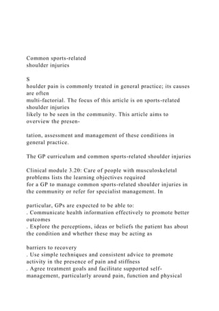
  • 82. veryuncomfortable and, therefore, could reduce patient compliance. Overall, careful consideration of the injury mechanism, patient demands, and concomitant injuries associated with anterior shoulder instability are crucial when deciding on nonopera- tive vs. operative intervention. Patients younger than 35 years of age should rarely be treated nonoperatively as the recurrence rate is unacceptably high. If treated nonoperatively, immobilization in internal rotation seems to be more comfortable and shows equal outcomes to immobilization in external rotation 92 Obere Extremität 2 · 2018 https://doi.org/10.1007/s11678penalty @M -hskip [email protected] 018penalty @M -hskip [email protected] 0449penalty @M -hskip [email protected] 1 Fig. 2 8 Images of a 46-year-oldman with right- sidedbicepstendonitis,diagnosedvia history,phys- icalexamination,andaT2- weightedmagneticresonanceimagingwithaclearhalosign(yellow circle) aroundthe long headof the biceps tendon indicating inflammation.The patient was treatedconser- vatively with physical therapyandNSAIDs butcontinuedto experiencesymptoms 6months later.He thusunderwentoperativemanagementasseeninbwiththelongheadof thebicepstendon(BT)and biceps reflection pulley visualizedthrough the
  • 83. standardposteriorviewing portal.HH humeral head and thus should be preferred, according to current literature findings. Biceps tendinitis Indications for nonoperative treatment of long head biceps tendinitis Inflammation of the long head biceps tendon (LHBT) can lead to damage and weakening of surrounding supporting structures, thereby causing LHBT in- stability. In turn, instability can place increased stresses on the LHBT, which subsequently increase inflammation. This cycle can predispose the LHBT to rupture. Given the potential success of non- operative management for most LHBT tendinopathies, a management strategy involving medications and physical ther- apy should be the first step in treating these conditions. After progressing a pa- tient through physical therapy, a course of nonsteroidal anti-inflammatory drugs (NSAIDs) and/or injections, it is impor- tanttore-evaluatethepatientforprogres- sion of pain, weakness, and mechanical symptoms. At that time, continuation of a home exercise program vs. consider- ation of additional interventions will be discussed based on symptom progres-
  • 84. sion. Ifapatientprogressesthroughallnon- operative treatment options and notes no improvement of pain or weakness, he or she should progress to surgical evalua- tion (. Fig. 2). This is also the case for patients suffering from biceps reflection pulley lesions because these lesions do nothealandsymptomsworsenovertime. In general, patients suitable for surgical evaluation include the following: young, highly motivated patients with instabil- ity or complete LHBT rupture; man- ual laborers with significant instability or complete LHBT rupture; elite-level ath- letes with instability or complete LHBT rupture; any individual with a complete LHBT rupture who is not agreeable to a potential loss of elbow flexion or fore- arm supination strength and long-stand- ing “Popeye” deformity; and any individ- ual whohas progressed throughall stages of nonoperative treatment and continues to have symptoms of pain and/or weak- ness that affects their quality of life. Treatment After identification of the underlying pathologic condition of the LHBT, treat- ment generally begins with activity modification, NSAIDs, and/or cortico- steroid injections [1, 53]. NSAIDs can provide short-term benefit for swelling
  • 85. and pain control. However, there is little evidence that they are efficacious in treating chronic tendon injuries [13]. Useofcorticosteroidinjectionsshould follow a similar treatment protocol to NSAIDs. Multiple case reports discuss the risk of tendon rupture with steroid injections, and caution should be exer- cised when injecting steroid around the LHBT [2, 13]. Corticosteroid injections alone will likely provide short-term anti- inflammatory effects for most LHBT dis- orders. However, they should be used for short-term pain relief and as an adjunct for the patient to initiate and tolerate a physical therapy program, rather than as a long-term treatment option. Be- cause these injections have the potential toreachtheglenohumeraljoint, theanes- theticofchoice, usedincombinationwith corticosteroid, should be ropivacaine, as it is found to be less chondrotoxic than bupivacaine [62]. Theinitiationofa3–6-monthphysical therapy program allows for progressive increase in muscle strength while pro- viding protection against further LHBT and associated structure injury during rehabilitation [1, 4, 19, 53, 67]. Other evolving nonoperative treat- mentoptionsforLHBTdisordersinclude prolotherapy (dextrose solution, sodium
  • 86. morrhuate), platelet-rich plasma (dif- fering concentrations of platelets, white blood cells, red blood cells, and activated and inactivated platelets), and stem cells (circulating stem cells, adipose-derived, bone marrow aspirate, bone marrow aspirate concentrate, amniotic mem- brane-derived). The choice to utilize one of these treatment options varies from patienttopatientand conditiontocondi- tion, and current research is beginning to thoroughly evaluate these interventions and to standardize treatment protocols [21, 23, 45, 46, 49]. Indications for these injections include pain impairing athletic performance, connective tissue laxity impairing athletic performance, and pain impairing rest and quality of life [49]. Future research is needed to de- termine which LHBT disorders respond best to, and what patient populations are the most suitable candidates for, such procedures. Acromioclavicular joint injuries Indications for nonoperative treatment of acromioclavicular joint injury Injuryclassificationisthesinglemostim- portant factor in determining the most appropriate treatment of acromioclavic- ular (AC) joint injuries. In 1989, Rock- wood and colleagues developed the clas-
  • 87. Obere Extremität 2 · 2018 93 Review article Fig. 3 8 Radiographs of a 26-year-oldmale patientafteradirectfallonto his rightshoulder.a Panoramic view after in- jury showing a probable Rockwoodtype II injury.b However,the Alexanderview demonstratestheclavicleoverriding the acromion, thus indicating horizontal instabilityanddefining thisasa RockwoodtypeIVinjury.Accordingly,the patient un- derwentoperativetherapywithtwodog- bonesinsteadofoneinordertobetteraddressthehorizontalinstability ,aspictured in c, the postoperative panoramicradiograph.d Postoperatively, the horizontal instability wasresolvedasdemonstratedon the Alexanderview 6 weeks aftersurgery sification system that is most widely used for AC joint injuries today [79]. No- tably, this system, which is based on the work of Tossy et al. [74], recognizes the importance of the coracoclavicular (CC) ligaments in joint stability [79]. Rockwood type I injuries are charac- terized by a sprain without rupture of the AC ligaments with no anatomic dis- location and intact trapezius and deltoid fascia. Type II injuries involve rupture of the AC joint ligaments but are otherwise similar to type I. Type III injuries are characterized by rupture of both the AC andCCligamentswithsuperiordisplace-
  • 88. ment of the clavicle of 25–100% com- pared with the contralateral shoulder; notably, the trapezius and deltoid fascia are disrupted with this injury. Type IV injuries generally present with additional horizontal instability (. Fig. 3). Type V injuries are similar to type-III injuries, but the clavicle is superiorly displaced more than 100% compared with the con- tralateral side. Type-VI injuries, which are rarely seen, involve rupture of both AC and CC ligaments with inferior dis- placement of the distal clavicle under- neath the acromion; the trapezius and deltoid fascia are disrupted [74, 79]. Although high-level studies are rare inthe orthopedic literature todefinitively guide optimal treatment, there is a com- mon consensus regarding the most ap- propriatetreatmentsbasedonRockwood type [6]. It is generally agreed that type I and II injuries should undergo initial nonop- erative treatment while types IV–VI re- quire surgery [6]. Optimal management of type III injuries has been controver- sial. In the highest-level study to date, the Canadian Orthopedic Trauma Soci- ety [16] recently completed a prospective randomized trial of 83 patients compar- ing nonoperative treatment of grade III, IV, or V AC joint injuries with operative intervention using a hook plate. Out- come scores at short-term follow-up as
  • 89. far as 2 years demonstrated no signifi- cant difference between the groups with the exception of superior radiographic results in the operative group [16]. Moreover, Petri and colleagues re- viewed 41 patients with Rockwood grade III AC joint injuries who were initially treated nonoperatively [60]. Nonoperative management consisted of formalphysicaltherapytwotothreetimes per week for at least 6 weeks using a pha- sic approach with progression dictated by patient tolerance and evidence of improved scapulohumeral kinematics. Nonoperative treatment failed in 12 pa- tients, who ultimately required surgery. Reasons cited for nonoperative failure included unremitting pain, weakness, instability, and dysfunction in spite of physical therapy. At a mean follow-up of 3.3 years, patient-reported outcome scores—including the American Shoul- der and Elbow Surgeons score (ASES), Quick Disabilities of the Arm, Shoul- der, and Hand score (QuickDASH), Single Assessment Numeric Evalua- tion score (SANE), and Short Form 12 Physical Component Summary (SF- 12 PCS)—did not significantly differ be- tween those who successfully completed nonoperative therapy and those who required eventual surgery [60]. In general, there is consensus that
  • 90. the horizontal stability of the clavicle is considered a potential key factor for a successful postoperative outcome. It is hypothesized that an unstable clavi- cle causes pain and functional deficits. Therefore, the ISAKOS shoulder com- mittee [7] recently proposed a modifica- tion to the classic Rockwood classifica- tion in which type III injuries may be fur- ther subdivided into types IIIA and IIIB; 94 Obere Extremität 2 · 2018 type IIIA injuries are horizontally stable and may respond well to conservative management, but type IIIB injuries are unstable and should therefore be treated surgically [7]. Treatment Typical nonoperative treatment consists of primary immobilization and subse- quent active rehabilitation [15]. How- ever, evidence to support the efficacy of specificrehabilitationprotocolsislimited [15]. Gladstoneetal. [25]publishedaphys- icaltherapyregimenforthenonoperative treatment of AC joint injuries types I, II, andIIIinathletes. Phase1lasts3–10days and focuses on elimination of pain and sling immobilization to protect the AC
  • 91. joint. Range-of-motion exercises begin in phase 2 with gradual progression of isotonic exercise for strengthening. Phase 3 involves advanced strengthen- ing, and phase 4 involves sports-specific training before full returntoactivity[25]. The total length of rehabilitation can last 3–6 months. Moreover, it is important to check on the scapula movement since a significant number of patients suffering from AC joint injuries also present with scapula dyskinesis. Overall, the general consensus re- garding management of AC joint in- juries is fairly straightforward: initial nonoperative treatment for Rockwood grades I–II, and operative intervention for grades IV–VI. For patients with grade III lesions, a closer look con- cerning the stability of the clavicle is necessary. Proximal humeral fracture Indications for nonoperative treatment of proximal humeral fractures The number of bone parts and concomi- tant displacement mainly influences the treatment strategy of proximal humeral fractures. Nonoperative treatment of two-part fractures with early rehabil- itation has been found to be at least as efficacious as surgical treatment in
  • 92. injuries with minimal displacement [29]. Betteroutcomesmaybeachievedwith surgical fixation in cases with signifi- cant displacement, a bony avulsion of the supraspinatus tendon, a block to range of motion, andinvolvementoftheanatomic neck. However, well-designed compar- ative studies of operative vs. nonoper- ative management of two-part fractures are lacking [26]. Some authors have found that greater tuberosity fractures with >5mm of dis- placement may benefit from surgical fix- ation to reduce the risk of subacromial impingement [58, 63]. Lesser tuberosity fractures with internal rotation impinge- ment may also benefit from surgery if nonoperative management fails [52]. In contrast to other parts of the proximal humerus, the anatomic neck is devoid of soft-tissue attachments and has a tenu- ous blood supply, which may result in an increased risk of osteonecrosis. Court-Brown et al. recommend 2 weeks of sling immobilization followed byphysical therapyforpatients with two- part surgical neck fractures and valgus- impacted fractures [17, 18]. Two-part proximal humeral fractures with >66% translation were treated with either sling immobilization or with internal fixation with flexible intramedullary nailing and tension-band wires [17, 18]. No statis-
  • 93. tical difference was reported between the groups with regard to Neer score, return to activities of daily living, and union rates [17, 18]. The data demon- strate that the Constant score diminishes with advancing age and degree of dis- placement. However, when calculated based on age-adjusted Constant score, the older patients actually had better scores than the younger patients [14, 17, 18, 34]. Therefore, sling immobilization is an appropriate treatment option for patients older than age 60 years with valgus-impacted, two-part surgical neck or two-part tuberosity fractures. Although three-part and four-part fractures often require surgical fixation, nonoperative management can be con- sidered for patients with poor baseline function and/or an inability to toler- ate surgery. In select three-part and four-part fractures, particularly valgus- impacted fractures with <1cm of dis- placement of the tuberosities in relation to the head fragment, nonsurgical treat- ment may yield good-to-excellent results [17]. Although surgical treatment of com- plex fracture patterns is generally advo- cated, theefficacyofoperativevs. nonop- erativemanagementremainstobeclearly delineated. In a study of 60 elderly pa- tients with a displaced three-part frac-
  • 94. ture of the proximal humerus, Olerud et al. found that surgical management with a locking plate resulted in better functional outcomes and health-related quality of life than did nonsurgical treat- ment, butatacostofadditionalsurgeryin 30% of patients [56]. By contrast, a meta- analysis of randomized controlled trials did not find improved functional out- comes with open reduction and internal fixation (ORIF) compared with nonsur- gical treatment in elderly patients with displaced three-part or four-part prox- imal humeral fractures [40]. The study concluded that these results must be con- sidered in the context of variable patient demographics. A systematic review supported the use of nonsurgical treatment of proximal humeral fractures and noted a 2% rate of osteonecrosis mainly associated with three-part and four-part fractures, high rates of radiographic union, and modest complication rates [32]. Ultimately, the patient’s baseline physiology and func- tion may help to quantify the potential advantages of nonsurgical management, even in the setting of complex fracture patterns. Treatment A number of proximal humeral fractures may be treated nonoperatively. However, patients must understand the expecta-
  • 95. tions with this treatment approach and comply with the accompanying restric- tions. In general, excellent results have been achieved with short-term immobi- lization (<2 weeks) in a sling and early physical therapy [28, 63, 72]. While the literature supports early mobilization, it is important to ensure that further frac- ture displacement does not occur. Sling immobilization with or without closed reduction also has a role in the man- Obere Extremität 2 · 2018 95 Review article agement of displaced proximal humeral fractures [69]. Practical conclusion 4 For rotator cuff tears, the best pos- sible outcomes with nonoperative therapyaregenerallyachievedforpa- tients presenting pain as the primary symptom of an atraumatic rotator cuff tear, largely intact coronal and axial force couples, and a willingness to trade functional deficits to avoid surgical risks. 4 In patients suffering from anterior shoulder instability, careful consid- eration of the injury mechanism,
  • 96. patient demands, and concomitant injuries associated with anterior shoulder instability are crucial when deciding on nonoperative vs. opera- tive intervention. Patients <35 years should rarely be treated nonopera- tively. 4 For tendinitis of the LHBT, treatment generally begins with a nonoperative treatment protocol including activity modification and NSAIDs. In patients with structural instability of the biceps tendon complex, or in any individual who continues to have symptoms of pain after nonoperative treatment, surgery is favored. 4 The general consensus regarding management of AC joint injuries sug- gests initial nonoperative treatment for Rockwood types I–II, and oper- ative intervention for types IV–VI. For patients with type III lesions, a pathologic instability of the clav- icle potentially requiring surgical stabilization should be considered. 4 Tuberosity fractures with >5mm of displacement may benefit from surgical fixation to reduce the risk of subacromial impingement as well as displaced multifragment fractures in young and active patients. Corresponding address
  • 97. P. J. Millett, M.D., M.Sc. The Steadman Clinic 181 West Meadow Drive suite 400, 81657 Vail, CO, USA [email protected] thesteadmanclinic.com Compliance with ethical guidelines Conflict of interest. A.B. Imhoffservesasaboardor committeememberforAGA,servesontheeditorial boardofArchives of Orthopaedic and Trauma Surgery, isapaidconsultantandreceivesroyaltiesandresearch supportfromArthrex, Inc.,servesontheeditorial boardofArthroskopie, isapaidconsultantandreceives royaltiesfromArthrosurface,servesasaboardor committeememberforDGOOC,servesasaboardor committeememberforDGOU,servesasaboardor committeememberforISAKOS,servesontheeditorial boardofKSSTA, isapaidconsultantformedi-bayreuth, servesontheeditorialboardofOOTR,andreceives royaltiesandfinancialsupportfromSpringerand Thieme. P.J.Millett isapaidconsultantforArthrex, Inc., receivesroyaltiesfromArthrex, Inc.,Medbridge, andSpringerPublishing,ownsstockorstockoptions inGameReadyandVuMedi,andreceivesresearch supportfromArthrex, Inc.,Ossur,Siemens,andSmith andNephew. J.Pogorzelski,E.M.Fritz,andJ.A.Godin declarethattheyhavenocompetinginterests. Thisarticledoesnotcontainanystudieswithhuman participantsoranimalsperformedbyanyoftheau- thors.
  • 98. OpenAccess. Thisarticleisdistributedundertheterms oftheCreativeCommonsAttribution4.0International License(http://creativecommons.org/licenses/by/ 4.0/),whichpermitsunrestricteduse,distribution, andreproductioninanymedium,providedyougive appropriatecredittotheoriginalauthor(s)andthe source,providealinktotheCreativeCommonslicense, andindicateifchangesweremade. References 1. Allen L (2013) Long head of biceps tendon: anatomy,biomechanics,pathology,diagnosisand management.UnivNMOrthopResJ2:21–23 2. Andres BM, Murrell GA (2008) Treatment of tendinopathy: what works, what does not, and what is on the horizon. Clin Orthop Relat Res 466:1539–1554 3. Balg F, Boileau P (2007) The instability severity index score. A simple pre-operative score to select patients for arthroscopic or open shoulder stabilisation.JBoneJointSurgBr89:1470–1477 4. Barber FA, Field LD, Ryu RK (2008) Biceps tendon and superior labrum injuries: decision making. InstrCourseLect57:527–538 5. Barnes LA, Kim HM, Caldwell JM et al (2017) Satisfaction, function and repair integrity after arthroscopic versus mini-open rotator cuff repair. BoneJointJ99-B:245–249 6. BeitzelK,CoteMP,ApostolakosJetal(2013)Current concepts in the treatment of acromioclavicular
  • 99. jointdislocations.Arthroscopy29:387–397 7. Beitzel K, Mazzocca AD, Bak K et al (2014) ISAKOS upper extremity committee consensus statement on the need for diversification of the Rockwood classification for acromioclavicular joint injuries. Arthroscopy30:271–278 8. Bishop J, Klepps S, Lo IK et al (2006) Cuff integrity after arthroscopic versus open rotator cuff repair: a prospective study. J Shoulder Elbow Surg 15:290–299 9. Bishop JA, Crall TS, Kocher MS (2011) Operative versus nonoperative treatment after primary traumatic anterior glenohumeral dislocation: expected-value decision analysis. J Shoulder ElbowSurg20:1087–1094 10. BoormanRS,MoreKD,HollinsheadRMetal(2014) The rotator cuff quality-of-life index predicts the outcome of nonoperative treatment of patients with a chronic rotator cuff tear. J Bone Joint Surg Am96:1883–1888 11. Boughebri O, Maqdes A, Moraiti C et al (2015) Results of 45 arthroscopic Bankart procedures: Does the ISIS remain a reliable prognostic assessment after 5 years? Eur J Orthop Surg Traumatol25:709–716 12. Buss DD, Lynch GP, Meyer CP et al (2004) Nonoperativemanagementforin-seasonathletes withanteriorshoulderinstability.AmJSportsMed 32:1430–1433
  • 100. 13. Childress MA, Beutler A (2013) Management of chronic tendon injuries. Am Fam Physician 87:486–490 14. ConstantCR,MurleyAH(1987)Aclinicalmethodof functionalassessmentoftheshoulder.ClinOrthop Relat Res. https://doi.org/10.1097/00003086- 198701000-00023 15. Cote MP, Wojcik KE, Gomlinski G et al (2010) Re- habilitationofacromioclavicularjointseparations: operative and nonoperative considerations. Clin SportsMed29:213–228(vii) 16. Cots(2015)Multicenterrandomizedclinicaltrialof nonoperativeversusoperativetreatmentofacute acromio-clavicular joint dislocation. J Orthop Trauma29:479–487 17. Court-Brown CM, Cattermole H, Mcqueen MM (2002) Impacted valgus fractures (B1.1) of the proximal humerus. The results of non-operative treatment.JBoneJointSurgBr84:504–508 18. Court-Brown CM, Garg A, Mcqueen MM (2001) The translated two-part fracture of the proximal humerus. Epidemiologyandoutcomeintheolder patient.JBoneJointSurgBr83:799–804 19. Eakin CL, Faber KJ, Hawkins RJ et al (1999) Biceps tendon disorders in athletes. J Am Acad Orthop Surg7:300–310 20. Finestone A, Milgrom C, Radeva-Petrova DR et al (2009) Bracing in external rotation for traumatic anterior dislocation of the shoulder. J Bone Joint
  • 101. SurgBr91:918–921 21. Finnoff JT, Fowler SP, Lai JK et al (2011) Treat- ment of chronic tendinopathy with ultrasound- guidedneedletenotomyandplatelet-richplasma injection.PMR3:900–911 22. FucenteseSF,VonRollAL,PfirrmannCWetal(2012) Evolution of nonoperatively treated symptomatic isolatedfull-thicknesssupraspinatustears. JBone JointSurgAm94:801–808 23. FullertonBD,ReevesKD(2010)Ultrasonographyin regenerative injection (prolotherapy) using dex- trose, platelet-rich plasma, and other injectants. PhysMedRehabilClinNAm21:585–605 24. Galatz LM, Ball CM, Teefey SA et al (2004) The outcome and repair integrity of completely arthroscopicallyrepairedlargeandmassiverotator cufftears.JBoneJointSurgAm86-a:219–224 96 Obere Extremität 2 · 2018 http://creativecommons.org/licenses/by/4.0/ http://creativecommons.org/licenses/by/4.0/ https://doi.org/10.1097/00003086-198701000-00023 https://doi.org/10.1097/00003086-198701000-00023 25. Gladstone JN, Wilk KE, Andrews JR (1997) Nonoperativetreatmentofacromioclavicular joint injuries.OperTechSportsMed5:78–87 26. Godin JA, Katthagen JC, Fritz EM et al (2017) Arthroscopic treatment of greater tuberosity
  • 102. avulsionfractures.ArthroscTech6:e777–e783 27. Greiwe RM, Galano G, Grantham J et al (2012) Arthroscopic stabilization for voluntary shoulder instability.JPediatrOrthop32:781–786 28. Hanson B, Neidenbach P, De Boer P et al (2009) Functional outcomes after nonoperative managementoffracturesoftheproximalhumerus. JShoulderElbowSurg18:612–621 29. Hodgson SA, Mawson SJ, Saxton JM et al (2007) Rehabilitation of two-part fractures of the neck of the humerus (two-year follow-up). J Shoulder ElbowSurg16:143–145 30. Hovelius L, Augustini BG, Fredin H et al (1996) Primary anterior dislocation of the shoulder in young patients. A ten-year prospective study. JBoneJointSurgAm78:1677–1684 31. Itoi E, Sashi R, Minagawa H et al (2001) Position of immobilization after dislocation of the gleno- humeral joint. A study with use of magnetic resonance imaging. J Bone Joint Surg Am 83- A:661–667 32. Iyengar JJ, Devcic Z, Sproul RC et al (2011) Nonoperative treatment of proximal humerus fractures: a systematic review. J Orthop Trauma 25:612–617 33. Jaggi A, Noorani A, Malone A et al (2012) Muscle activation patterns in patients with recurrent shoulderinstability. IntJShoulderSurg6:101–107
  • 103. 34. Katolik LI, Romeo AA, Cole BJ et al (2005) Normalization of the constant score. J Shoulder ElbowSurg14:279–285 35. Kirkley A, Griffin S, Richards C et al (1999) Prospective randomized clinical trial comparing theeffectivenessofimmediatearthroscopicstabi- lizationversusimmobilizationandrehabilitationin firsttraumaticanteriordislocationsoftheshoulder. Arthroscopy15:507–514 36. Kirkley A, Werstine R, Ratjek A et al (2005) Prospective randomized clinical trial comparing theeffectivenessofimmediatearthroscopicstabi- lization versus immobilization and rehabilitation in first traumatic anterior dislocations of the shoulder: long-term evaluation. Arthroscopy 21:55–63 37. Kuhn JE, Dunn WR, Sanders R et al (2013) Effectiveness of physical therapy in treating atraumatic full-thickness rotator cuff tears: amulticenterprospectivecohortstudy. JShoulder ElbowSurg22:1371–1379 38. Kukkonen J, Joukainen A, Lehtinen J et al (2014) Treatment of non-traumatic rotator cuff tears: a randomised controlled trial with one-year clinical results.BoneJointJ96-B:75–81 39. Kukkonen J, Joukainen A, Lehtinen J et al (2015) Treatment of nontraumatic rotator cuff tears: a randomized controlled trial with two years of clinical and imaging follow-up. J Bone Joint Surg Am97:1729–1737
  • 104. 40. Li Y, Zhao L, Zhu L et al (2013) Internal fixation versus nonoperative treatment for displaced 3-part or 4-part proximal humeral fractures in elderly patients: a meta-analysis of randomized controlledtrials.PLoSONE8:e75464 41. LinJC,WeintraubN,AragakiDR(2008)Nonsurgical treatmentforrotatorcuffinjuryintheelderly.JAm MedDirAssoc9:626–632 42. Lo IK, Burkhart SS (2003) Current concepts in arthroscopic rotator cuff repair. Am J Sports Med 31:308–324 43. Mather RC, Koenig L, Acevedo D et al (2013) The societal and economic valueof rotator cuff repair. JBoneJointSurgAm95:1993–2000 44. MatherRC,OrlandoLA,HendersonRAetal(2011)A predictivemodelofshoulderinstabilityafterafirst- time anterior shoulder dislocation. J Shoulder ElbowSurg20:259–266 45. Mautner K, Blazuk J (2015) Where do injectable stemcelltreatmentsapplyintreatmentofmuscle, tendon,andligamentinjuries? PMR7:S33–S40 46. Mautner K, Malanga GA, Smith J et al (2015) A call for a standard classification system for future biologic research: the rationale for new PRP nomenclature.PMR7:S53–S59 47. Meislin RJ, Sperling JW, Stitik TP (2005) Persistent shoulder pain: epidemiology, pathophysiology, anddiagnosis.AmJOrthop(BelleMead,NJ)34:5–9
  • 105. 48. Millett PJ, Wilcox RB 3rd, O’holleran JD et al (2006) Rehabilitation of the rotator cuff: an evaluation-based approach. J Am Acad Orthop Surg14:599–609 49. Moon YL, Ha SH, Lee YK et al (2011) Comparative studiesofplatelet-richplasma(PRP)andprolother- apy for proximal biceps tendinitis. Clin Shoulder Elbow14:153–158 50. Moosmayer S, Lund G, Seljom US et al (2014) Tendon repair compared with physiotherapy in the treatment of rotator cuff tears: a randomized controlled study in 103 cases with a five-year follow-up.JBoneJointSurgAm96:1504–1514 51. Moroder P, Minkus M, Bohm E et al (2017) Use of shoulder pacemaker for treatment of functional shoulder instability: proof of concept. Obere Extremitat12:103–108 52. Neer CS 2nd, Craig EV, Fukuda H (1983) Cuff-tear arthropathy.JBoneJointSurgAm65:1232–1244 53. Nho SJ, Strauss EJ, Lenart BA et al (2010) Long head of the biceps tendinopathy: diagnosis and management.JAmAcadOrthopSurg18:645–656 54. NorteGE,WestA,GnacinskiMetal(2011)On-field management of the acute anteriorglenohumeral dislocation.PhysSportsmed39:151–162 55. Oh LS, Wolf BR, Hall MP et al (2007) Indications for rotatorcuffrepair:asystematicreview.ClinOrthop RelatRes455:52–63
  • 106. 56. Olerud P, Ahrengart L, Ponzer S et al (2011) Internal fixation versus nonoperative treatment of displaced 3-part proximal humeral fractures in elderly patients: a randomized controlled trial. J ShoulderElbowSurg20:747–755 57. Owens BD, Dickens JF, Kilcoyne KG et al (2012) Management of mid-season traumatic anterior shoulder instability in athletes. J Am Acad Orthop Surg20:518–526 58. Park TS, Choi IY, Kim YH et al (1997) A new suggestion for the treatment of minimally displacedfracturesofthegreatertuberosityofthe proximalhumerus.BullHospJtDis56:171–176 59. Petri M, Ettinger M, Brand S et al (2016) Non- operativemanagementofrotatorcufftears. Open OrthopJ10:349–356 60. Petri M, Warth RJ, Greenspoon JA et al (2016) Clinical results after conservative management for grade III acromioclavicular joint injuries: does eventual surgery affect overall outcomes? Arthroscopy32:740–746 61. Phadnis J, Arnold C, Elmorsy A et al (2015) Utility oftheinstabilityseverityindexscoreinpredicting failure after arthroscopic anterior stabilization of theshoulder.AmJSportsMed43:1983–1988 62. PiperSL,KimHT(2008)Comparisonofropivacaine and bupivacaine toxicity in human articular chondrocytes.JBoneJointSurgAm90:986–991 63. PlatzerP,Kutscha-LissbergF,LehrSetal(2005)The
  • 107. influenceofdisplacementonshoulderfunctionin patients with minimally displaced fractures of the greatertuberosity. Injury36:1185–1189 64. Pope D, Croft P, Pritchard C et al (1997) Prevalence of shoulder pain in the community: the influence ofcasedefinition.AnnRheumDis56:308–312 65. Rouleau DM, Hébert-Davies J, Djahangiri A et al (2013) Validation of the instability shoulder index score in a multicenter reliability study in 114 consecutivecases.AmJSportsMed41:278–282 66. Rowe CR, Pierce DS, Clark JG (1973) Voluntary dislocation of the shoulder. A preliminary report on a clinical, electromyographic, and psychiatric studyoftwenty-sixpatients. JBoneJointSurgAm 55:445–460 67. Ryu JH, Pedowitz RA (2010) Rehabilitation of biceps tendon disorders in athletes. Clin Sports Med29:229–246(vii–viii) 68. SafranO,SchroederJ,BloomRetal(2011)Natural history of nonoperatively treated symptomatic rotator cuff tears in patients 60 years old or younger.AmJSportsMed39:710–714 69. SchlegelTF,HawkinsRJ(1994)Displacedproximal humeralfractures: evaluationandtreatment.JAm AcadOrthopSurg2:54–66 70. Takwale VJ, Calvert P, Rattue H (2000) Involuntary positionalinstabilityoftheshoulderinadolescents and young adults. Is there any benefit from treatment? JBoneJointSurgBr82:719–723
  • 108. 71. Taylor DC, Arciero RA (1997) Pathologic changes associated with shoulder dislocations. Arthro- scopic and physical examination findings in first- time, traumatic anterior dislocations. Am J Sports Med25:306–311 72. Tejwani NC, Liporace F, Walsh M et al (2008) Functional outcome following one-part proximal humeralfractures: aprospectivestudy. JShoulder ElbowSurg17:216–219 73. Thomazeau H, Courage O,Barth J et al (2010) Can weimprovetheindicationforBankartarthroscopic repair? A preliminary clinical study using the ISIS score.OrthopTraumatolSurgRes96:S77–83 74. Tossy JD, Mead NC, Sigmond HM (1963) Acromio- clavicular separations: useful and practical classification for treatment. Clin Orthop Relat Res 28:111–119 75. Van Der Meijden OA, Westgard P, Chandler Z et al (2012) Rehabilitation after arthroscopic rotator cuffrepair: currentconceptsreviewandevidence- basedguidelines. IntJSportsPhysTher7:197–218 76. Vavken P, Sadoghi P, Quidde J et al (2014) Immobilization in internal or external rotation does not change recurrence rates after traumatic anterior shoulder dislocation. J Shoulder Elbow Surg23:13–19 77. Whelan DB, Kletke SN, Schemitsch G et al (2016) Immobilization in external rotation versus internal rotation after primary anterior shoulder
  • 109. dislocation: a meta-analysis of randomized controlledtrials.AmJSportsMed44:521–532 78. Whelan DB, Litchfield R, Wambolt E et al (2014) External rotation immobilization for primary shoulder dislocation: a randomized controlled trial.ClinOrthopRelatRes472:2380–2386 79. Williams G, Nguyen V, Rockwood C (1989) Classifi- cationandradiographicanalysisofacromioclavic- ulardislocations.ApplRadiol18:29–34 80. Yamaguchi K, Tetro AM, Blam O et al (2001) Naturalhistoryofasymptomaticrotatorcufftears: a longitudinal analysis of asymptomatic tears detected sonographically. J Shoulder Elbow Surg 10:199–203 Obere Extremität 2 · 2018 97 Nonoperative treatment of five common shoulder injuriesAbstractZusammenfassungIntroductionRotator cuff tearsIndications for nonoperative treatment of symptomatic full- thickness rotator cuff tearsTreatmentAnterior shoulder instabilityIndications for nonoperative treatment of anterior shoulder instabilityTreatmentBiceps tendinitisIndications for nonoperative treatment of long head biceps tendinitisTreatmentAcromioclavicular joint injuriesIndications for nonoperative treatment of acromioclavicular joint injuryTreatmentProximal humeral fractureIndications for nonoperative treatment of proximal humeral fracturesTreatmentPractical conclusionReferences