SlideShare ist ein Scribd-Unternehmen logo
1 von 75
Downloaden Sie, um offline zu lesen
Male Reproductive System
• The reproductive
system in men has
components in the
abdomen, pelvis,
and perineum.
• The major components
are a testis, epididymis,
ductus deferens, and
ejaculatory duct on each
side, and the urethra
and penis in the midline.
• Three types of
accessory glands are
associated with the
system:
– A single prostate;
– A pair of seminal
vesicles;
– A pair of bulbourethral
glands.
Male Reproductive System
The scrotum
• The scrotum is an
outpouching of the
lower part of the
anterior abdominal
wall.
• It contains the testes,
the epididymides,
and the lower ends of
the spermatic cords.
The scrotum
• It is divided on its
surface into two
compartments by a
raphé, which is
continued forward to the
under surface of the
penis, and
backward, along the middle
line of the perineum to
the anus.
• Each compartment
contains one of the two
testes, and one of the
epididymides.
The scrotum
• The wall of the scrotum
has the following layers:
– Skin
– Superficial fascia
– Spermatic fasciae
– Tunica vaginalis
The scrotum
Skin
•The skin of the scrotum is
thin, wrinkled, and
pigmented and forms a
single pouch. A slightly
raised ridge in the midline
indicates the line of fusion
of the two lateral
labioscrotal swellings.
Superficial fascia
•This is continuous with the fatty
and membranous layers of the
anterior abdominal wall.
•The fat is replaced by smooth
muscle called the dartos muscle.
•This is innervated by
sympathetic nerve fibers and is
responsible for the wrinkling of
the overlying skin.
The scrotum
Spermatic fasciae
•It has three layers which lie
beneath the superficial fascia and
are derived from the three layers of
the anterior abdominal wall on each
side.
•The external spermatic fascia is
derived from the aponeurosis of the
external oblique muscle; the
cremasteric fascia is derived from
the internal oblique muscle; and,
finally, the internal spermatic fascia
is derived from the fascia
transversalis.
Tunica vaginalis
This lies within the spermatic
fasciae and covers the
anterior, medial, and lateral
surfaces of each testis.
Lymph Drainage of the
Scrotum
• Lymph from the skin
and fascia, including
the tunica vaginalis,
drains into the
superficial inguinal
lymph nodes .
Testes
• Testis has ellipsoid-
shaped.
• Testes develop in the
abdomen and move
before birth into the
scrotum.
• The left testis usually
lies at a lower level
than the right.
Testes
• The testis are covered
by:
• A closed sac of
peritoneum (the tunica
vaginalis), which
originally connected to
the abdominal cavity.
Normally after testicular
descent, the connection
closes, leaving a fibrous
remnant.
• It is covered by a fibrous
capsule called the tunica
albuginea.
Testes
• In the inner surface of the
capsule is a series of fibrous
septa that divide the interior
of the organ into lobules.
• Lying within each lobule are 1
to 3 coiled seminiferous
tubules.
• The tubules open into a
network of channels called
the rete testis.
• Small efferent ductules
connect the rete testis to the
upper end of the epididymis.
Testes
Epididymis
• The epididymis is a
single, long coiled
duct that courses
along the
posterolateral side of
the testis.
• The tunica vaginalis
covers the epididymis
with the exception of
the posterior border.
Epididymis
• Structurally, epididymis
divided into :
– Head
– Body
– tail
Epididymis
It has two distinct components:
–The efferent ductules, which form an enlarged coiled
mass that sits on the posterior superior pole of the testis
and forms the head of the epididymis;
–The true epididymis, which is a single, long coiled duct
into which the efferent ductules all drain, and which
continues inferiorly along the posterolateral margin of the
testis as the body of epididymis and enlarges to form the
tail of epididymis at the inferior pole of the testis.
Arterial Blood Supply of the Testis
and Epididymis
• The testicular artery is
a branch of the
abdominal aorta.
• The testicular veins emerge
from the testis and the
epididymis as a venous
network, the pampiniform
plexus.
• This becomes reduced to a
single vein as it ascends
through the inguinal canal.
• The right testicular vein
drains into the inferior
vena cava, and the left vein
joins the left renal vein.
Lymphatic Drainage of The
Testes
• Lymphatic drainage of
the testes is to the
para- aortic lymph
nodes.
Ductus deferens
• (Latin: "carrying-away
vessel"), also called vas
deferens.
• The ductus deferens is
a long muscular duct
that transports
spermatozoa from the
tail of the epididymis to
the ejaculatory duct.
Ductus deferens course
• The vas arises from the tail of
the epididymis and traverses
the inguinal canal to the deep
ring, passes downwards on
the lateral wall of the pelvis
almost to the ischial
tuberosity and turns medially
to cross the ureter posterior
to the bladder.
• It continues inferomedially
along the base of the bladder,
anterior to the rectum, almost
to the midline, where it is
joined by the duct of the
seminal vesicle to form the
ejaculatory duct.
Ductus deferens course
• The terminal part of
the vas deferens is
dilated to form the
ampulla of the vas
deferens.
• The ejaculatory duct
penetrates through the
prostate gland to
connect with the
prostatic urethra.
Seminal vesicle
• The seminal vesicles
are an accessory gland
of the male
reproductive system .
• The seminal vesicles
are two lobulated
organs about 2 in. (5
cm) long lying on the
posterior surface of the
bladder .
Seminal vesicle
• On the medial side of
each vesicle lies the
terminal part of the vas
deferens.
• Posteriorly, the seminal
vesicles are related to
the rectum.
• Inferiorly, each seminal
vesicle narrows and joins
the vas deferens of the
same side to form the
ejaculatory duct.
Blood Supply of Seminal
vesicle
Arteries
•The arterial blood supply
from, the inferior vesicle and
middle rectal arteries.
Veins
• The veins drain into
the internal iliac veins.
Ejaculatory Ducts
• The two ejaculatory ducts are
each less than 1 in. (2.5 cm)
long and are formed by the
union of the vas deferens and
the duct of the seminal vesicle.
• The ejaculatory ducts pierce
the posterior surface of the
prostate and open into the
prostatic part of the urethra,
close to the margins of the
prostatic utricle; their function
is to drain the seminal fluid
into the prostatic urethra.
Semen
• Semen is a milky white, somewhat sticky mixture of sperm,
testicular fluid, and accessory gland secretions. The liquid
provides a transport medium and nutrients and contains
chemicals that protect and activate the sperm and facilitate
their movement.
Semen contains several substances that
play many roles:
• Prostaglandins decrease the viscosity of mucus guarding the
entry (cervix) of the uterus and stimulate reverse peristalsis in
the uterus, facilitating sperm movement through the female
reproductive tract.
• The hormone relaxin and certain enzymes in semen enhance
sperm motility.
• Contained ATP provides energy.
• Certain ingredients suppress the immune response in the
female’s reproductive tract.
• Antibiotic chemicals destroy some bacteria.
• Clotting factors in semen coagulate it just after it is ejaculated.
Coagulation causes the sperm to stick to the walls of the
vagina and prevents the initially immobile sperm from
draining out of the vagina. Soon after semen coagulates,
fibrinolysin liquefies the sticky mass and the sperm swim out
and begin their journey through the female duct system
The Bulbo-Urethral Glands
• The bulbo-urethral glands are pea-sized glands located
inferior to the prostate in the urogenital diaphragm
• They produce a thick, clear mucus, some of which drains into
the spongy urethra and lubricates the glans penis when
sexually excited.
• The mucus neutralizes traces of acidic urine and lubricates the
urethra just prior to ejaculation.
Prostate
• The prostate is an unpaired
accessory structure of the
male reproductive system
that surrounds the urethra
in the pelvic cavity .
• It lies immediately inferior
to the bladder, above the
urogenital diaphragm,
posterior to the pubic
symphysis, and anterior to
the rectum.
Prostate
• The prostate is shaped like
an inverted rounded cone
with a larger base, which
is continuous above with
the neck of the bladder,
and a narrower apex,
which rests below on the
pelvic floor.
• The inferolateral surfaces
of the prostate are in
contact with the levator
ani muscles that together
cradle the prostate
between them.
Prostate
Prostate
• The two ejaculatory
ducts pierce the upper
part of the posterior
surface of the prostate
to open into the
prostatic urethra at
the lateral margins of
the prostatic utricle.
Relations of Prostate
Superiorly
•The base of the prostate is
continuous with the neck of
the bladder.
•The urethra enters the center
of the base of the prostate.
Inferiorly
• The apex of the prostate
lies on the upper surface of
the urogenital diaphragm.
• The urethra leaves the
prostate just above the
apex on the anterior
surface
Relations of Prostate
Anteriorly
•The prostate is related to the
symphysis pubis.
•The prostate is connected to
the posterior aspect of the
pubic bones by the
puboprostatic ligaments.
Laterally
•The prostate is embraced by
the anterior fibers of the
levator ani.
Posteriorly
•The prostate is closely
related to the anterior
surface of the rectal ampulla
and is separated from it by
the rectovesical septum
(fascia of Denonvilliers).
Structure of the Prostate
• Enclosed within thin dense fibrous capsule
• Inner loose sheath derived from pelvic fascia is prostatic
sheath͟
– Continuous inferiorly with superior fascia of urogenital
diaphragm
– Posteriorly it is part of rectovesical septum
– Separates bladder, seminal vesicles and prostate from
rectum
• Prostatic venous plexus lies between fibrous capsule and
prostatic sheath.
Structure of the Prostate
Prostate divided into:
– Two lateral lobes
– One median lobe
– Anterior and posterior
lobes
Structure of the Prostate
• Anterior
– Tissue lying anterior to urethra
– No glands; fibromuscular tissue only
• Median
– Cone-shaped region between ejaculatory ducts and
urethra
• Lateral (left & right)
– Main mass of gland, continuous posteriorly
– Separated by prostatic urethra
• Posterior
– Describes postero-medial part of lateral lobes
palpable through rectum on DRE.
Functions of Prostate
• During ejaculation, prostatic smooth muscle contracts,
squeezing the prostatic secretion into the prostatic urethra via
several ducts. This fluid plays a role in activating sperm and
accounts for up to one-third of the semen volume. It is a
milky, slightly acid fluid that contains citrate (a nutrient
source), several enzymes (including fibrinolysin,
hyaluronidase, and acid phosphatase), and prostate-specific
antigen (PSA).
Blood Supply of The Prostate
• Arterial supply
– Arteries derived from internal pudenal, inferior vesical and
middle rectal arteries (branches of internal iliac)
• Venous drainage
– Veins form prostatic venous plexus around sides and base
of prostate – located between capsule and sheath
– Drains into internal iliac veins
– Also communicates with vesical venous plexus and
vertebral venous plexuses.
Lymphatics and innervation of
Prostate gland
• Lymphatic drainage
– Lymph vessels terminate in internal iliac and sacral
lymph
• nodes
– Some vessels from posterior surface pass with lymph
• vessels froŵ ďladder to exterŶal iliaĐ LN’s
• Innervation
– Parasympathetic fibres arise from pelvic splanchnic
nerves
– Sympathetic fibres from inferior hypogastric plexuses
Penis
• The penis is a
pendulous organ
suspended from the
front and sides of the
pubic arch and
containing the
greater part of the
urethra.
Penis
• It consists of internal
root, external shaft, &
glans.
– Root: the portion of the
penis that extends
internally into the pelvic
cavity.
– Shaft: the length of the
penis between the glans
and the body.
– Glans: the head of the
penis; has many nerve
endings.
• Foreskin: a covering of
skin over the penile glans.
Penis
Penis
• The root of penis
consists of the two crura,
which are proximal parts
of the corpora cavernosa
attached to the pubic
arch, and the bulb of
penis, which is the
proximal part of the
corpus spongiosum
anchored to the perineal
membrane.
Penis
• The body of the penis is essentially
composed of three cylinders of
erectile tissue enclosed in a tubular
sheath of fascia (Buck's fascia).
• The erectile tissue is made up of
two dorsally placed corpora
cavernosa and a single corpus
spongiosum applied to their ventral
surface .
• At its distal extremity, the corpus
spongiosum expands to form the
glans penis, which covers the distal
ends of the corpora cavernosa.
• On the tip of the glans penis is the
slitlike orifice of the urethra, called
the external urethral meatus.
Penis
External penile structures
• Corona: the rim of the
penile glans.
• Frenulum: thin strip of
skin connecting the
glans to the shaft on
the underside of the
penis.
Both are highly sensitive
areas to the touch
Blood Supply of The Penis
Arteries
•The corpora cavernosa
are supplied by the deep
arteries of the penis ; the
corpus spongiosum is
supplied by the artery of
the bulb.
•In addition, there is the
dorsal artery of the
penis.
•All the above arteries
are branches of the
internal pudendal artery.
Veins
• The veins drain into the
internal pudendal
veins.
Lymphatics and innervation of
The Penis
Nerve Supply
Sensation
•The nerve supply is from
the pudendal nerve and
the pelvic plexuses.
Erectile function
•Parasympathetic(excitat
ory)
•Sympathetic (inhibitory)
Lymph Drainage
•The skin of the penis is
drained into the medial group
of superficial inguinal nodes.
•The deep structures of the
penis are drained into the
internal iliac nodes.
Analagous structures in male
and female sexual anatomy
Male
Glans
Foreskin
Shaft
Clitoris
Clitoral hood
Scrotal sac
Testes
Female
Labia minora
Labia majora
Ovaries
Physiology of the Male
Reproductive System
The chief phases of the male sexual response are
1. Erection of the penis, which allows it to penetrate the female
vagina
2. Ejaculation, which expels semen into the vagina
Erection
• Erection, enlargement and stiffening of the penis, results
from engorgement of the erectile bodies with blood. It
happens because of the parasympathetic function of arteriole.
• Sexual excitement triggers a parasympathetic reflex that
promotes release of nitric oxide (NO) locally.
• Nitric oxide relaxes smooth muscle in the penile blood vessel
walls, dilating these arterioles, and the erectile bodies fill with
blood.
• Expansion of the corpora cavernosa of the penis compresses
their drainage veins, retarding blood outflow and maintaining
engorgement.
• The corpus spongiosum expands but not nearly as much as
the cavernosa. Its function is to keep the urethra open during
ejaculation.
• The erect penis not bend or fold during intercourse. The
longitudinal and circular arrangement of collagen fibers
surrounding the penis prevents this problem.
• Ejaculation
• Ejaculation is the propulsion of semen from the male duct
system.
• Although erection is under parasympathetic control,
ejaculation is under sympathetic control.
• When impulses provoking erection reach a critical level, a
spinal reflex is initiated, and a massive discharge of nerve
impulses occurs over the sympathetic nerves serving the
genital organs
1. The bladder sphincter muscle constricts, preventing
expulsion of urine or reflux of semen into the bladder.
2. The reproductive ducts and accessory glands
contract, emptying their contents into the urethra.
3. Semen in the urethra triggers a spinal reflex through
somatic motor neurons. The bulbospongiosus muscles of
the penis undergo a rapid series of contractions,
propelling semen from the urethra at a speed of up to
500 cm/s (200 inches/s, close to 11 mi/h). These
rhythmic contractions are accompanied by intense
pleasure and many systemic changes, such as generalized
muscle contraction, rapid heartbeat, and elevated blood
pressure.
Spermatogenesis.
• The process of spermatogenesis encompasses the phases
through the production of sperm. Conversion of the haploid
spermatids to sperm cells is specifically called spermiogenesis.
• Throughout spermatogenesis, descendants of the same
spermatogonium remain closely attached to one another by
cytoplasmic bridges. They are also surrounded by and
connected to nonreplicating supporting cells called
sustentocytes or Sertoli cells, which extend from the basal
lamina to the tubule lumen.
PHASES OF SPERMATOGENESIS
• Spermatogonial Phase (Mitosis)
• Spermatocyte Phase/Spermatocytosis (Meiosis)
• Spermatid Phase (Spermiogenesis)
64
PHASE I-Spermatogonial Phase
65
PHASE II-SPERMATOCYTOSIS
66
Spermatids
PHASE II- MEOSIS
67
Prophase-22 days
Largest cell
PHASE III-(SPERMIOGENESIS)
68
SPERMATOGENESIS
Cytokinesis is incomplete
Spermatogonial
Phase
Spermatocyte
phase
Spermiogenesis
phase
69
SPERMATOGENESIS
Cytokinesis is incomplete
Spermatogonial
Phase
Spermatocyte
phase
Spermiogenesis
phase
70
The sustentocytes:
• Provide nutrients and essential signals to the dividing cells,
even telling them to live or die
• Move the cells along to the lumen
• Secrete testicular fluid (rich in androgens and metabolic
bacids) that provides the transport medium for sperm in the
lumen
• Phagocytize faulty germ cells and the excess cytoplasm
sloughed off as the spermatids transform into sperm
• Produce chemical mediators (inhibin and androgen-binding
protein) that help regulate spermatogenesis
Transformation of a spermatid intoa
functionalsperm.
The stepwise process of spermiogenesis:
1. The Golgi apparatus packages the acrosomal enzymes,
2. The acrosome forms at the anterior end of the nucleus and
the centrioles gather at the opposite end of the nucleus,
3. Microtubules elaborate to form the flagellum,
4. Mitochondria multiply and take up position around the
proximal portion of the flagellum, and
5. Excess cytoplasm sloughs off.
6. Structure of an immature sperm than has just been released
from a sustentocyte.
7. Structure of a fully mature sperm.
Male reproductive system

Weitere ähnliche Inhalte

Was ist angesagt?

Anatomy of female reproductive organs
Anatomy of female reproductive organsAnatomy of female reproductive organs
Anatomy of female reproductive organs
amrit kaur
 
Female reproductive system presentation
Female reproductive system presentationFemale reproductive system presentation
Female reproductive system presentation
VanessaHamilton
 

Was ist angesagt? (20)

Male reproductive system
Male reproductive systemMale reproductive system
Male reproductive system
 
The male reproductive system
The male reproductive systemThe male reproductive system
The male reproductive system
 
Male reproductive system
Male reproductive systemMale reproductive system
Male reproductive system
 
Human Reproductive system: Anatomy and physiology
Human Reproductive system: Anatomy and physiologyHuman Reproductive system: Anatomy and physiology
Human Reproductive system: Anatomy and physiology
 
Female reproductive system
Female reproductive systemFemale reproductive system
Female reproductive system
 
Female reproductive system
Female reproductive systemFemale reproductive system
Female reproductive system
 
Female reproductive system Physiology
Female reproductive system  PhysiologyFemale reproductive system  Physiology
Female reproductive system Physiology
 
Anatomy & Physiology of the Male Reproductive System
Anatomy & Physiology of the Male Reproductive SystemAnatomy & Physiology of the Male Reproductive System
Anatomy & Physiology of the Male Reproductive System
 
Anatomy of female reproductive organs
Anatomy of female reproductive organsAnatomy of female reproductive organs
Anatomy of female reproductive organs
 
Reproductive system
Reproductive systemReproductive system
Reproductive system
 
Female reproductive system presentation
Female reproductive system presentationFemale reproductive system presentation
Female reproductive system presentation
 
Anatomy and physiology of male reproductive system
Anatomy and physiology of male reproductive systemAnatomy and physiology of male reproductive system
Anatomy and physiology of male reproductive system
 
Spermatogenesis (Male Reproductive System-2)
Spermatogenesis (Male Reproductive System-2)Spermatogenesis (Male Reproductive System-2)
Spermatogenesis (Male Reproductive System-2)
 
Testis by Dr Pandian M
Testis by Dr Pandian M Testis by Dr Pandian M
Testis by Dr Pandian M
 
Malereproductivesystem
MalereproductivesystemMalereproductivesystem
Malereproductivesystem
 
Reproductive system
Reproductive systemReproductive system
Reproductive system
 
Anatomy and Physiology of Male reproductive system
Anatomy and Physiology of Male reproductive systemAnatomy and Physiology of Male reproductive system
Anatomy and Physiology of Male reproductive system
 
Female reproductive system
Female reproductive system Female reproductive system
Female reproductive system
 
Reproductive system Notes
Reproductive system NotesReproductive system Notes
Reproductive system Notes
 
Male reproductive system
Male reproductive systemMale reproductive system
Male reproductive system
 

Ähnlich wie Male reproductive system

2. Male reproductive (Acessory glands, Urethra and penis)
2. Male reproductive (Acessory glands, Urethra  and penis)2. Male reproductive (Acessory glands, Urethra  and penis)
2. Male reproductive (Acessory glands, Urethra and penis)
Dr. Mohammad Mahmoud
 
malereproductivesystemnew-190314170104.pdf
malereproductivesystemnew-190314170104.pdfmalereproductivesystemnew-190314170104.pdf
malereproductivesystemnew-190314170104.pdf
Dennis Agyemfra
 
reproductinnnnnnnnnvesystem-1703240714mmm45.pptx
reproductinnnnnnnnnvesystem-1703240714mmm45.pptxreproductinnnnnnnnnvesystem-1703240714mmm45.pptx
reproductinnnnnnnnnvesystem-1703240714mmm45.pptx
Subi Babu
 
female reproductive organ 28-Jan-2022.pptx
female reproductive organ 28-Jan-2022.pptxfemale reproductive organ 28-Jan-2022.pptx
female reproductive organ 28-Jan-2022.pptx
SavitaHanamsagar
 
Male reproductive system
Male reproductive systemMale reproductive system
Male reproductive system
MBBS IMS MSU
 

Ähnlich wie Male reproductive system (20)

malereproductivesystem
malereproductivesystemmalereproductivesystem
malereproductivesystem
 
2. Male reproductive (Acessory glands, Urethra and penis)
2. Male reproductive (Acessory glands, Urethra  and penis)2. Male reproductive (Acessory glands, Urethra  and penis)
2. Male reproductive (Acessory glands, Urethra and penis)
 
2. Anatomy-1.pptx
2. Anatomy-1.pptx2. Anatomy-1.pptx
2. Anatomy-1.pptx
 
prostateAnatomyand lobeswith agechanges.pptx
prostateAnatomyand lobeswith agechanges.pptxprostateAnatomyand lobeswith agechanges.pptx
prostateAnatomyand lobeswith agechanges.pptx
 
malereproductivesystemnew-190314170104.pdf
malereproductivesystemnew-190314170104.pdfmalereproductivesystemnew-190314170104.pdf
malereproductivesystemnew-190314170104.pdf
 
female reproductive organ 28-Jan-2022.pptx
female reproductive organ 28-Jan-2022.pptxfemale reproductive organ 28-Jan-2022.pptx
female reproductive organ 28-Jan-2022.pptx
 
Basics, derma
Basics, dermaBasics, derma
Basics, derma
 
The reproductive system
The reproductive systemThe reproductive system
The reproductive system
 
male reproductive organs
male reproductive organsmale reproductive organs
male reproductive organs
 
L4- PELVIC CAVITY PART 2-1.pptx
L4- PELVIC CAVITY PART 2-1.pptxL4- PELVIC CAVITY PART 2-1.pptx
L4- PELVIC CAVITY PART 2-1.pptx
 
Urinary system by ismail surchi
Urinary system by ismail surchiUrinary system by ismail surchi
Urinary system by ismail surchi
 
Male reproductive system
Male reproductive systemMale reproductive system
Male reproductive system
 
reproductinnnnnnnnnvesystem-1703240714mmm45.pptx
reproductinnnnnnnnnvesystem-1703240714mmm45.pptxreproductinnnnnnnnnvesystem-1703240714mmm45.pptx
reproductinnnnnnnnnvesystem-1703240714mmm45.pptx
 
female reproductive organ 28-Jan-2022.pptx
female reproductive organ 28-Jan-2022.pptxfemale reproductive organ 28-Jan-2022.pptx
female reproductive organ 28-Jan-2022.pptx
 
Male reproductive system
Male reproductive systemMale reproductive system
Male reproductive system
 
External genitalia.pptx
External genitalia.pptxExternal genitalia.pptx
External genitalia.pptx
 
Lect -male reproductive organs
Lect -male reproductive organsLect -male reproductive organs
Lect -male reproductive organs
 
Anatomy of Female Reproductive System.pptx
Anatomy of Female Reproductive System.pptxAnatomy of Female Reproductive System.pptx
Anatomy of Female Reproductive System.pptx
 
reproductivesystem-170324071445 (1).pptx
reproductivesystem-170324071445 (1).pptxreproductivesystem-170324071445 (1).pptx
reproductivesystem-170324071445 (1).pptx
 
REPRODUCTIVE SYSTEM anatomyyyyyyyyyyyyyyyyy
REPRODUCTIVE SYSTEM anatomyyyyyyyyyyyyyyyyyREPRODUCTIVE SYSTEM anatomyyyyyyyyyyyyyyyyy
REPRODUCTIVE SYSTEM anatomyyyyyyyyyyyyyyyyy
 

Mehr von Dr. Binu Babu Nursing Lectures Incredibly Easy

Mehr von Dr. Binu Babu Nursing Lectures Incredibly Easy (20)

PEPLAU’S THEORY 1.pptx
PEPLAU’S THEORY 1.pptxPEPLAU’S THEORY 1.pptx
PEPLAU’S THEORY 1.pptx
 
Literature Review.ppsx
Literature Review.ppsxLiterature Review.ppsx
Literature Review.ppsx
 
Research Problems.ppsx
Research Problems.ppsxResearch Problems.ppsx
Research Problems.ppsx
 
Drugs used in CPR and Emergency.ppsx
Drugs used in CPR and Emergency.ppsxDrugs used in CPR and Emergency.ppsx
Drugs used in CPR and Emergency.ppsx
 
Anti - Venoum.pptx
Anti - Venoum.pptxAnti - Venoum.pptx
Anti - Venoum.pptx
 
Evidence Based Practice
Evidence Based PracticeEvidence Based Practice
Evidence Based Practice
 
Critical Review.pptx
Critical Review.pptxCritical Review.pptx
Critical Review.pptx
 
TNM Staging of tumor
TNM Staging of tumorTNM Staging of tumor
TNM Staging of tumor
 
Drug Dosage Calculations
Drug Dosage CalculationsDrug Dosage Calculations
Drug Dosage Calculations
 
Virtual Learning Environment
Virtual Learning EnvironmentVirtual Learning Environment
Virtual Learning Environment
 
The special senses - Touch
The special senses - TouchThe special senses - Touch
The special senses - Touch
 
Pain pathway
Pain pathwayPain pathway
Pain pathway
 
Immunity
ImmunityImmunity
Immunity
 
The special senses - Taste
The special senses - TasteThe special senses - Taste
The special senses - Taste
 
The special senses - smell
The special senses - smellThe special senses - smell
The special senses - smell
 
The special senses - hearing
The special senses - hearingThe special senses - hearing
The special senses - hearing
 
Scope of nurse led clinic in oncology
Scope of nurse led clinic in oncologyScope of nurse led clinic in oncology
Scope of nurse led clinic in oncology
 
Bone Marrow Transplantation
Bone Marrow TransplantationBone Marrow Transplantation
Bone Marrow Transplantation
 
Immunotherapy, gene therapy and phototherapy
Immunotherapy, gene therapy and phototherapyImmunotherapy, gene therapy and phototherapy
Immunotherapy, gene therapy and phototherapy
 
Cancer in a glance
Cancer in a glanceCancer in a glance
Cancer in a glance
 

Kürzlich hochgeladen

Call Girls in Gagan Vihar (delhi) call me [🔝 9953056974 🔝] escort service 24X7
Call Girls in Gagan Vihar (delhi) call me [🔝  9953056974 🔝] escort service 24X7Call Girls in Gagan Vihar (delhi) call me [🔝  9953056974 🔝] escort service 24X7
Call Girls in Gagan Vihar (delhi) call me [🔝 9953056974 🔝] escort service 24X7
9953056974 Low Rate Call Girls In Saket, Delhi NCR
 
Russian Call Girls Lucknow Just Call 👉👉7877925207 Top Class Call Girl Service...
Russian Call Girls Lucknow Just Call 👉👉7877925207 Top Class Call Girl Service...Russian Call Girls Lucknow Just Call 👉👉7877925207 Top Class Call Girl Service...
Russian Call Girls Lucknow Just Call 👉👉7877925207 Top Class Call Girl Service...
adilkhan87451
 
🌹Attapur⬅️ Vip Call Girls Hyderabad 📱9352852248 Book Well Trand Call Girls In...
🌹Attapur⬅️ Vip Call Girls Hyderabad 📱9352852248 Book Well Trand Call Girls In...🌹Attapur⬅️ Vip Call Girls Hyderabad 📱9352852248 Book Well Trand Call Girls In...
🌹Attapur⬅️ Vip Call Girls Hyderabad 📱9352852248 Book Well Trand Call Girls In...
Call Girls In Delhi Whatsup 9873940964 Enjoy Unlimited Pleasure
 

Kürzlich hochgeladen (20)

Call Girls Service Jaipur {9521753030} ❤️VVIP RIDDHI Call Girl in Jaipur Raja...
Call Girls Service Jaipur {9521753030} ❤️VVIP RIDDHI Call Girl in Jaipur Raja...Call Girls Service Jaipur {9521753030} ❤️VVIP RIDDHI Call Girl in Jaipur Raja...
Call Girls Service Jaipur {9521753030} ❤️VVIP RIDDHI Call Girl in Jaipur Raja...
 
8980367676 Call Girls In Ahmedabad Escort Service Available 24×7 In Ahmedabad
8980367676 Call Girls In Ahmedabad Escort Service Available 24×7 In Ahmedabad8980367676 Call Girls In Ahmedabad Escort Service Available 24×7 In Ahmedabad
8980367676 Call Girls In Ahmedabad Escort Service Available 24×7 In Ahmedabad
 
Top Rated Bangalore Call Girls Majestic ⟟ 9332606886 ⟟ Call Me For Genuine S...
Top Rated Bangalore Call Girls Majestic ⟟  9332606886 ⟟ Call Me For Genuine S...Top Rated Bangalore Call Girls Majestic ⟟  9332606886 ⟟ Call Me For Genuine S...
Top Rated Bangalore Call Girls Majestic ⟟ 9332606886 ⟟ Call Me For Genuine S...
 
Call Girls Tirupati Just Call 8250077686 Top Class Call Girl Service Available
Call Girls Tirupati Just Call 8250077686 Top Class Call Girl Service AvailableCall Girls Tirupati Just Call 8250077686 Top Class Call Girl Service Available
Call Girls Tirupati Just Call 8250077686 Top Class Call Girl Service Available
 
💕SONAM KUMAR💕Premium Call Girls Jaipur ↘️9257276172 ↙️One Night Stand With Lo...
💕SONAM KUMAR💕Premium Call Girls Jaipur ↘️9257276172 ↙️One Night Stand With Lo...💕SONAM KUMAR💕Premium Call Girls Jaipur ↘️9257276172 ↙️One Night Stand With Lo...
💕SONAM KUMAR💕Premium Call Girls Jaipur ↘️9257276172 ↙️One Night Stand With Lo...
 
VIP Hyderabad Call Girls Bahadurpally 7877925207 ₹5000 To 25K With AC Room 💚😋
VIP Hyderabad Call Girls Bahadurpally 7877925207 ₹5000 To 25K With AC Room 💚😋VIP Hyderabad Call Girls Bahadurpally 7877925207 ₹5000 To 25K With AC Room 💚😋
VIP Hyderabad Call Girls Bahadurpally 7877925207 ₹5000 To 25K With AC Room 💚😋
 
Call Girls in Gagan Vihar (delhi) call me [🔝 9953056974 🔝] escort service 24X7
Call Girls in Gagan Vihar (delhi) call me [🔝  9953056974 🔝] escort service 24X7Call Girls in Gagan Vihar (delhi) call me [🔝  9953056974 🔝] escort service 24X7
Call Girls in Gagan Vihar (delhi) call me [🔝 9953056974 🔝] escort service 24X7
 
Most Beautiful Call Girl in Bangalore Contact on Whatsapp
Most Beautiful Call Girl in Bangalore Contact on WhatsappMost Beautiful Call Girl in Bangalore Contact on Whatsapp
Most Beautiful Call Girl in Bangalore Contact on Whatsapp
 
Top Quality Call Girl Service Kalyanpur 6378878445 Available Call Girls Any Time
Top Quality Call Girl Service Kalyanpur 6378878445 Available Call Girls Any TimeTop Quality Call Girl Service Kalyanpur 6378878445 Available Call Girls Any Time
Top Quality Call Girl Service Kalyanpur 6378878445 Available Call Girls Any Time
 
Russian Call Girls Lucknow Just Call 👉👉7877925207 Top Class Call Girl Service...
Russian Call Girls Lucknow Just Call 👉👉7877925207 Top Class Call Girl Service...Russian Call Girls Lucknow Just Call 👉👉7877925207 Top Class Call Girl Service...
Russian Call Girls Lucknow Just Call 👉👉7877925207 Top Class Call Girl Service...
 
Call Girls Raipur Just Call 9630942363 Top Class Call Girl Service Available
Call Girls Raipur Just Call 9630942363 Top Class Call Girl Service AvailableCall Girls Raipur Just Call 9630942363 Top Class Call Girl Service Available
Call Girls Raipur Just Call 9630942363 Top Class Call Girl Service Available
 
Best Rate (Patna ) Call Girls Patna ⟟ 8617370543 ⟟ High Class Call Girl In 5 ...
Best Rate (Patna ) Call Girls Patna ⟟ 8617370543 ⟟ High Class Call Girl In 5 ...Best Rate (Patna ) Call Girls Patna ⟟ 8617370543 ⟟ High Class Call Girl In 5 ...
Best Rate (Patna ) Call Girls Patna ⟟ 8617370543 ⟟ High Class Call Girl In 5 ...
 
Premium Call Girls In Jaipur {8445551418} ❤️VVIP SEEMA Call Girl in Jaipur Ra...
Premium Call Girls In Jaipur {8445551418} ❤️VVIP SEEMA Call Girl in Jaipur Ra...Premium Call Girls In Jaipur {8445551418} ❤️VVIP SEEMA Call Girl in Jaipur Ra...
Premium Call Girls In Jaipur {8445551418} ❤️VVIP SEEMA Call Girl in Jaipur Ra...
 
Call Girls Hosur Just Call 9630942363 Top Class Call Girl Service Available
Call Girls Hosur Just Call 9630942363 Top Class Call Girl Service AvailableCall Girls Hosur Just Call 9630942363 Top Class Call Girl Service Available
Call Girls Hosur Just Call 9630942363 Top Class Call Girl Service Available
 
Call Girls Rishikesh Just Call 8250077686 Top Class Call Girl Service Available
Call Girls Rishikesh Just Call 8250077686 Top Class Call Girl Service AvailableCall Girls Rishikesh Just Call 8250077686 Top Class Call Girl Service Available
Call Girls Rishikesh Just Call 8250077686 Top Class Call Girl Service Available
 
Premium Bangalore Call Girls Jigani Dail 6378878445 Escort Service For Hot Ma...
Premium Bangalore Call Girls Jigani Dail 6378878445 Escort Service For Hot Ma...Premium Bangalore Call Girls Jigani Dail 6378878445 Escort Service For Hot Ma...
Premium Bangalore Call Girls Jigani Dail 6378878445 Escort Service For Hot Ma...
 
Call Girls Visakhapatnam Just Call 8250077686 Top Class Call Girl Service Ava...
Call Girls Visakhapatnam Just Call 8250077686 Top Class Call Girl Service Ava...Call Girls Visakhapatnam Just Call 8250077686 Top Class Call Girl Service Ava...
Call Girls Visakhapatnam Just Call 8250077686 Top Class Call Girl Service Ava...
 
Top Rated Hyderabad Call Girls Chintal ⟟ 9332606886 ⟟ Call Me For Genuine Se...
Top Rated  Hyderabad Call Girls Chintal ⟟ 9332606886 ⟟ Call Me For Genuine Se...Top Rated  Hyderabad Call Girls Chintal ⟟ 9332606886 ⟟ Call Me For Genuine Se...
Top Rated Hyderabad Call Girls Chintal ⟟ 9332606886 ⟟ Call Me For Genuine Se...
 
🌹Attapur⬅️ Vip Call Girls Hyderabad 📱9352852248 Book Well Trand Call Girls In...
🌹Attapur⬅️ Vip Call Girls Hyderabad 📱9352852248 Book Well Trand Call Girls In...🌹Attapur⬅️ Vip Call Girls Hyderabad 📱9352852248 Book Well Trand Call Girls In...
🌹Attapur⬅️ Vip Call Girls Hyderabad 📱9352852248 Book Well Trand Call Girls In...
 
Call Girls Vasai Virar Just Call 9630942363 Top Class Call Girl Service Avail...
Call Girls Vasai Virar Just Call 9630942363 Top Class Call Girl Service Avail...Call Girls Vasai Virar Just Call 9630942363 Top Class Call Girl Service Avail...
Call Girls Vasai Virar Just Call 9630942363 Top Class Call Girl Service Avail...
 

Male reproductive system

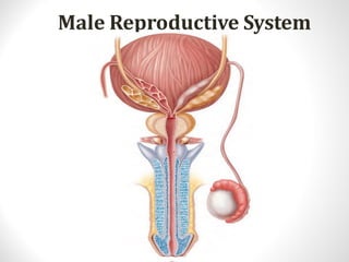
  • 2. Male Reproductive System • The reproductive system in men has components in the abdomen, pelvis, and perineum.
  • 3. • The major components are a testis, epididymis, ductus deferens, and ejaculatory duct on each side, and the urethra and penis in the midline. • Three types of accessory glands are associated with the system: – A single prostate; – A pair of seminal vesicles; – A pair of bulbourethral glands.
  • 5. The scrotum • The scrotum is an outpouching of the lower part of the anterior abdominal wall. • It contains the testes, the epididymides, and the lower ends of the spermatic cords.
  • 6. The scrotum • It is divided on its surface into two compartments by a raphé, which is continued forward to the under surface of the penis, and backward, along the middle line of the perineum to the anus. • Each compartment contains one of the two testes, and one of the epididymides.
  • 7. The scrotum • The wall of the scrotum has the following layers: – Skin – Superficial fascia – Spermatic fasciae – Tunica vaginalis
  • 8. The scrotum Skin •The skin of the scrotum is thin, wrinkled, and pigmented and forms a single pouch. A slightly raised ridge in the midline indicates the line of fusion of the two lateral labioscrotal swellings. Superficial fascia •This is continuous with the fatty and membranous layers of the anterior abdominal wall. •The fat is replaced by smooth muscle called the dartos muscle. •This is innervated by sympathetic nerve fibers and is responsible for the wrinkling of the overlying skin.
  • 9. The scrotum Spermatic fasciae •It has three layers which lie beneath the superficial fascia and are derived from the three layers of the anterior abdominal wall on each side. •The external spermatic fascia is derived from the aponeurosis of the external oblique muscle; the cremasteric fascia is derived from the internal oblique muscle; and, finally, the internal spermatic fascia is derived from the fascia transversalis. Tunica vaginalis This lies within the spermatic fasciae and covers the anterior, medial, and lateral surfaces of each testis.
  • 10.
  • 11. Lymph Drainage of the Scrotum • Lymph from the skin and fascia, including the tunica vaginalis, drains into the superficial inguinal lymph nodes .
  • 12. Testes • Testis has ellipsoid- shaped. • Testes develop in the abdomen and move before birth into the scrotum. • The left testis usually lies at a lower level than the right.
  • 13.
  • 14. Testes • The testis are covered by: • A closed sac of peritoneum (the tunica vaginalis), which originally connected to the abdominal cavity. Normally after testicular descent, the connection closes, leaving a fibrous remnant. • It is covered by a fibrous capsule called the tunica albuginea.
  • 15. Testes • In the inner surface of the capsule is a series of fibrous septa that divide the interior of the organ into lobules. • Lying within each lobule are 1 to 3 coiled seminiferous tubules. • The tubules open into a network of channels called the rete testis. • Small efferent ductules connect the rete testis to the upper end of the epididymis.
  • 17. Epididymis • The epididymis is a single, long coiled duct that courses along the posterolateral side of the testis. • The tunica vaginalis covers the epididymis with the exception of the posterior border.
  • 18. Epididymis • Structurally, epididymis divided into : – Head – Body – tail
  • 19. Epididymis It has two distinct components: –The efferent ductules, which form an enlarged coiled mass that sits on the posterior superior pole of the testis and forms the head of the epididymis; –The true epididymis, which is a single, long coiled duct into which the efferent ductules all drain, and which continues inferiorly along the posterolateral margin of the testis as the body of epididymis and enlarges to form the tail of epididymis at the inferior pole of the testis.
  • 20. Arterial Blood Supply of the Testis and Epididymis • The testicular artery is a branch of the abdominal aorta.
  • 21. • The testicular veins emerge from the testis and the epididymis as a venous network, the pampiniform plexus. • This becomes reduced to a single vein as it ascends through the inguinal canal. • The right testicular vein drains into the inferior vena cava, and the left vein joins the left renal vein.
  • 22. Lymphatic Drainage of The Testes • Lymphatic drainage of the testes is to the para- aortic lymph nodes.
  • 23. Ductus deferens • (Latin: "carrying-away vessel"), also called vas deferens. • The ductus deferens is a long muscular duct that transports spermatozoa from the tail of the epididymis to the ejaculatory duct.
  • 24. Ductus deferens course • The vas arises from the tail of the epididymis and traverses the inguinal canal to the deep ring, passes downwards on the lateral wall of the pelvis almost to the ischial tuberosity and turns medially to cross the ureter posterior to the bladder. • It continues inferomedially along the base of the bladder, anterior to the rectum, almost to the midline, where it is joined by the duct of the seminal vesicle to form the ejaculatory duct.
  • 25. Ductus deferens course • The terminal part of the vas deferens is dilated to form the ampulla of the vas deferens. • The ejaculatory duct penetrates through the prostate gland to connect with the prostatic urethra.
  • 26. Seminal vesicle • The seminal vesicles are an accessory gland of the male reproductive system . • The seminal vesicles are two lobulated organs about 2 in. (5 cm) long lying on the posterior surface of the bladder .
  • 27. Seminal vesicle • On the medial side of each vesicle lies the terminal part of the vas deferens. • Posteriorly, the seminal vesicles are related to the rectum. • Inferiorly, each seminal vesicle narrows and joins the vas deferens of the same side to form the ejaculatory duct.
  • 28. Blood Supply of Seminal vesicle Arteries •The arterial blood supply from, the inferior vesicle and middle rectal arteries. Veins • The veins drain into the internal iliac veins.
  • 29. Ejaculatory Ducts • The two ejaculatory ducts are each less than 1 in. (2.5 cm) long and are formed by the union of the vas deferens and the duct of the seminal vesicle. • The ejaculatory ducts pierce the posterior surface of the prostate and open into the prostatic part of the urethra, close to the margins of the prostatic utricle; their function is to drain the seminal fluid into the prostatic urethra.
  • 30.
  • 31. Semen • Semen is a milky white, somewhat sticky mixture of sperm, testicular fluid, and accessory gland secretions. The liquid provides a transport medium and nutrients and contains chemicals that protect and activate the sperm and facilitate their movement.
  • 32. Semen contains several substances that play many roles: • Prostaglandins decrease the viscosity of mucus guarding the entry (cervix) of the uterus and stimulate reverse peristalsis in the uterus, facilitating sperm movement through the female reproductive tract. • The hormone relaxin and certain enzymes in semen enhance sperm motility. • Contained ATP provides energy. • Certain ingredients suppress the immune response in the female’s reproductive tract.
  • 33. • Antibiotic chemicals destroy some bacteria. • Clotting factors in semen coagulate it just after it is ejaculated. Coagulation causes the sperm to stick to the walls of the vagina and prevents the initially immobile sperm from draining out of the vagina. Soon after semen coagulates, fibrinolysin liquefies the sticky mass and the sperm swim out and begin their journey through the female duct system
  • 34. The Bulbo-Urethral Glands • The bulbo-urethral glands are pea-sized glands located inferior to the prostate in the urogenital diaphragm • They produce a thick, clear mucus, some of which drains into the spongy urethra and lubricates the glans penis when sexually excited. • The mucus neutralizes traces of acidic urine and lubricates the urethra just prior to ejaculation.
  • 35. Prostate • The prostate is an unpaired accessory structure of the male reproductive system that surrounds the urethra in the pelvic cavity . • It lies immediately inferior to the bladder, above the urogenital diaphragm, posterior to the pubic symphysis, and anterior to the rectum.
  • 36. Prostate • The prostate is shaped like an inverted rounded cone with a larger base, which is continuous above with the neck of the bladder, and a narrower apex, which rests below on the pelvic floor. • The inferolateral surfaces of the prostate are in contact with the levator ani muscles that together cradle the prostate between them.
  • 38. Prostate • The two ejaculatory ducts pierce the upper part of the posterior surface of the prostate to open into the prostatic urethra at the lateral margins of the prostatic utricle.
  • 39. Relations of Prostate Superiorly •The base of the prostate is continuous with the neck of the bladder. •The urethra enters the center of the base of the prostate. Inferiorly • The apex of the prostate lies on the upper surface of the urogenital diaphragm. • The urethra leaves the prostate just above the apex on the anterior surface
  • 40. Relations of Prostate Anteriorly •The prostate is related to the symphysis pubis. •The prostate is connected to the posterior aspect of the pubic bones by the puboprostatic ligaments. Laterally •The prostate is embraced by the anterior fibers of the levator ani. Posteriorly •The prostate is closely related to the anterior surface of the rectal ampulla and is separated from it by the rectovesical septum (fascia of Denonvilliers).
  • 41. Structure of the Prostate • Enclosed within thin dense fibrous capsule • Inner loose sheath derived from pelvic fascia is prostatic sheath͟ – Continuous inferiorly with superior fascia of urogenital diaphragm – Posteriorly it is part of rectovesical septum – Separates bladder, seminal vesicles and prostate from rectum • Prostatic venous plexus lies between fibrous capsule and prostatic sheath.
  • 42. Structure of the Prostate Prostate divided into: – Two lateral lobes – One median lobe – Anterior and posterior lobes
  • 43. Structure of the Prostate • Anterior – Tissue lying anterior to urethra – No glands; fibromuscular tissue only • Median – Cone-shaped region between ejaculatory ducts and urethra • Lateral (left & right) – Main mass of gland, continuous posteriorly – Separated by prostatic urethra • Posterior – Describes postero-medial part of lateral lobes palpable through rectum on DRE.
  • 44. Functions of Prostate • During ejaculation, prostatic smooth muscle contracts, squeezing the prostatic secretion into the prostatic urethra via several ducts. This fluid plays a role in activating sperm and accounts for up to one-third of the semen volume. It is a milky, slightly acid fluid that contains citrate (a nutrient source), several enzymes (including fibrinolysin, hyaluronidase, and acid phosphatase), and prostate-specific antigen (PSA).
  • 45. Blood Supply of The Prostate • Arterial supply – Arteries derived from internal pudenal, inferior vesical and middle rectal arteries (branches of internal iliac) • Venous drainage – Veins form prostatic venous plexus around sides and base of prostate – located between capsule and sheath – Drains into internal iliac veins – Also communicates with vesical venous plexus and vertebral venous plexuses.
  • 46. Lymphatics and innervation of Prostate gland • Lymphatic drainage – Lymph vessels terminate in internal iliac and sacral lymph • nodes – Some vessels from posterior surface pass with lymph • vessels froŵ ďladder to exterŶal iliaĐ LN’s • Innervation – Parasympathetic fibres arise from pelvic splanchnic nerves – Sympathetic fibres from inferior hypogastric plexuses
  • 47.
  • 48. Penis • The penis is a pendulous organ suspended from the front and sides of the pubic arch and containing the greater part of the urethra.
  • 49. Penis • It consists of internal root, external shaft, & glans. – Root: the portion of the penis that extends internally into the pelvic cavity. – Shaft: the length of the penis between the glans and the body. – Glans: the head of the penis; has many nerve endings. • Foreskin: a covering of skin over the penile glans.
  • 50. Penis
  • 51. Penis • The root of penis consists of the two crura, which are proximal parts of the corpora cavernosa attached to the pubic arch, and the bulb of penis, which is the proximal part of the corpus spongiosum anchored to the perineal membrane.
  • 52. Penis • The body of the penis is essentially composed of three cylinders of erectile tissue enclosed in a tubular sheath of fascia (Buck's fascia). • The erectile tissue is made up of two dorsally placed corpora cavernosa and a single corpus spongiosum applied to their ventral surface . • At its distal extremity, the corpus spongiosum expands to form the glans penis, which covers the distal ends of the corpora cavernosa. • On the tip of the glans penis is the slitlike orifice of the urethra, called the external urethral meatus.
  • 53. Penis
  • 54. External penile structures • Corona: the rim of the penile glans. • Frenulum: thin strip of skin connecting the glans to the shaft on the underside of the penis. Both are highly sensitive areas to the touch
  • 55. Blood Supply of The Penis Arteries •The corpora cavernosa are supplied by the deep arteries of the penis ; the corpus spongiosum is supplied by the artery of the bulb. •In addition, there is the dorsal artery of the penis. •All the above arteries are branches of the internal pudendal artery. Veins • The veins drain into the internal pudendal veins.
  • 56. Lymphatics and innervation of The Penis Nerve Supply Sensation •The nerve supply is from the pudendal nerve and the pelvic plexuses. Erectile function •Parasympathetic(excitat ory) •Sympathetic (inhibitory) Lymph Drainage •The skin of the penis is drained into the medial group of superficial inguinal nodes. •The deep structures of the penis are drained into the internal iliac nodes.
  • 57. Analagous structures in male and female sexual anatomy Male Glans Foreskin Shaft Clitoris Clitoral hood Scrotal sac Testes Female Labia minora Labia majora Ovaries
  • 58. Physiology of the Male Reproductive System The chief phases of the male sexual response are 1. Erection of the penis, which allows it to penetrate the female vagina 2. Ejaculation, which expels semen into the vagina
  • 59. Erection • Erection, enlargement and stiffening of the penis, results from engorgement of the erectile bodies with blood. It happens because of the parasympathetic function of arteriole. • Sexual excitement triggers a parasympathetic reflex that promotes release of nitric oxide (NO) locally. • Nitric oxide relaxes smooth muscle in the penile blood vessel walls, dilating these arterioles, and the erectile bodies fill with blood.
  • 60. • Expansion of the corpora cavernosa of the penis compresses their drainage veins, retarding blood outflow and maintaining engorgement. • The corpus spongiosum expands but not nearly as much as the cavernosa. Its function is to keep the urethra open during ejaculation. • The erect penis not bend or fold during intercourse. The longitudinal and circular arrangement of collagen fibers surrounding the penis prevents this problem.
  • 61. • Ejaculation • Ejaculation is the propulsion of semen from the male duct system. • Although erection is under parasympathetic control, ejaculation is under sympathetic control. • When impulses provoking erection reach a critical level, a spinal reflex is initiated, and a massive discharge of nerve impulses occurs over the sympathetic nerves serving the genital organs
  • 62. 1. The bladder sphincter muscle constricts, preventing expulsion of urine or reflux of semen into the bladder. 2. The reproductive ducts and accessory glands contract, emptying their contents into the urethra. 3. Semen in the urethra triggers a spinal reflex through somatic motor neurons. The bulbospongiosus muscles of the penis undergo a rapid series of contractions, propelling semen from the urethra at a speed of up to 500 cm/s (200 inches/s, close to 11 mi/h). These rhythmic contractions are accompanied by intense pleasure and many systemic changes, such as generalized muscle contraction, rapid heartbeat, and elevated blood pressure.
  • 63. Spermatogenesis. • The process of spermatogenesis encompasses the phases through the production of sperm. Conversion of the haploid spermatids to sperm cells is specifically called spermiogenesis. • Throughout spermatogenesis, descendants of the same spermatogonium remain closely attached to one another by cytoplasmic bridges. They are also surrounded by and connected to nonreplicating supporting cells called sustentocytes or Sertoli cells, which extend from the basal lamina to the tubule lumen.
  • 64. PHASES OF SPERMATOGENESIS • Spermatogonial Phase (Mitosis) • Spermatocyte Phase/Spermatocytosis (Meiosis) • Spermatid Phase (Spermiogenesis) 64
  • 67. PHASE II- MEOSIS 67 Prophase-22 days Largest cell
  • 71. The sustentocytes: • Provide nutrients and essential signals to the dividing cells, even telling them to live or die • Move the cells along to the lumen • Secrete testicular fluid (rich in androgens and metabolic bacids) that provides the transport medium for sperm in the lumen • Phagocytize faulty germ cells and the excess cytoplasm sloughed off as the spermatids transform into sperm • Produce chemical mediators (inhibin and androgen-binding protein) that help regulate spermatogenesis
  • 72.
  • 73.
  • 74. Transformation of a spermatid intoa functionalsperm. The stepwise process of spermiogenesis: 1. The Golgi apparatus packages the acrosomal enzymes, 2. The acrosome forms at the anterior end of the nucleus and the centrioles gather at the opposite end of the nucleus, 3. Microtubules elaborate to form the flagellum, 4. Mitochondria multiply and take up position around the proximal portion of the flagellum, and 5. Excess cytoplasm sloughs off. 6. Structure of an immature sperm than has just been released from a sustentocyte. 7. Structure of a fully mature sperm.