
Empfohlen
Weitere ähnliche Inhalte
Was ist angesagt?
Was ist angesagt? (20)
Andere mochten auch
Ähnlich wie Endoplasmic reticulum
Ähnlich wie Endoplasmic reticulum (20)
Kürzlich hochgeladen
Kürzlich hochgeladen (20)
Endoplasmic reticulum
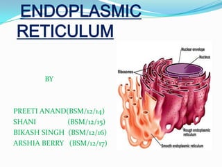
- 1. ENDOPLASMIC RETICULUM BY PREETI ANAND(BSM/12/14) SHANI (BSM/12/15) BIKASH SINGH (BSM/12/16) ARSHIA BERRY (BSM/12/17)
- 2. INTRODUCTION In the year 1945- The lace like membranes of the endoplasmic reticulum were first seen in the cytoplasm of chick embryo cells. These are membrane bound channels, seen in the form of a network of delicate strands and vesicles in the cytoplasm. These are single membrane cell organelles. These form an interconnected network of tubules, vesicles and cisternae with in cells. ER are considered as one of the components of cytoskeleton along with microtubules,microfilaments and intermediate filaments. These are first of all observed by Porter, Claude and Full am in (1945) as a network. The term ”Endoplasmic reticulum” was first used by Porter and Fullman (1952)
- 3. Location Present in almost all eukaryotic cell. These are found to be absent in mature erythrocytes,ova,embryonic cells and prokaryotes. The ER often occupies most of the cytoplasm. Amount • The ER varies in amount from cell to cell. In spermatocytes, it is represented by a few vacuoles only. • In the cells of adipose tissue, it is quite simple, having the form of a few tubules. • The cells that are actively synthesizing proteins, such as liver and pancreatic cells and fibroblast, have abundant ER. • Endoplasmic reticulum forms 30-60 % of the total membrane in a cell.
- 4. Origin of endoplasmic reticulum At present manner of origin of the endoplasmic is not definitely known. The most concrete hypothesis is that the ER is “budded” off from the nuclear envelope (wischnitzer, 1974). The ER appears to arise from the outer membrane of the nuclear envelope by out folding , or from the plasma membrane by in folding. The smooth ER seem to arise from the rough ER by detachment of ribosomes.
- 5. There are two basic morphological types of ER namely RER and SER. The ER membrane is thinner (50 Ǻ) than that of plasma membrane (80-100Ǻ thick)
- 6. PHYSICAL STRUCTURE The ER is 3-dimensional network of intracellular. It is formed of three types of element: 1-Cisternae 2-Tubules 3-Vesicles Cisternae These are flattened , unbranched, sac-like element. They lie in stacks parallel to one another. They bear ribosomes on the surface that, therefore, appears rough. It contain glycoproteins named ribophorin-I & ribophorin-II that bind the ribosomes.
- 7. Tubules These are irregular branching element which form a network along with other element. These are often free of ribosomes. Vesicles These are oval and rounded ,vacuole like element. These are also free of ribosomes. All the element of ER freely communicates with one another, and contain a fluid called endoplasmic matrix, in the ER lumen. These matrix is different from cytoplasmic matrix outside the ER The ER may pass from one cell to another through the plasmodesmata in the form of desmotubules.
- 10. Molecular structure The membrane of ER are composed of two layers of phospholipid molecules sandwiched by two layers of proteins molecules like other membrane in the cell wall. Types The endoplasmic reticulum is of two types: 1-Smooth endoplasmic reticulum (SER) 2-Rough endoplasmic reticulum (RER)
- 11. Endoplasmic Matrix The space inside the tubules and vesicles is filled with a watery medium that is different from the fluid in the cytosol outside the ER. Their walls are constructed of lipid bilayers membranes that contains large amount of proteins , similar to the cell membrane.
- 12. Smooth ER Smooth ER is an arrangement of tubules,vesicles and sacs. The size and structure of the SER varies between the cells. The SER can change within a cells lifetime to allow the cell to adapt to changes in its function and requirements. There are no ribosome’s attached to the membrane surface. The SER is connected to the nuclear envelope
- 13. The network of the SER allows there to be enough surface area for the action or storage of key enzymes or the products of the enzymes. The SER is less stable. The SER is characteristic of cells in which synthesis of non-protein substances takes place.
- 14. ROUGH ENDOPLASMIC RETICULIM (RER) The surface of the RER is studded with ribosome, giving it a rough appearance. It mainly consists of cisternae. The membrane of the RER forms large double membrane sheets Which is located near and continuous with the outer layer of the nuclear envelope. RER is very imp. in the synthesis and packaging of proteins e:g, Russell’s bodies of plasma, nissel’s granules of nerve cell Binding site of the ribosome on the RER is the translocon .
- 15. The ribosomes bound to the RER at any one time are not a stable part of this organelles structure Because ribosomes are constantly being bound and released from the membranes. Ribosomes only binds to the RER once a specific protein-nucleic acid complex forms in the cytosol. This special complex forms when a free ribosome begins translating the mRNA of a protein destined for the secretory pathway. The first 5-30 amino acid polymerized encode a single peptide, a molecular message that is recognized and bound by a single recognition particle (SRP).
- 16. The ribosomes that become attached to the endoplasmic reticulum synthesize all trans membrane proteins. Most secreted proteins that are stored in the Golgi apparatus, lysosomes, and endosomes. Translation pauses and the ribosomes complex binds to the RER translocon
- 17. Protein Transport As proteins are formed in the endoplasmic reticulum, they are transported through the tubules toward proteins of the SER that lie nearest to Golgi apparatus. At this point, small transport vesicles composed of small envelopes of smooth ER continually break away and diffuse to the deepest layer of Golgi apparatus. Inside this vesicles are the synthesized proteins and other product from the ER present.
- 18. Transport vesicles They are surrounded by coating protein called COP I, COP II.(Coat Protein complex) COP II targets vesicles to the Golgi apparatus. Transparent proteins from the RER to Golgi apparatus. This process is termed as anterograde transport. COP I transports proteins from the cis end of the Golgi complex back to the RER. This process is termed as retrograde transport.
- 21. Second method of transport out of the endoplasmic reticulum involves areas called membrane contact sites. Where membranes of the endoplasmic reticulum and other organelles are held together , allowing the transfer of lipid and other small molecules.
- 22. FUNCTION OF ERROUGH ENDOPLASMIC RETICULUM (RER) SMOOTH ENDOPLASMIC RETICULUM (SER) SACROPLASMIC RETICULUM (SR)
- 23. FUNCTION OF RER Surface for Ribosomes- The RER provides space and ribophorins for the attachment of ribosomes to itself. Surface for protein synthesis Formation of Glycoprotein- Linking of sugars to for glycoprotein starts in the RER and is completed in Golgi complex. Synthesis of precursors- The RER produce enzyme precursors for the formation of lysosomes by Golgi Complex. Smooth ER formation- The RER gives rise to the smooth ER by loss of ribosomes.
- 24. FUNCTION OF SER The smooth endoplasmic reticulum lacks ribosomes and functions in lipid metabolism, carbohydrate metabolism, and detoxification and is especially abundant in mammalian liver and gonad cells. It also synthesizes phospholipids. Cells which secrete these products, such as those in the testes, ovaries, and skin oil glands have a great deal of smooth endoplasmic reticulum.
- 25. Detoxification-The SER brings about detoxification in the liver , i.e., converts harmful materials(drugs, poisons) into harmless ones for excretion by the cell. Formation of organelles- The SER produces Golgi apparatus , lysosomes and vacuoles. It also carries out the attachment of receptors on cell membrane proteins and steroid metabolism. In muscle cells, it regulates calcium ion concentration The smooth endoplasmic reticulum also contains the enzyme glucose-6-phosphatase, which converts glucose-6-phosphate to glucose, a step in gluconeogenesis.
- 26. SR The sarcoplasmic reticulum (SR) is smooth ER found in smooth and striated muscle. The only structural difference between this organelle and the smooth endoplasmic reticulum is the medley of proteins they have, both bound to their membranes and drifting within the confines of their lumens. This fundamental difference is indicative of their functions. The endoplasmic reticulum synthesizes molecules, while the sarcoplasmic reticulum stores and pumps calcium ions.
- 27. The sarcoplasmic reticulum contains large stores of calcium, which it sequesters and then releases when the muscle cell is stimulated. It plays a major role in excitation-contraction coupling in muscles cells.
- 28. Sarcoplasmic reticulum in the excitationcontraction coupling in muscle cells.
- 29. THANK YOU