
Empfohlen
Weitere ähnliche Inhalte
Was ist angesagt?
Was ist angesagt? (20)
Ähnlich wie Cerebrovascular accident
Ähnlich wie Cerebrovascular accident (20)
Kürzlich hochgeladen
Kürzlich hochgeladen (20)
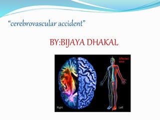
Cerebrovascular accident
- 5. BLOOD SUPPLY TO THE BRAIN Blood is supplied to the brain by two major pairs of arteries-Internal carotid &Vertebral arteries
- 7. INTRODUCTION CVA is a term used to described neurological changes caused by interruption in the blood supply to a part of the brain. A stroke, or brain attack or cerebrovascular accident, is the rapid loss of brain function due to disturbance in the blood supply to the brain. A stroke is a medical emergency and can cause permanent neurological damage and death.
- 8. DEFINITION Stroke or CVA as the rapid development of clinical signs and symptoms of a focal neurological disturbance lasting more than 24 hours or leading to death with no apparent cause other than vascular origin (WHO, 2005).
- 9. EPIDEMIOLOGY Stroke is the: Third most common cause of death First leading cause of disability 25% with initial stroke die within 1 year 50-75% will be functionally independent 25% will live with permanent disability
- 10. CAUSES OF CVA A stroke occurs when the blood supply to the brain are interrupted or reduced, this deprives brain of oxygen and nutrients which cause brain cell to die. The major causes of CVA are: Ischemia thrombosis Embolism Haemorrhage Transient ischemic attack
- 11. RISK FACTORS (Modifiable) (1) Atherosclerosis (narrowing of the arteries)
- 13. (4) Untreated Irregular Heart Beats
- 14. (5) Smoking (6) Diabetes
- 15. Other risk factors are: Obesity Physical inactivity Use of ilicit drugs,such as: coccaine Drinking alcohal Oral contraceptives use High cholesterol hypercoagulability
- 16. TYPES OF CVA Stroke can be divided into two major categories: ischemic stroke (85%) in which vascular occlusion and significant hypoperfusion occur and hemorrhagic (15%), in which there is extravasations of blood into the brain or subarachnoid space
- 17. Ischemic stroke results from inadequate blood supply to the part of the brain from a partial or complete occlusion of an artery that supply blood to the brain. This accounts for approximately 85% of all the stroke. Ischemic stroke are further classified into : Thrombolytic stroke: a thrombolytic stroke occurs from injury to the blood vessels walls and formation of blood clots . A clots may be caused by fatty deposit that builds up in artery (atherosclerosis), the lumen of the blood vessels may become narrowed and occluded and then infraction may occur
- 18. Embolic stroke: it occurs when a blood clots and other debris forms away from the brain, commonly in the heart and travels through the blood stem and lodges in narrower brain artery resulting in occlusion and infraction.
- 19. Ischemic stroke
- 20. Both result in a lack of blood flow to the brain and buildup of blood that puts too much pressure on the brain.
- 21. Hemorrhagic stroke A hemorrhagic stroke occurs when a blood vessel on the brain's surface ruptures and fills the space between the brain and skull with blood (subarachnoid hemorrhage) or when a defective artery in the brain bursts and fills the surrounding tissue with blood (cerebral hemorrhage).
- 22. Types of hemorrhagic stroke are: Intracerebral hemorrhage: in this type of hemorrhagic stroke, an artery on and near the surface of the brain brust and spills into the surface of the surrounding brain tissue and damaging brain cells. Subarachnoid hemorrhage: in subarachnoid hemorrhage an artery on and near the brain brust and spills into the space between brain and skull.
- 25. PATHOPHYSIOLOGY Normally the brain received a constant flow of blood for a normal function as it is unable to store oxygen & glucose. The blood flow is important for the removal of metabolic waste like CO2. otherwise due to lack of blood flow, the brain may be damaged within a short period. The brain is hypoxia/anoxia due to different causes Cerebral ischemia Alter cerebral metabolism Permanent damage of brain cell within 3-5ms Neurological deficit seen in the patient
- 27. CLINICAL FEATURES Stroke symptoms typically starts suddenly over a second to a minute. The symptoms depends on the area of brain affected. The most extensive the area of the brain affected more functions are likely to be lost. Some forms of stroke can cause additional symptoms for e.g, in an intracranial hemorrhage ,the affected area may compress other structure. The symptoms associated with strokes are:
- 28. sudden onset of a severe headache – “worst headache of one’s life” (specially in hemorrhagic and embolic stroke) Change or Loss Of Conscious nausea, vomiting, seizures, stiff neck Cushing triad: hypertension, bradycardia,respiratory changes
- 29. Hemiparesis & hemiplesia Aphasia & Dysphasia Visual changes Honer’s syndrome(paralysis of nerve of the one eye) Agnosia (disturbances in ability to recognize familiar object) Sensory deficit & behaviour changes Incontinence & Unilateral neglate • Homonymous hemianopsia – defective vision or blindness right or left halves of visual fields of both eyes
- 30. o Unilateral weakness, Numbness, Tingling Swallowing difficulty Bladder/bowel incontinence Pupillary abnormalities, Nystagmus Hemiplegia, Hemi paresis Facial droop, Seizures Confusion, Coma Hypertonicity
- 32. Clinical Manifestations Right Brain – Left Brain Damage
- 33. DIAGNOSIS WORK UP Laboratory CBC-for infection Platelets-identify haemorrhagic risk PT-identify coagulopathy Electrolytes Imaging CT scan-site of haemorrhage and ischemia MRI Angiography Carotid ultrasonography Arteriography
- 34. MANAGEMENT OF CVA Goals of management: Identify stroke early Maintain cerebral oxygenation Restore cerebral blood flow Prevent complication Rehabilitation after stroke
- 35. Identify the stroke early: identify the stroke using FAST treatment technique. FAST stands for: F: means face: if one side of the face drops it is the possible signs of CVA A: means Arms: If the person can not hold the both arms out it is the another possible stroke sign S: means speech: slurring of speech and poor understanding of simple language is another possible for stroke. T: means time if any of FAS signs are positive seek an attention. T in fast also means longer time the blockage of blood to the brain the more damage can occur.
- 37. MEDICAL MANAGEMENT Acute care: Proper positioning strictly at 30 degree head elevated Perform endotracheal intubation if necessary Cautiously lower blood pressure Avoid excessive hypotension Hyperventilation Give mannitol to minimize cerebral edema Give isotonic fluid eg normal saline Avoid hyperthermia Anticonvulsant drug
- 38. Specific pharmacological management :treatment of CVA depends up on the types of Stroke: Ischemic stroke: Anticoagulant medicine (warfarin) Antipletelet aggregating medicine (aspirin) Thrombolytic medicine tPA: (tissue plasminogen Activator) produce localized fibrinolysis by binding to the fibrin in the thrombi Embolic stroke: treat other underlying cause
- 39. SURGICAL MANAGEMENT A carotid endarterectomy Craniotomy coiling , binding and clipping of aneurysm
- 42. Transluminal angioplasty: insertion of the balloon to open the stenosed artery and improve blood flow. The balloon is threaded up into the carotid artery via the catheter inserted into the femoral artery.
- 43. NURSING MANAGEMENT Assessment: Change in the level of consciousness or responsiveness by using Glasgow coma scale Presence or absence of voluntary or involuntary movements of the extremities; muscle tone; body posture; and position of the head assess for any alteration in vital signs Eye opening, comparative size of pupils and pupillary reactions to light.
- 44. Assess for cranial nerve dysfunction Assess for skin breakdown, contracture, and other complications of immobility Assess for stiffness and flaccidity of neck Monitor for effectiveness of anticoagulant Assess for bowel and bladder function Assess for facial drop
- 45. Nursing Diagnosis Impaired physical mobility related to hemi paresis ,loss of balance ,and coordination and brain injury Acute pain related to hemiplegia and disuse Self care deficit related to hemiplegia. Disturbed sensory perception related to altered sensory reception transmission and integration. Impaired swallowing related to cranial nerve dysfunction
- 46. Incontinence of urine related to flaccid bladder Disturbed thought process related to brain damage Risk for develop contracture related to physical immobility Risk for impaired skin integrity related to hemi paresis and decreased movements Interrupted family coping process related to catastrophic illness and care giving burden Imbalance nutrition less than body requirements related to impaired self feeding chewing and swallowing
- 47. Nursing intervention: Preventing complication of immobility: Position to prevent contractures, use measures to relief pressure. Place the affected extremities slightly flexed on pillow with each joined positioned higher than the preceding one to prevent edema. Prevent foot drop by using foot boards Change position every 2 hourly Range of motion exercises
- 48. Use of stocking in legs to prevent deep vein thrombosis. Chest physiotherapy and suctioning to prevent chest infection Teach patient to use unaffected parts to move an affected parts Frequently assess for contractures Assist patient in ambulation if required.
- 49. Preventing shoulder pain: Never lift the patient by flaccid shoulder Use of sling when ambulating Range of motion exercise Elevate the arm and hand to prevent dependant edema Administer analgesic as prescribed
- 50. Optimizing cognitive abilities Be aware of the patients cognitive alertness and adjust environment accordingly Participation in cognitive retraining programs: reality orientation, visual imagery Focus on patient strength and give positive feedback In patient with increased awareness interact patient with is familiar objects and persons.
- 51. Preventing risk of injury: Keep the side rails of bed Frequent skin inspection for manifestation of complication Remind client to walk slowly, rest adequately between the interval of walking.
- 52. Improving nutritional status: Carefully assess the client diet to ensure adequate nutrition Encourage small frequent feeding and allow enough time to chew Remind patient to chew in unaffected side Feed the patient in upright position Give supplemental food as needed Inspect mouth for food collection and pocketing before entry of each bolus food
- 53. Reduce environmental distraction to improve patient concentration Provide frequent oral care If the patient can not swallow at all provide NG tube feeding
- 54. Facilitating communication: Speak at slower rate and give time to patient to respond Speak by using verbal cues and gesture and be consistent and repeat as necessary Listen and watch carefully when aphasic patient attempts to communicate Give planty of time to response and reinforce attempts as well as correct response Help the family to communicate with the patient and minimize distraction.
- 55. Fostering independence: Teach the patient to use unaffected side for activity of daily living but not to neglect affected side. Make sure personal care items are nearby patients obtain assistance with transfer and other activities as needed Encourage patient in positive achievement.
- 56. Attaining bladder control: Insert indwelling catheter during an acute phase for accurate fluid management and remove catheter as soon as status stabilization Establishing regular voiding schedule Strictly maintain urine intake and output recording Assist with standing or sitting to void
- 57. Strengthening family coping: Encourage the family to maintain outside interest Teach stress management technique such as relaxation technique, exercise Involves as many family and friends to participate in patients care Provide information about stroke and prognosis Teach the family that stroke survivors may show depression in the first 3 months of recovery
- 58. Patient education and health maintainence: Teach the patient and family to adopt home environment for safety and ease of use Instruct the patient of the need for rest provides throughout the day Reassure the family that it is common for post stroke patient to experience emotional liability and depression; treatment can be given Encourage consistency in environment without distraction
- 59. Assist the family to obtain self help aid for patient Instruct family in management of aphasia: e,g using alternate means of communication Educate those at risk of stroke about life style modification therapy can lower the risk.
- 60. COMPLICATIONS Aspiration pneumonia Dysphagia Deep vein thrombosis Pulmonary embolism Post stroke depression Brain stem herniation
- 61. keys of prevention.. 1. If you STOP….. 2. Reduce your intake of …. 3. Maintain your BP 4.
- 62. Remember..the key is prevention
- 63. PROGNOSIS Recovery rates vary depending on age and the part of the brain affected and the extent of the stroke. Function will be restored in more than half of individuals with moderate to severe paralysis on one side of the body Stroke (all types) has an overall mortality rate of 39.1 per 100,000 individuals. About 5% to 10% of stroke survivors have a second stroke within a year. But there's reason for hope: Although up to 30 percent of stroke survivors suffer some permanent disability, more than half recover functional independence after a stroke. Control of risk factors such as hypertension, atrial fibrillation, atherosclerosis, obesity, and high lipid levels is important to prevent additional strokes. Rehabilitation is a significant factor in stroke outcomes
- 64. Rehabilitation The sooner the better ! Consists of medical & nursing care Physical & occupational therapy Use of adaptive aids Begins in the hospital and continue at an outpatient clinic or at home. One of the most import factors in successful recovery is the person’s own motivation!
- 65. The goal of a stroke rehabilitation program is to help to relearn skills that lost when stroke affected part of brain. Stroke rehabilitation can help to regain independence and improve the quality of life. The severity of stroke complications and each person's ability to recover lost abilities varies widely. Researchers have found that the central nervous system is adaptive and can recover some functions. They also have found that it's necessary to keep practicing regained skills.
- 66. It's common for stroke rehabilitation to start as soon 24 to 48 hours after stroke, during acute hospital stay The duration of the stroke rehabilitation depends on the severity of the stroke and related complications. Although some stroke survivors recover quickly, most need some form of stroke rehabilitation long term, possibly months or years after their stroke.
- 73. Thank you !