SlideShare ist ein Scribd-Unternehmen logo
1 von 10
Downloaden Sie, um offline zu lesen
Ojvensha E learning Resources-Prepared by Dr.B.B.Gosai
Classification of Joints (Viva, Short notes, Short Questions)
Structural classification:
Fibrous joints: (***short note)
In this type of joints the bones are joined together by fibrous tissue.
Due to the presence of fibrous tissue these joints are either immovable or permit movement
to a very little extent.
Fibrous joints are further divided into the following subtypes;
1. Sutures: These are peculiar to skull and are immovable. According to the shape of
the bony margin the sutures may be;
 Plane sutures
 Serrate sutures
 Denticulate sutures
 Squamous sutures
 Limbous sutures
 Schindylesis
Types of Sutures
Ojvensha E learning Resources-Prepared by Dr.B.B.Gosai
2. Syndesmosis: In this type of fibrous joints the bones are connected with
interosseus ligament for example the inferior tibiofibular joint and Interosseous
membrane between Radius and Ulna, Tibia and Fibula.
Inferior tibiofibular joint
3. Gomphosis: These are also known as peg and socket joints. Examples are tooth in
the socket.
Gomphosis (Teeth in their Sockets)
Ojvensha E learning Resources-Prepared by Dr.B.B.Gosai
Cartilaginous joints: (***short note)
In this type of joints there is a piece of cartilage between the bones which hold the bones
together and makes a joint.
Cartilaginous joints are further divided into the following subtypes;
1. Primary cartilaginous joints: (synchondrosis): In this subtype the bones are
united by a plate of hyaline cartilage so that the joint is immovable and strong.
These joints are temporary in nature because after a certain age the cartilaginous
plate is replaced by the bone. Examples of this type of joints are joint between the
epiphyses and diaphysis of a growing long bone (Epiphyseal Plate), the
costochondral joint and the first chondrosternal joint.
Joint between epiphysis and diaphysis with intervening epiphyseal plate of
cartilage
2. Secondary cartilaginous joints: (symphysis): These are also known as fibro-
cartilaginous joints. There articular surface is covered by a thin layer of hyaline
cartilage and the bones are united by fibro-cartilage.
These joints are permanent and persist throughout the life of an individual.
Typically the secondary cartilaginous joints occur in the median plane of the body
and permit limited movements because of compressible pad of cartilage in them.
The thickness of the fibro-cartilage in these joints is directly related to the range of
movement the joint offers. Examples of this type of joints are; symphysis pubis,
manubrio-sternal joint and intervertebral joints between the vertebral bodies.
Ojvensha E learning Resources-Prepared by Dr.B.B.Gosai
Symphysis Pubis
Synovial joints:
Synovial joints are most evolved and therefore most mobile type of joints.
Functional classification:
Immovable joints: (Synarthroses)
They are fixed joints at which there is no movement. The articular surfaces are joined by tough
fibrous tissue. Often the edges of the bones are dovetailed into one another as in the sutures of
the skull.
Partially moveable joints: (Amphiarthroses)
They are joints in which slight movement is possible. A pad of cartilage lies between the bone
surface and there is a fibrous capsule to hold the bone and cartilage in place. The cartilages of
such joints also act as shock absorbers for example the intervertebral discs between the bodies
of vertebrae where the cartilage is strengthened by extra collagen fibers.
Freely moveable joints: (Diarthroses)
They are the synovial joints of structural classification. They are freely moveable though at
some of them the movement is restricted by the shape of the articulating surfaces and by the
ligaments which hold them together. These ligaments are of elastic connective tissue.
Ojvensha E learning Resources-Prepared by Dr.B.B.Gosai
Structure of Synovial Joint (***short note)
A typical Synovial Joint (Image source: Madhero88/Wikipedia)
Synovial joints are most evolved and therefore most mobile type of joints. They possess the
following characteristic features;
 There articular surfaces are covered with hyaline cartilage. This articular cartilage
is avascular, non nervous and elastic. Lubricated with synovial fluid, the cartilage
forms slippery surfaces for free movements.
 Between the articular surfaces there is a joint cavity filled with synovial fluid. The
cavity may be partially or completely subdivided by an articular disc known as
meniscus.
 The joint is surrounded by an articular capsule which is fibrous in nature and is
lined by synovial membrane. Because of its rich nerve supply the fibrous capsule is
sensitive to stretches imposed by movements.
 The synovial membrane lines the entire joint except the articular surfaces
covered by hyaline cartilage. It is this membrane that secretes the slimy fluid called
synovial fluid which lubricates the joint and nourishes the articular cartilage.
 Varying degrees of movements are always permitted by the synovial joints.
Ojvensha E learning Resources-Prepared by Dr.B.B.Gosai
Types of synovial joints: (***short note, viva and Short Questions)
Scheme of Types of Synovial Joints (Image Source: Produnis/Wikipedia)
Synovial joints are of the following types;
Plane synovial joints:
The articular surfaces of plane synovial joints are more or less plane.
These joints permit gliding movements in various directions.
Examples are intercarpal joints, intertarsal joints, and joints between the articular processes of
vertebrae.
Ojvensha E learning Resources-Prepared by Dr.B.B.Gosai
Plane Synovial Joints of Carpus
Hinge joints:
In these joints the articular surfaces are pulley shaped.
There are strong collateral ligaments to provide stability to the joint.
Movements are permitted in one plane around a transverse axis.
Examples are elbow joint, ankle joint, interphaleangeal joint.
Hinge Joint (Elbow Joint)
Pivot joints:
Pivot joints are formed by a central bony pivot surrounded by an osteo-ligamentous ring.
Movements are permitted in one plane around a vertical axis. Examples of this type are
superior and inferior radioulnar joints and the median atlantoaxial joint.
Proximal Radioulnar Joint
Ojvensha E learning Resources-Prepared by Dr.B.B.Gosai
Condylar joints:
These are also known as bicondylar joints. There articular surfaces consist of two distinct
condyles in which one is convex surface (called the male surface) fitting into a concave
surface (called the female surface) of the other bone. These joints mainly permit the
movement in plane around a transverse axis. Example of this type of joints is knee joint, TM
joint.
Knee Joint (Posterior View)
Ellipsoid joints:
In this case the articular surfaces include an oval convex male surface fitting into an ellipsoid
female surface.
The movements are permitted around two axis; flexion and extension around the transverse
axis and adduction and abduction round antero-posterior axis.
Combination of these movements produces Circumduction.
Typical rotation around a third vertical axis does not occur.
Examples of this type of joints are wrist joint, metacarpophalangeal joint and atlanto-occipital
joint.
Ojvensha E learning Resources-Prepared by Dr.B.B.Gosai
Wrist Joint (ellipsoid Joint)
Saddle joints:
Articular surfaces are reciprocally concavo-convex.
Movements are similar to those permitted by ellipsoid joint with addition of some rotation
(conjunct rotation (rotation which accompany other movements)) around a third axis which
occurs independently.
Examples of this type of joints are first carpometacarpal joint, sternoclavicular joint,
calcaneocuboid joint.
Saddle Joint
Ojvensha E learning Resources-Prepared by Dr.B.B.Gosai
Ball and socket joints:
These are also called spheroidal joints.
There articular surfaces include a globular head fitting into a cup shaped socket.
Movement occurs around an indefinite number of axes which have common center.
Flexion, extension, abduction, adduction, rotation, Circumduction all occur quite freely.
Examples of this type of joints are shoulder joint, hip joint and talo-calcaneonavicular joint.
Ball and Socket Joint (Hip Joint)
==================X================

Weitere ähnliche Inhalte

Was ist angesagt?

Was ist angesagt? (20)

Cartilage
CartilageCartilage
Cartilage
 
Synovial joints
Synovial jointsSynovial joints
Synovial joints
 
Skeletal System 1
Skeletal System 1Skeletal System 1
Skeletal System 1
 
Joints and its classification.
Joints and its classification.Joints and its classification.
Joints and its classification.
 
Classification of joints
Classification of jointsClassification of joints
Classification of joints
 
Cartilage
Cartilage Cartilage
Cartilage
 
General anatomy of muscles
General anatomy of muscles General anatomy of muscles
General anatomy of muscles
 
Arthrology
ArthrologyArthrology
Arthrology
 
basics of physiology
basics of physiologybasics of physiology
basics of physiology
 
Synovial joint
Synovial jointSynovial joint
Synovial joint
 
Axial skeleton parts 1 2
Axial skeleton parts 1  2Axial skeleton parts 1  2
Axial skeleton parts 1 2
 
The Joints
The JointsThe Joints
The Joints
 
Structure of synovial joint and muscle attachment
Structure of synovial joint and muscle attachmentStructure of synovial joint and muscle attachment
Structure of synovial joint and muscle attachment
 
Structure of long bone
Structure of long boneStructure of long bone
Structure of long bone
 
Joints (arthrology)
Joints (arthrology)Joints (arthrology)
Joints (arthrology)
 
Skeletal system
Skeletal systemSkeletal system
Skeletal system
 
Joints articulations
Joints  articulationsJoints  articulations
Joints articulations
 
Musculoskeletal System
Musculoskeletal System Musculoskeletal System
Musculoskeletal System
 
Veins arteries online
Veins arteries onlineVeins arteries online
Veins arteries online
 
Chap07 Bone Tissue
Chap07 Bone TissueChap07 Bone Tissue
Chap07 Bone Tissue
 

Ähnlich wie Classification of joints dr.gosai

Ähnlich wie Classification of joints dr.gosai (20)

JOINt.pptx
JOINt.pptxJOINt.pptx
JOINt.pptx
 
JOINTS.pptx
JOINTS.pptxJOINTS.pptx
JOINTS.pptx
 
joints.pdf
joints.pdfjoints.pdf
joints.pdf
 
mbbs ims msu
mbbs ims msumbbs ims msu
mbbs ims msu
 
anatomy of joints.pptx
anatomy of joints.pptxanatomy of joints.pptx
anatomy of joints.pptx
 
anatomy of joints
anatomy of jointsanatomy of joints
anatomy of joints
 
1 точно ушул .pdf
1 точно ушул .pdf1 точно ушул .pdf
1 точно ушул .pdf
 
Connection of bones
Connection of bonesConnection of bones
Connection of bones
 
THE JOINTS.pptx
THE JOINTS.pptxTHE JOINTS.pptx
THE JOINTS.pptx
 
Joints in human body
Joints in human bodyJoints in human body
Joints in human body
 
Lecture 1. (introduction to joint)
Lecture 1. (introduction to joint)Lecture 1. (introduction to joint)
Lecture 1. (introduction to joint)
 
joints of human beings with different types
joints of human beings with different typesjoints of human beings with different types
joints of human beings with different types
 
Nikku pdf on joints
Nikku pdf on jointsNikku pdf on joints
Nikku pdf on joints
 
Share_784_Joints.pdf
Share_784_Joints.pdfShare_784_Joints.pdf
Share_784_Joints.pdf
 
08 Articulations General Features
08 Articulations   General Features08 Articulations   General Features
08 Articulations General Features
 
JOINTS.pptx
JOINTS.pptxJOINTS.pptx
JOINTS.pptx
 
Joints anatomy and physiology
Joints anatomy and physiologyJoints anatomy and physiology
Joints anatomy and physiology
 
08 Articulations General Features
08 Articulations   General Features08 Articulations   General Features
08 Articulations General Features
 
Joints_Skeleton system.pptx
Joints_Skeleton system.pptxJoints_Skeleton system.pptx
Joints_Skeleton system.pptx
 
Cartilage & joints
Cartilage & jointsCartilage & joints
Cartilage & joints
 

Mehr von Dr.B.B. Gosai

AN 1.1 introduction anatomical terminology-Dr.Gosai
AN 1.1 introduction anatomical terminology-Dr.GosaiAN 1.1 introduction anatomical terminology-Dr.Gosai
AN 1.1 introduction anatomical terminology-Dr.GosaiDr.B.B. Gosai
 
Sexual differentiation-Dr.B.B.Gosai
Sexual differentiation-Dr.B.B.GosaiSexual differentiation-Dr.B.B.Gosai
Sexual differentiation-Dr.B.B.GosaiDr.B.B. Gosai
 
Vestibular and auditory apparatus-Dr.B.B.Gosai
Vestibular and auditory apparatus-Dr.B.B.GosaiVestibular and auditory apparatus-Dr.B.B.Gosai
Vestibular and auditory apparatus-Dr.B.B.GosaiDr.B.B. Gosai
 
Brachial and lumbosacral plexus-Dr.B.B.Gosai
Brachial and lumbosacral plexus-Dr.B.B.GosaiBrachial and lumbosacral plexus-Dr.B.B.Gosai
Brachial and lumbosacral plexus-Dr.B.B.GosaiDr.B.B. Gosai
 
Lower limb bones-joints-muscles-Dr.B.B.Gosai
Lower limb bones-joints-muscles-Dr.B.B.GosaiLower limb bones-joints-muscles-Dr.B.B.Gosai
Lower limb bones-joints-muscles-Dr.B.B.GosaiDr.B.B. Gosai
 
Upper limb bones-joints-muscles-Dr.B.B.Gosai
Upper limb bones-joints-muscles-Dr.B.B.GosaiUpper limb bones-joints-muscles-Dr.B.B.Gosai
Upper limb bones-joints-muscles-Dr.B.B.GosaiDr.B.B. Gosai
 
Introduction human movement apparatus-Dr.B.B.Gosai
Introduction human movement apparatus-Dr.B.B.GosaiIntroduction human movement apparatus-Dr.B.B.Gosai
Introduction human movement apparatus-Dr.B.B.GosaiDr.B.B. Gosai
 
Spinal cord dr.gosai
Spinal cord dr.gosaiSpinal cord dr.gosai
Spinal cord dr.gosaiDr.B.B. Gosai
 
White matter of cerebrum dr.gosai
White matter of cerebrum dr.gosaiWhite matter of cerebrum dr.gosai
White matter of cerebrum dr.gosaiDr.B.B. Gosai
 
Ventricles of brain dr.gosai
Ventricles of brain dr.gosaiVentricles of brain dr.gosai
Ventricles of brain dr.gosaiDr.B.B. Gosai
 
Anatomy of Breast in clinical perspective-Dr.Gosai
Anatomy of Breast in clinical perspective-Dr.GosaiAnatomy of Breast in clinical perspective-Dr.Gosai
Anatomy of Breast in clinical perspective-Dr.GosaiDr.B.B. Gosai
 
Pericardium and heart
Pericardium and heartPericardium and heart
Pericardium and heartDr.B.B. Gosai
 
Extraocular muscles dr.gosai
Extraocular muscles dr.gosaiExtraocular muscles dr.gosai
Extraocular muscles dr.gosaiDr.B.B. Gosai
 
Blood supply of brain dr.gosai
Blood supply of brain dr.gosaiBlood supply of brain dr.gosai
Blood supply of brain dr.gosaiDr.B.B. Gosai
 
Basal ganglia dr.gosai
Basal ganglia dr.gosaiBasal ganglia dr.gosai
Basal ganglia dr.gosaiDr.B.B. Gosai
 

Mehr von Dr.B.B. Gosai (20)

AN 1.1 introduction anatomical terminology-Dr.Gosai
AN 1.1 introduction anatomical terminology-Dr.GosaiAN 1.1 introduction anatomical terminology-Dr.Gosai
AN 1.1 introduction anatomical terminology-Dr.Gosai
 
Sexual differentiation-Dr.B.B.Gosai
Sexual differentiation-Dr.B.B.GosaiSexual differentiation-Dr.B.B.Gosai
Sexual differentiation-Dr.B.B.Gosai
 
Vestibular and auditory apparatus-Dr.B.B.Gosai
Vestibular and auditory apparatus-Dr.B.B.GosaiVestibular and auditory apparatus-Dr.B.B.Gosai
Vestibular and auditory apparatus-Dr.B.B.Gosai
 
Brachial and lumbosacral plexus-Dr.B.B.Gosai
Brachial and lumbosacral plexus-Dr.B.B.GosaiBrachial and lumbosacral plexus-Dr.B.B.Gosai
Brachial and lumbosacral plexus-Dr.B.B.Gosai
 
Lower limb bones-joints-muscles-Dr.B.B.Gosai
Lower limb bones-joints-muscles-Dr.B.B.GosaiLower limb bones-joints-muscles-Dr.B.B.Gosai
Lower limb bones-joints-muscles-Dr.B.B.Gosai
 
Upper limb bones-joints-muscles-Dr.B.B.Gosai
Upper limb bones-joints-muscles-Dr.B.B.GosaiUpper limb bones-joints-muscles-Dr.B.B.Gosai
Upper limb bones-joints-muscles-Dr.B.B.Gosai
 
Introduction human movement apparatus-Dr.B.B.Gosai
Introduction human movement apparatus-Dr.B.B.GosaiIntroduction human movement apparatus-Dr.B.B.Gosai
Introduction human movement apparatus-Dr.B.B.Gosai
 
Spinal cord dr.gosai
Spinal cord dr.gosaiSpinal cord dr.gosai
Spinal cord dr.gosai
 
White matter of cerebrum dr.gosai
White matter of cerebrum dr.gosaiWhite matter of cerebrum dr.gosai
White matter of cerebrum dr.gosai
 
Ventricles of brain dr.gosai
Ventricles of brain dr.gosaiVentricles of brain dr.gosai
Ventricles of brain dr.gosai
 
Anatomy of Breast in clinical perspective-Dr.Gosai
Anatomy of Breast in clinical perspective-Dr.GosaiAnatomy of Breast in clinical perspective-Dr.Gosai
Anatomy of Breast in clinical perspective-Dr.Gosai
 
Pericardium and heart
Pericardium and heartPericardium and heart
Pericardium and heart
 
Larynx
LarynxLarynx
Larynx
 
Pharynx dr.gosai
Pharynx dr.gosaiPharynx dr.gosai
Pharynx dr.gosai
 
Extraocular muscles dr.gosai
Extraocular muscles dr.gosaiExtraocular muscles dr.gosai
Extraocular muscles dr.gosai
 
Blood supply of brain dr.gosai
Blood supply of brain dr.gosaiBlood supply of brain dr.gosai
Blood supply of brain dr.gosai
 
Cerebellum dr.gosai
Cerebellum dr.gosaiCerebellum dr.gosai
Cerebellum dr.gosai
 
Basal ganglia dr.gosai
Basal ganglia dr.gosaiBasal ganglia dr.gosai
Basal ganglia dr.gosai
 
Brain stem dr.gosai
Brain stem dr.gosaiBrain stem dr.gosai
Brain stem dr.gosai
 
Cerebrum
CerebrumCerebrum
Cerebrum
 

Kürzlich hochgeladen

Alper Gobel In Media Res Media Component
Alper Gobel In Media Res Media ComponentAlper Gobel In Media Res Media Component
Alper Gobel In Media Res Media ComponentInMediaRes1
 
Paris 2024 Olympic Geographies - an activity
Paris 2024 Olympic Geographies - an activityParis 2024 Olympic Geographies - an activity
Paris 2024 Olympic Geographies - an activityGeoBlogs
 
Accessible design: Minimum effort, maximum impact
Accessible design: Minimum effort, maximum impactAccessible design: Minimum effort, maximum impact
Accessible design: Minimum effort, maximum impactdawncurless
 
Presiding Officer Training module 2024 lok sabha elections
Presiding Officer Training module 2024 lok sabha electionsPresiding Officer Training module 2024 lok sabha elections
Presiding Officer Training module 2024 lok sabha electionsanshu789521
 
Arihant handbook biology for class 11 .pdf
Arihant handbook biology for class 11 .pdfArihant handbook biology for class 11 .pdf
Arihant handbook biology for class 11 .pdfchloefrazer622
 
microwave assisted reaction. General introduction
microwave assisted reaction. General introductionmicrowave assisted reaction. General introduction
microwave assisted reaction. General introductionMaksud Ahmed
 
A Critique of the Proposed National Education Policy Reform
A Critique of the Proposed National Education Policy ReformA Critique of the Proposed National Education Policy Reform
A Critique of the Proposed National Education Policy ReformChameera Dedduwage
 
Introduction to ArtificiaI Intelligence in Higher Education
Introduction to ArtificiaI Intelligence in Higher EducationIntroduction to ArtificiaI Intelligence in Higher Education
Introduction to ArtificiaI Intelligence in Higher Educationpboyjonauth
 
mini mental status format.docx
mini    mental       status     format.docxmini    mental       status     format.docx
mini mental status format.docxPoojaSen20
 
POINT- BIOCHEMISTRY SEM 2 ENZYMES UNIT 5.pptx
POINT- BIOCHEMISTRY SEM 2 ENZYMES UNIT 5.pptxPOINT- BIOCHEMISTRY SEM 2 ENZYMES UNIT 5.pptx
POINT- BIOCHEMISTRY SEM 2 ENZYMES UNIT 5.pptxSayali Powar
 
Software Engineering Methodologies (overview)
Software Engineering Methodologies (overview)Software Engineering Methodologies (overview)
Software Engineering Methodologies (overview)eniolaolutunde
 
URLs and Routing in the Odoo 17 Website App
URLs and Routing in the Odoo 17 Website AppURLs and Routing in the Odoo 17 Website App
URLs and Routing in the Odoo 17 Website AppCeline George
 
18-04-UA_REPORT_MEDIALITERAСY_INDEX-DM_23-1-final-eng.pdf
18-04-UA_REPORT_MEDIALITERAСY_INDEX-DM_23-1-final-eng.pdf18-04-UA_REPORT_MEDIALITERAСY_INDEX-DM_23-1-final-eng.pdf
18-04-UA_REPORT_MEDIALITERAСY_INDEX-DM_23-1-final-eng.pdfssuser54595a
 
Organic Name Reactions for the students and aspirants of Chemistry12th.pptx
Organic Name Reactions  for the students and aspirants of Chemistry12th.pptxOrganic Name Reactions  for the students and aspirants of Chemistry12th.pptx
Organic Name Reactions for the students and aspirants of Chemistry12th.pptxVS Mahajan Coaching Centre
 
CARE OF CHILD IN INCUBATOR..........pptx
CARE OF CHILD IN INCUBATOR..........pptxCARE OF CHILD IN INCUBATOR..........pptx
CARE OF CHILD IN INCUBATOR..........pptxGaneshChakor2
 
Introduction to AI in Higher Education_draft.pptx
Introduction to AI in Higher Education_draft.pptxIntroduction to AI in Higher Education_draft.pptx
Introduction to AI in Higher Education_draft.pptxpboyjonauth
 
Kisan Call Centre - To harness potential of ICT in Agriculture by answer farm...
Kisan Call Centre - To harness potential of ICT in Agriculture by answer farm...Kisan Call Centre - To harness potential of ICT in Agriculture by answer farm...
Kisan Call Centre - To harness potential of ICT in Agriculture by answer farm...Krashi Coaching
 
PSYCHIATRIC History collection FORMAT.pptx
PSYCHIATRIC   History collection FORMAT.pptxPSYCHIATRIC   History collection FORMAT.pptx
PSYCHIATRIC History collection FORMAT.pptxPoojaSen20
 
Separation of Lanthanides/ Lanthanides and Actinides
Separation of Lanthanides/ Lanthanides and ActinidesSeparation of Lanthanides/ Lanthanides and Actinides
Separation of Lanthanides/ Lanthanides and ActinidesFatimaKhan178732
 

Kürzlich hochgeladen (20)

Staff of Color (SOC) Retention Efforts DDSD
Staff of Color (SOC) Retention Efforts DDSDStaff of Color (SOC) Retention Efforts DDSD
Staff of Color (SOC) Retention Efforts DDSD
 
Alper Gobel In Media Res Media Component
Alper Gobel In Media Res Media ComponentAlper Gobel In Media Res Media Component
Alper Gobel In Media Res Media Component
 
Paris 2024 Olympic Geographies - an activity
Paris 2024 Olympic Geographies - an activityParis 2024 Olympic Geographies - an activity
Paris 2024 Olympic Geographies - an activity
 
Accessible design: Minimum effort, maximum impact
Accessible design: Minimum effort, maximum impactAccessible design: Minimum effort, maximum impact
Accessible design: Minimum effort, maximum impact
 
Presiding Officer Training module 2024 lok sabha elections
Presiding Officer Training module 2024 lok sabha electionsPresiding Officer Training module 2024 lok sabha elections
Presiding Officer Training module 2024 lok sabha elections
 
Arihant handbook biology for class 11 .pdf
Arihant handbook biology for class 11 .pdfArihant handbook biology for class 11 .pdf
Arihant handbook biology for class 11 .pdf
 
microwave assisted reaction. General introduction
microwave assisted reaction. General introductionmicrowave assisted reaction. General introduction
microwave assisted reaction. General introduction
 
A Critique of the Proposed National Education Policy Reform
A Critique of the Proposed National Education Policy ReformA Critique of the Proposed National Education Policy Reform
A Critique of the Proposed National Education Policy Reform
 
Introduction to ArtificiaI Intelligence in Higher Education
Introduction to ArtificiaI Intelligence in Higher EducationIntroduction to ArtificiaI Intelligence in Higher Education
Introduction to ArtificiaI Intelligence in Higher Education
 
mini mental status format.docx
mini    mental       status     format.docxmini    mental       status     format.docx
mini mental status format.docx
 
POINT- BIOCHEMISTRY SEM 2 ENZYMES UNIT 5.pptx
POINT- BIOCHEMISTRY SEM 2 ENZYMES UNIT 5.pptxPOINT- BIOCHEMISTRY SEM 2 ENZYMES UNIT 5.pptx
POINT- BIOCHEMISTRY SEM 2 ENZYMES UNIT 5.pptx
 
Software Engineering Methodologies (overview)
Software Engineering Methodologies (overview)Software Engineering Methodologies (overview)
Software Engineering Methodologies (overview)
 
URLs and Routing in the Odoo 17 Website App
URLs and Routing in the Odoo 17 Website AppURLs and Routing in the Odoo 17 Website App
URLs and Routing in the Odoo 17 Website App
 
18-04-UA_REPORT_MEDIALITERAСY_INDEX-DM_23-1-final-eng.pdf
18-04-UA_REPORT_MEDIALITERAСY_INDEX-DM_23-1-final-eng.pdf18-04-UA_REPORT_MEDIALITERAСY_INDEX-DM_23-1-final-eng.pdf
18-04-UA_REPORT_MEDIALITERAСY_INDEX-DM_23-1-final-eng.pdf
 
Organic Name Reactions for the students and aspirants of Chemistry12th.pptx
Organic Name Reactions  for the students and aspirants of Chemistry12th.pptxOrganic Name Reactions  for the students and aspirants of Chemistry12th.pptx
Organic Name Reactions for the students and aspirants of Chemistry12th.pptx
 
CARE OF CHILD IN INCUBATOR..........pptx
CARE OF CHILD IN INCUBATOR..........pptxCARE OF CHILD IN INCUBATOR..........pptx
CARE OF CHILD IN INCUBATOR..........pptx
 
Introduction to AI in Higher Education_draft.pptx
Introduction to AI in Higher Education_draft.pptxIntroduction to AI in Higher Education_draft.pptx
Introduction to AI in Higher Education_draft.pptx
 
Kisan Call Centre - To harness potential of ICT in Agriculture by answer farm...
Kisan Call Centre - To harness potential of ICT in Agriculture by answer farm...Kisan Call Centre - To harness potential of ICT in Agriculture by answer farm...
Kisan Call Centre - To harness potential of ICT in Agriculture by answer farm...
 
PSYCHIATRIC History collection FORMAT.pptx
PSYCHIATRIC   History collection FORMAT.pptxPSYCHIATRIC   History collection FORMAT.pptx
PSYCHIATRIC History collection FORMAT.pptx
 
Separation of Lanthanides/ Lanthanides and Actinides
Separation of Lanthanides/ Lanthanides and ActinidesSeparation of Lanthanides/ Lanthanides and Actinides
Separation of Lanthanides/ Lanthanides and Actinides
 

Classification of joints dr.gosai

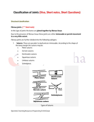
  • 1. Ojvensha E learning Resources-Prepared by Dr.B.B.Gosai Classification of Joints (Viva, Short notes, Short Questions) Structural classification: Fibrous joints: (***short note) In this type of joints the bones are joined together by fibrous tissue. Due to the presence of fibrous tissue these joints are either immovable or permit movement to a very little extent. Fibrous joints are further divided into the following subtypes; 1. Sutures: These are peculiar to skull and are immovable. According to the shape of the bony margin the sutures may be;  Plane sutures  Serrate sutures  Denticulate sutures  Squamous sutures  Limbous sutures  Schindylesis Types of Sutures
  • 2. Ojvensha E learning Resources-Prepared by Dr.B.B.Gosai 2. Syndesmosis: In this type of fibrous joints the bones are connected with interosseus ligament for example the inferior tibiofibular joint and Interosseous membrane between Radius and Ulna, Tibia and Fibula. Inferior tibiofibular joint 3. Gomphosis: These are also known as peg and socket joints. Examples are tooth in the socket. Gomphosis (Teeth in their Sockets)
  • 3. Ojvensha E learning Resources-Prepared by Dr.B.B.Gosai Cartilaginous joints: (***short note) In this type of joints there is a piece of cartilage between the bones which hold the bones together and makes a joint. Cartilaginous joints are further divided into the following subtypes; 1. Primary cartilaginous joints: (synchondrosis): In this subtype the bones are united by a plate of hyaline cartilage so that the joint is immovable and strong. These joints are temporary in nature because after a certain age the cartilaginous plate is replaced by the bone. Examples of this type of joints are joint between the epiphyses and diaphysis of a growing long bone (Epiphyseal Plate), the costochondral joint and the first chondrosternal joint. Joint between epiphysis and diaphysis with intervening epiphyseal plate of cartilage 2. Secondary cartilaginous joints: (symphysis): These are also known as fibro- cartilaginous joints. There articular surface is covered by a thin layer of hyaline cartilage and the bones are united by fibro-cartilage. These joints are permanent and persist throughout the life of an individual. Typically the secondary cartilaginous joints occur in the median plane of the body and permit limited movements because of compressible pad of cartilage in them. The thickness of the fibro-cartilage in these joints is directly related to the range of movement the joint offers. Examples of this type of joints are; symphysis pubis, manubrio-sternal joint and intervertebral joints between the vertebral bodies.
  • 4. Ojvensha E learning Resources-Prepared by Dr.B.B.Gosai Symphysis Pubis Synovial joints: Synovial joints are most evolved and therefore most mobile type of joints. Functional classification: Immovable joints: (Synarthroses) They are fixed joints at which there is no movement. The articular surfaces are joined by tough fibrous tissue. Often the edges of the bones are dovetailed into one another as in the sutures of the skull. Partially moveable joints: (Amphiarthroses) They are joints in which slight movement is possible. A pad of cartilage lies between the bone surface and there is a fibrous capsule to hold the bone and cartilage in place. The cartilages of such joints also act as shock absorbers for example the intervertebral discs between the bodies of vertebrae where the cartilage is strengthened by extra collagen fibers. Freely moveable joints: (Diarthroses) They are the synovial joints of structural classification. They are freely moveable though at some of them the movement is restricted by the shape of the articulating surfaces and by the ligaments which hold them together. These ligaments are of elastic connective tissue.
  • 5. Ojvensha E learning Resources-Prepared by Dr.B.B.Gosai Structure of Synovial Joint (***short note) A typical Synovial Joint (Image source: Madhero88/Wikipedia) Synovial joints are most evolved and therefore most mobile type of joints. They possess the following characteristic features;  There articular surfaces are covered with hyaline cartilage. This articular cartilage is avascular, non nervous and elastic. Lubricated with synovial fluid, the cartilage forms slippery surfaces for free movements.  Between the articular surfaces there is a joint cavity filled with synovial fluid. The cavity may be partially or completely subdivided by an articular disc known as meniscus.  The joint is surrounded by an articular capsule which is fibrous in nature and is lined by synovial membrane. Because of its rich nerve supply the fibrous capsule is sensitive to stretches imposed by movements.  The synovial membrane lines the entire joint except the articular surfaces covered by hyaline cartilage. It is this membrane that secretes the slimy fluid called synovial fluid which lubricates the joint and nourishes the articular cartilage.  Varying degrees of movements are always permitted by the synovial joints.
  • 6. Ojvensha E learning Resources-Prepared by Dr.B.B.Gosai Types of synovial joints: (***short note, viva and Short Questions) Scheme of Types of Synovial Joints (Image Source: Produnis/Wikipedia) Synovial joints are of the following types; Plane synovial joints: The articular surfaces of plane synovial joints are more or less plane. These joints permit gliding movements in various directions. Examples are intercarpal joints, intertarsal joints, and joints between the articular processes of vertebrae.
  • 7. Ojvensha E learning Resources-Prepared by Dr.B.B.Gosai Plane Synovial Joints of Carpus Hinge joints: In these joints the articular surfaces are pulley shaped. There are strong collateral ligaments to provide stability to the joint. Movements are permitted in one plane around a transverse axis. Examples are elbow joint, ankle joint, interphaleangeal joint. Hinge Joint (Elbow Joint) Pivot joints: Pivot joints are formed by a central bony pivot surrounded by an osteo-ligamentous ring. Movements are permitted in one plane around a vertical axis. Examples of this type are superior and inferior radioulnar joints and the median atlantoaxial joint. Proximal Radioulnar Joint
  • 8. Ojvensha E learning Resources-Prepared by Dr.B.B.Gosai Condylar joints: These are also known as bicondylar joints. There articular surfaces consist of two distinct condyles in which one is convex surface (called the male surface) fitting into a concave surface (called the female surface) of the other bone. These joints mainly permit the movement in plane around a transverse axis. Example of this type of joints is knee joint, TM joint. Knee Joint (Posterior View) Ellipsoid joints: In this case the articular surfaces include an oval convex male surface fitting into an ellipsoid female surface. The movements are permitted around two axis; flexion and extension around the transverse axis and adduction and abduction round antero-posterior axis. Combination of these movements produces Circumduction. Typical rotation around a third vertical axis does not occur. Examples of this type of joints are wrist joint, metacarpophalangeal joint and atlanto-occipital joint.
  • 9. Ojvensha E learning Resources-Prepared by Dr.B.B.Gosai Wrist Joint (ellipsoid Joint) Saddle joints: Articular surfaces are reciprocally concavo-convex. Movements are similar to those permitted by ellipsoid joint with addition of some rotation (conjunct rotation (rotation which accompany other movements)) around a third axis which occurs independently. Examples of this type of joints are first carpometacarpal joint, sternoclavicular joint, calcaneocuboid joint. Saddle Joint
  • 10. Ojvensha E learning Resources-Prepared by Dr.B.B.Gosai Ball and socket joints: These are also called spheroidal joints. There articular surfaces include a globular head fitting into a cup shaped socket. Movement occurs around an indefinite number of axes which have common center. Flexion, extension, abduction, adduction, rotation, Circumduction all occur quite freely. Examples of this type of joints are shoulder joint, hip joint and talo-calcaneonavicular joint. Ball and Socket Joint (Hip Joint) ==================X================