SlideShare ist ein Scribd-Unternehmen logo
1 von 20
Downloaden Sie, um offline zu lesen
Ojvensha E learning Resources-Prepared by Dr.B.B.Gosai
Brain stem
Introduction:
 It is part of central nervous system connecting spinal cord with the forebrain.
Location:
 Posterior cranial fossa on basilar part of occipital bone.
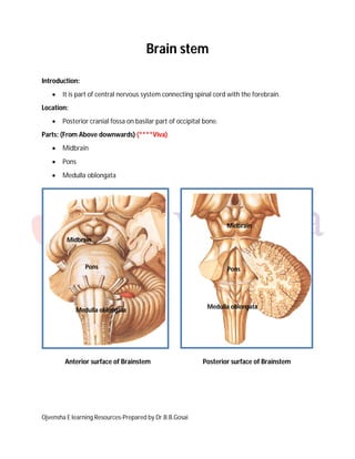
Parts: (From Above downwards) (****Viva)
 Midbrain
 Pons
 Medulla oblongata
Anterior surface of Brainstem Posterior surface of Brainstem
Midbrain
Pons
Medulla oblongata
Pons
Midbrain
Medulla oblongata
Ojvensha E learning Resources-Prepared by Dr.B.B.Gosai
Midbrain:
 Upper most part of brain stem.
 It connects brainstem to diencephalon and cerebrum.
External Features: (Midbrain) (***viva)
Anterior surface of Brainstem Posterior surface of Brainstem
Features on Anterior surface:
 Cerebral peduncles (Crus cerebri): Diverging peduncles are bundle of fibers seen on
anterior surface.
Features on Posterior surface:
 Colliculi: (All four colliculi together also known as Corpora Quadrigemina or tectum of
midbrain)
1. Two superior colliculi: (Concerned with Visuospinal reflex)
2. Two inferior colliculi: (Relay station of Auditory pathway)
Cerebral peduncle
Occulomotor nerve Trochlear nerve
Inferior colliculus
Superior colliculus
Ojvensha E learning Resources-Prepared by Dr.B.B.Gosai
Nerves emerging from Midbrain: (***Viva)
 Occulomotor nerve (III cranial nerve): emerging from medial aspect of cerebral
peduncles on anterior surface supplies all muscles of eyeball except Superior oblique
and Lateral rectus.
 Trochlear nerve (IV cranial nerve): emerging from posterior surface of midbrain below
inferior colliculus. (ONLY CRANIAL NERVE EMERGING FROM POSTERIOR SURFACE)
Internal Features: Internal features studied by two sections of midbrain:
1. Transverse Section (T.S.) at level of superior colliculus
2. Transverse Section (T.S.) at level of inferior colliculus
Transverse Section (T.S.) at level of superior colliculus:
Ojvensha E learning Resources-Prepared by Dr.B.B.Gosai
 Gray matter seen in the section: (Nuclei seen at this level) (***viva)
1. Occulomotor nucleus: in the periaqueductal region supplies all muscles of
eyeball except superior oblique and lateral rectus. It is also contain fibers of
Edinger Westphal nucleus (Parasympathetic nucleus) for light reflex and
accommodation reflex.
2. Red nucleus: red colous nucleus in tegmentum of midbrain concerned with
motor activity.
3. Substantia Nigra: Purple colour nucleus between crus cerebri and tegmentum.
They are dopaminergic neurons and damage to this neurons leads to Parkinson’s
disease.
4. Nucleus of superior colliculs: Center for Visuospinal reflex (Movement of
eyeball, head and neck and trunk while following a moving object for example
looking at the flying objects like plane).
 White matter seen in the section: (***viva)
1. Crus cerebri: containing corticopontine fibers from cortex to pontine nuclei and
corticonuclear and corticospinal tracts.
2. Spinal lemniscus: Bundle of fibers containing spinothalamic tracts to thalamus.
3. Trigeminal lemniscus: Bundle of fibers containing sensory fibers from trigeminal
nerve to thalamus.
4. Medial lemniscus: Bundle of fibers containing posterior column fibers from
spinal cord to thalamus.
Ojvensha E learning Resources-Prepared by Dr.B.B.Gosai
Transverse Section (T.S.) at level of inferior colliculus:
 Gray matter seen in the section: (Nuclei seen at this level) (***viva)
1. Troclear nucleus: in the periaqueductal region supplies superior oblique muscle
of eye
2. Substantia Nigra: Purple colour nucleus between crus cerebri and tegmentum.
They are dopaminergic neurons and damage to this neurons leads to Parkinson’s
disease.
3. Nucleus of inferior colliculs: Relay station of Auditory pathway of hearing.
 White matter seen in the section: (***viva)
1. Crus cerebri: containing corticopontine fibers from cortex to pontine nuclei and
corticonuclear and corticospinal tracts.
2. Superior cerebellar peduncle: Fibers from cerebellum cross in midbrain at this
level.
Ojvensha E learning Resources-Prepared by Dr.B.B.Gosai
3. Spinal lemniscus, Trigeminal lemniscus and Medial lemniscus same as section
at level of superior colliculus).
4. Lateral lemniscus: Bundle of fibers containing auditory pathway to inferior
colliculus for relay.
Applied anatomy of Midbrain: (***viva)
 Trauma to midbrain: due to sharpe edge of tentorium cerebelli.
 Ipsilateral paralysis of muscles of eye
 Dilated pupil and loss of accommodation reflex and light reflex
 Blockage of cerebral aqueduct:
 It leads to Hydrocephalus.
 Weber’s Syndrome:
 Occlusion of Posterior cerebral artery
 Involve Occulomotor nerve and crus cerebri.
 Ipsilateral ophthalmopegia
 Contralateral paralysis of lower part of face, tongue, arm and leg
 Eyeball deviated laterally (Paralysis of medial rectus)
 Drooping of eyelid (ptosis)
 Pupil is dilated and fixedto light and accomodation reflex
Ojvensha E learning Resources-Prepared by Dr.B.B.Gosai
 Benedict’s Syndrome:

 Occlusion of Posterior cerebral artery
 Similar to Weber’s syndrome with following differences:
 Involve Medial lemniscus and Red nucleus
 Contralateral hemianesthesia
 Involuntary movements of limbs on opposite side.
Ojvensha E learning Resources-Prepared by Dr.B.B.Gosai
Pons:
 Middle and widest part of brain stem.
 It is between medulla oblongata and midbrain.
Anterior surface of Brainstem Posterior surface of Brainstem
External Features: (***viva)
Features on Anterior surface:
 Basilar sulcus: Midline sulcus which is occupied by Basilar Artery.
 Transverse running fibres on the surface: Pontocerebellar fibres which continues as
Mddle cerebellar peduncle
 Trigeminal nerve emerge at lateral part of pons
Basilar Sulcus
Trigeminal nerve
Facial colliculus
Ojvensha E learning Resources-Prepared by Dr.B.B.Gosai
Features on Posterior surface: (***viva)
 Median sulcus in the midline
 Facial colliculus: Paramedian elevation raised by underlying abducent nucleus covered
by winding fibres of facial nerve
Nerves emerging from Pons: (***viva)
 Trigeminal nerve (V cranial nerve): emerging from lateral part. It demarcates between
pons and middle cerebellar peduncle.
Internal Features: Internal features studied by two sections of Pons:
 It is divided into two parts by transversely running fibers known as Trapezoid body:
 Basilar part: Anterior part
 Tegmentum: Posterior part
 Basilar part is same at both level of sections:
 Basilar part contains: Pontocerebellar tract (transversely running) & Corticospinal tract
(Vertically running) and dispersed Pontine nuclei
1. Transverse Section (T.S.) at level of Trigeminal nuclei
2. Transverse Section (T.S.) at level of Facial colliculus
Ojvensha E learning Resources-Prepared by Dr.B.B.Gosai
Transverse Section (T.S.) at level of Trigeminal nuclei:
 Gray matter seen in the section: (Nuclei seen at this level) (**viva)
1. Motor nucleus of trigeminal nerve: supply muscles of mastication.
2. Principal sensory nucleus of trigeminal nerve: Lateral to motor nucleus (Touch
and Pressure sensations from Face) (**viva)
 White matter seen in the section:
1. Trapezoid body (Fibers derived from cochlear nuclei and run transversely)
2. Medial lemniscus: Continuation of posterior column fibers
3. Spinal lemniscus: Spinothalamic tracts
4. Lateral Lemniscus: Continuation of auditory pathway
Ojvensha E learning Resources-Prepared by Dr.B.B.Gosai
Transverse Section (T.S.) at level of Facial colliculus:
 Gray matter seen in the section: (Nuclei seen at this level) (****Viva)
1. Facial nucleus: Supply muscles of facial expression. Facial nerve from facial
nucleus winds around the abducent nucleus.(Neurobiotaxis)
2. Abducent nucleus: supply Lateral rectus muscle of eye.
3. Vestibular and cochlear nuclei
 White matter seen in the section:
1. Trapezoid body
2. Medial longitudinal fasciculus
3. Spinal lemniscus, Medial Lemniscus and Lateral Lemniscus: (Same as above
section )
Ojvensha E learning Resources-Prepared by Dr.B.B.Gosai
Applied Anatomy of Pons
Tumors of pons:
 Occuring in childhood
 Weakness of facial muscles, Lateral rectus paralysis (Medial squint of eye) and
contralateral hemiparesis is characteristic of involvement of Pons.
 Nystagmus
 Impairment of hearing
 Weakness of jaw muscles
Pontine hemorrhage: (****viva)
 Hemorrhage from basilar artery
 “Pinpoint” pupil
 Facial paralysis same side
 Paralysis of muscles of opposite side
Ojvensha E learning Resources-Prepared by Dr.B.B.Gosai
Medulla Oblongata:
 Lower part of brain stem
 Continues as Spinal cord at foramen magnum
 Pyramidal in shape
Anterior surface of Brainstem Posterior surface of Brainstem
External Features: (Medulla Oblongata) (****Very important –Short note and viva)
Features on Anterior surface: (***viva)
 Anterior Median fissure-same like spinal cord
 Pyramidal dicussation-crossing over of corticospinal tract
 Pyramid-an paramedian elevation raised by corticospinal tract
 Olive- An oval shaped elevation lateral to pyramid
 Inferior Cerebellar peduncle: Bundle of fibres lateral to olive
Pyramid
VII
Vagal triangle
Hypoglossal triangle
VI
Olive Gracile and cuneate
tubercles
SCP
ICP
MCP
VIII
X
IX
XI
XII
Pyramidal decussation
Ojvensha E learning Resources-Prepared by Dr.B.B.Gosai
Features on Posterior surface: (***viva)
 Closed Part: lower part with posterior median sulcus, tractus gracilis & cuneatus,
Gracile & cuneate tubercles
 Open part: Vagal triangle & Hypoglossal triangle
Nerves emerging from Medulla oblongata: (***viva)
 Abducent nerve (VI cranial nerve): at ponto-medullary junction near pyramid
 Facial nerve (VII Cranial nerve) & Vestibulocochlear nerve (VIII Cranial nerve): at
pontomedullary junction laterally
 Glossopharyngeal (IX cranial nerve) , Vagus (X cranial nerve)and cranial part of
Accessory nerve (XI cranial nerve): Behind the olive
 Hypoglossal nerve XII Cranial nerve): Between the pyramid & olive
Internal Features: Internal features studied by three sections of Pons:
1. Transverse Section (T.S.) at level of Pyramidal decussation
2. Transverse Section (T.S.) at level of Sensory decussation
3. Transverse Section (T.S.) at the level of Open part of Medulla oblongata
Ojvensha E learning Resources-Prepared by Dr.B.B.Gosai
Transverse Section (T.S.) at level of Pyramidal decussation:
 Gray matter seen in the section:
1. Supraspinal nucleus continuation of Substantia gelatinosa (Nucleus for 1st
cervical nerve)
2. Spinal Accessory Nucleus (Supply Sternocleidomastoid and trapezius)
 White matter seen in the section:
1. Pyramidal Tracts
2. Pyamidal deccussation where 75 % fibres of Pyramidal tract (Corticospinal
teract) crosses to opposite side (Motor tract) and form lateral corticospinal
tract. (***viva)
3. Other ascending tracts
Ojvensha E learning Resources-Prepared by Dr.B.B.Gosai
Transverse Section (T.S.) at level of Sensory decussation:
 Gray matter seen in the section: (Nuclei at this level) (**viva)
1. Nucleus Gracilis & Cuneatus (Relay station for tectile descriminations, vibration
and conscious joint sensation)
2. Nucleus of Spinal Tract of trigeminal nerve (Pain and temperature from face)
 White matter seen in the section:
1. Pyramidal Tracts
2. Sensory deccussation where fibres of Gracile & Cuneate tract crosses to
opposite side by internal arcuate fibres and forms Medial Lemniscus (Posterior
column fibers) (***viva)
3. Lateral and Anterior spinothalamic tract combine to form Spinal Lemniscus
4. Other ascending tracts
Ojvensha E learning Resources-Prepared by Dr.B.B.Gosai
Transverse Section (T.S.) at level of Open part of Medulla oblongata (At the level of Olive):
(****Very important Section)
 Gray matter seen in the section: (Nuclei seen at this level) (***viva)
1. Hypoglossal Nucleus (Supply muscles of tongue)
2. Dorsal Vagal Nucleus (Parasympathetic to heart, lung and abdomen)
3. Vestibular nuclei(Balance) & Cochlear Nuclei (Hearing)
4. Inferior Olivary Nucleus (extrapyramidal motor)
5. Nucleus Ambiguus (Combine nucleus of 9th
,10th
and Cranial accessory nerve-
Pharynx, soft palate Larynx)
6. Nucleus of tractus solitarius (Taste sensation)
7. Nucleus of Spinal Tract of trigeminal nerve
Ojvensha E learning Resources-Prepared by Dr.B.B.Gosai
8. Arcuate nuclei: Displaced pontine nuclei
 White matter seen in the section:
1. Pyramidal Tracts
2. Medial Lemniscus
3. Tectospinal tract
4. Medial Longitudinal Fasciculus (Paramedian bundle- coordinate activities of
different nuclei)
5. Inferior cerebellar peduncle
6. Spinal lemniscus
7. Other ascending tracts
Applied Anatomy (Medulla oblongata):
Medulla oblongata contain dorsal vagal nucleus which is cardiorespiratory centre and
damage to this leads to Death. Hence it is also known as vital center.
Arnold-Chiari Malformation is congenital anomaly.
1. Herniation of tonsil of cerebellum and Medulla oblongata through Foramen
magnum.
2. Blockage of roof of fourth ventricle.
3. Internal Hydrocephalus.
4. Associated other anomalies like spina bifida.
Ojvensha E learning Resources-Prepared by Dr.B.B.Gosai
 Lateral Medullary Syndrome: (PICA Syndrome) (**viva)
 Blockage of Posterior Inferior Cerebellar Artery
 Damage to lateral part of Medulla
 Paralysis of palatal and laryngeal muscle
 Loss of balance
 Vertigo, nausea, vomiting, Nystagmus
 Dysphagia-difficulty in swallowing
 Dysarthria-difficulty in speech
 Loss of pain (Analgesia) and temperature sensations on face
 Ipsilateral Horner’s syndrome
Ojvensha E learning Resources-Prepared by Dr.B.B.Gosai
Medial Medullary Syndrome:
 Blockage of Vertebral artery
 Damage to Medial part
 Contralateral hemiplegia
 Ipsilateral Paralysis of muscles of tongue-tip pf tungue deviates to the
paralysed side.
 Contralateral loss of vibration, joint position and tectile discrimination
sensations
==================X================

Weitere ähnliche Inhalte

Was ist angesagt? (20)

Pectoral muscles
Pectoral musclesPectoral muscles
Pectoral muscles
 
Anatomy of cerebellum
Anatomy of cerebellumAnatomy of cerebellum
Anatomy of cerebellum
 
Brachial Plexus Anatomy
Brachial Plexus AnatomyBrachial Plexus Anatomy
Brachial Plexus Anatomy
 
The cerebellum
The cerebellumThe cerebellum
The cerebellum
 
The brainstem
The brainstemThe brainstem
The brainstem
 
Third ventricle
Third ventricleThird ventricle
Third ventricle
 
Cerebrum
CerebrumCerebrum
Cerebrum
 
The ribs
The ribsThe ribs
The ribs
 
Corpus callosum
Corpus callosumCorpus callosum
Corpus callosum
 
NEUROANATOMY OF BRAINSTEM
NEUROANATOMY OF BRAINSTEMNEUROANATOMY OF BRAINSTEM
NEUROANATOMY OF BRAINSTEM
 
Pyramidal tract by Sunita.M.Tiwale,Prof. Dept of physiology,D.Y.Patil Medical...
Pyramidal tract by Sunita.M.Tiwale,Prof. Dept of physiology,D.Y.Patil Medical...Pyramidal tract by Sunita.M.Tiwale,Prof. Dept of physiology,D.Y.Patil Medical...
Pyramidal tract by Sunita.M.Tiwale,Prof. Dept of physiology,D.Y.Patil Medical...
 
cervical and brachial plexus
cervical and brachial plexuscervical and brachial plexus
cervical and brachial plexus
 
Corticospinal tract (Pyramidal tract)
Corticospinal tract (Pyramidal tract)Corticospinal tract (Pyramidal tract)
Corticospinal tract (Pyramidal tract)
 
Spinal cord (2)
Spinal cord (2)Spinal cord (2)
Spinal cord (2)
 
Spinal cord
Spinal cordSpinal cord
Spinal cord
 
Cerebrum
CerebrumCerebrum
Cerebrum
 
Anatomy of basal ganglia
Anatomy of basal gangliaAnatomy of basal ganglia
Anatomy of basal ganglia
 
Lateral ventricle of Brain. By Dr.N.Mugunthan.M.S
Lateral ventricle of Brain. By Dr.N.Mugunthan.M.SLateral ventricle of Brain. By Dr.N.Mugunthan.M.S
Lateral ventricle of Brain. By Dr.N.Mugunthan.M.S
 
Cerebellum physiology
Cerebellum  physiologyCerebellum  physiology
Cerebellum physiology
 
Anatomy of anterior compartment of arm
Anatomy of anterior compartment of armAnatomy of anterior compartment of arm
Anatomy of anterior compartment of arm
 

Andere mochten auch (8)

Labelled Brain sections
Labelled Brain sectionsLabelled Brain sections
Labelled Brain sections
 
Pons by DR.TATHEER
Pons by DR.TATHEERPons by DR.TATHEER
Pons by DR.TATHEER
 
The Cerebral Cortex by Dr. NIda Kanwal, Neurosrugery LNH, Karachi
The Cerebral Cortex by Dr. NIda Kanwal, Neurosrugery LNH, KarachiThe Cerebral Cortex by Dr. NIda Kanwal, Neurosrugery LNH, Karachi
The Cerebral Cortex by Dr. NIda Kanwal, Neurosrugery LNH, Karachi
 
cerebrum sections for revision
cerebrum sections for revisioncerebrum sections for revision
cerebrum sections for revision
 
Chapt14 lecture (4)
Chapt14 lecture (4)Chapt14 lecture (4)
Chapt14 lecture (4)
 
Anatomy Of The Cerebellum Questions
Anatomy Of The Cerebellum QuestionsAnatomy Of The Cerebellum Questions
Anatomy Of The Cerebellum Questions
 
1 basal ganglia
1 basal ganglia1 basal ganglia
1 basal ganglia
 
blood supply of heart
blood supply of heartblood supply of heart
blood supply of heart
 

Ähnlich wie Brain stem anatomy

Ähnlich wie Brain stem anatomy (20)

Cerebellum dr.gosai
Cerebellum dr.gosaiCerebellum dr.gosai
Cerebellum dr.gosai
 
10-cerebellum.ppt
10-cerebellum.ppt10-cerebellum.ppt
10-cerebellum.ppt
 
Cerebellum
CerebellumCerebellum
Cerebellum
 
Nervous systems in vertebrates: T.Y.B.Sc. Sem VI Notes
Nervous systems in vertebrates: T.Y.B.Sc. Sem VI NotesNervous systems in vertebrates: T.Y.B.Sc. Sem VI Notes
Nervous systems in vertebrates: T.Y.B.Sc. Sem VI Notes
 
Ch11 Ppt Lect
Ch11 Ppt LectCh11 Ppt Lect
Ch11 Ppt Lect
 
Cerebellum and basal ganglia
Cerebellum and basal gangliaCerebellum and basal ganglia
Cerebellum and basal ganglia
 
Anatomy of brainstem and its clinical significance
Anatomy of brainstem and its clinical significanceAnatomy of brainstem and its clinical significance
Anatomy of brainstem and its clinical significance
 
Neuroanatomy Cerebellum-5 (Muhadharaty).pdf
Neuroanatomy Cerebellum-5 (Muhadharaty).pdfNeuroanatomy Cerebellum-5 (Muhadharaty).pdf
Neuroanatomy Cerebellum-5 (Muhadharaty).pdf
 
Cerebellum by Prof Soumya Chakraborty Bhattacharya
Cerebellum   by Prof Soumya Chakraborty BhattacharyaCerebellum   by Prof Soumya Chakraborty Bhattacharya
Cerebellum by Prof Soumya Chakraborty Bhattacharya
 
Anatomy of cerebellum and 4th ventricle
Anatomy of cerebellum and 4th ventricleAnatomy of cerebellum and 4th ventricle
Anatomy of cerebellum and 4th ventricle
 
Cerebellum
CerebellumCerebellum
Cerebellum
 
Cerebellum.ppt
Cerebellum.pptCerebellum.ppt
Cerebellum.ppt
 
Cerebellum by dr.gourav thakre 20 03-2012
Cerebellum by dr.gourav thakre 20 03-2012Cerebellum by dr.gourav thakre 20 03-2012
Cerebellum by dr.gourav thakre 20 03-2012
 
Anatomy of Orbit
Anatomy of Orbit Anatomy of Orbit
Anatomy of Orbit
 
Neuroanatomy (generally)
Neuroanatomy (generally)Neuroanatomy (generally)
Neuroanatomy (generally)
 
Cerebrum
CerebrumCerebrum
Cerebrum
 
Tracts of spinal cord (1)
Tracts of spinal cord (1)Tracts of spinal cord (1)
Tracts of spinal cord (1)
 
Cerebellum ppt
Cerebellum pptCerebellum ppt
Cerebellum ppt
 
Anatomy of Cerebellum 1
Anatomy of Cerebellum  1Anatomy of Cerebellum  1
Anatomy of Cerebellum 1
 
Midbrain
MidbrainMidbrain
Midbrain
 

Mehr von Dr.B.B. Gosai

AN 1.1 introduction anatomical terminology-Dr.Gosai
AN 1.1 introduction anatomical terminology-Dr.GosaiAN 1.1 introduction anatomical terminology-Dr.Gosai
AN 1.1 introduction anatomical terminology-Dr.GosaiDr.B.B. Gosai
 
Sexual differentiation-Dr.B.B.Gosai
Sexual differentiation-Dr.B.B.GosaiSexual differentiation-Dr.B.B.Gosai
Sexual differentiation-Dr.B.B.GosaiDr.B.B. Gosai
 
Vestibular and auditory apparatus-Dr.B.B.Gosai
Vestibular and auditory apparatus-Dr.B.B.GosaiVestibular and auditory apparatus-Dr.B.B.Gosai
Vestibular and auditory apparatus-Dr.B.B.GosaiDr.B.B. Gosai
 
Brachial and lumbosacral plexus-Dr.B.B.Gosai
Brachial and lumbosacral plexus-Dr.B.B.GosaiBrachial and lumbosacral plexus-Dr.B.B.Gosai
Brachial and lumbosacral plexus-Dr.B.B.GosaiDr.B.B. Gosai
 
Lower limb bones-joints-muscles-Dr.B.B.Gosai
Lower limb bones-joints-muscles-Dr.B.B.GosaiLower limb bones-joints-muscles-Dr.B.B.Gosai
Lower limb bones-joints-muscles-Dr.B.B.GosaiDr.B.B. Gosai
 
Upper limb bones-joints-muscles-Dr.B.B.Gosai
Upper limb bones-joints-muscles-Dr.B.B.GosaiUpper limb bones-joints-muscles-Dr.B.B.Gosai
Upper limb bones-joints-muscles-Dr.B.B.GosaiDr.B.B. Gosai
 
Introduction human movement apparatus-Dr.B.B.Gosai
Introduction human movement apparatus-Dr.B.B.GosaiIntroduction human movement apparatus-Dr.B.B.Gosai
Introduction human movement apparatus-Dr.B.B.GosaiDr.B.B. Gosai
 
Spinal cord dr.gosai
Spinal cord dr.gosaiSpinal cord dr.gosai
Spinal cord dr.gosaiDr.B.B. Gosai
 
Ventricles of brain dr.gosai
Ventricles of brain dr.gosaiVentricles of brain dr.gosai
Ventricles of brain dr.gosaiDr.B.B. Gosai
 
Classification of joints dr.gosai
Classification of joints dr.gosaiClassification of joints dr.gosai
Classification of joints dr.gosaiDr.B.B. Gosai
 
Anatomy of Breast in clinical perspective-Dr.Gosai
Anatomy of Breast in clinical perspective-Dr.GosaiAnatomy of Breast in clinical perspective-Dr.Gosai
Anatomy of Breast in clinical perspective-Dr.GosaiDr.B.B. Gosai
 
Pericardium and heart
Pericardium and heartPericardium and heart
Pericardium and heartDr.B.B. Gosai
 
Extraocular muscles dr.gosai
Extraocular muscles dr.gosaiExtraocular muscles dr.gosai
Extraocular muscles dr.gosaiDr.B.B. Gosai
 
Blood supply of brain dr.gosai
Blood supply of brain dr.gosaiBlood supply of brain dr.gosai
Blood supply of brain dr.gosaiDr.B.B. Gosai
 
Basal ganglia dr.gosai
Basal ganglia dr.gosaiBasal ganglia dr.gosai
Basal ganglia dr.gosaiDr.B.B. Gosai
 
Introduction abdomen-dr.gosai
Introduction abdomen-dr.gosaiIntroduction abdomen-dr.gosai
Introduction abdomen-dr.gosaiDr.B.B. Gosai
 
Esophagus stomach-dr.gosai
Esophagus stomach-dr.gosaiEsophagus stomach-dr.gosai
Esophagus stomach-dr.gosaiDr.B.B. Gosai
 
Anatomical correlation of local anesthesia in dentistry
Anatomical correlation of local anesthesia in dentistryAnatomical correlation of local anesthesia in dentistry
Anatomical correlation of local anesthesia in dentistryDr.B.B. Gosai
 

Mehr von Dr.B.B. Gosai (20)

AN 1.1 introduction anatomical terminology-Dr.Gosai
AN 1.1 introduction anatomical terminology-Dr.GosaiAN 1.1 introduction anatomical terminology-Dr.Gosai
AN 1.1 introduction anatomical terminology-Dr.Gosai
 
Sexual differentiation-Dr.B.B.Gosai
Sexual differentiation-Dr.B.B.GosaiSexual differentiation-Dr.B.B.Gosai
Sexual differentiation-Dr.B.B.Gosai
 
Vestibular and auditory apparatus-Dr.B.B.Gosai
Vestibular and auditory apparatus-Dr.B.B.GosaiVestibular and auditory apparatus-Dr.B.B.Gosai
Vestibular and auditory apparatus-Dr.B.B.Gosai
 
Brachial and lumbosacral plexus-Dr.B.B.Gosai
Brachial and lumbosacral plexus-Dr.B.B.GosaiBrachial and lumbosacral plexus-Dr.B.B.Gosai
Brachial and lumbosacral plexus-Dr.B.B.Gosai
 
Lower limb bones-joints-muscles-Dr.B.B.Gosai
Lower limb bones-joints-muscles-Dr.B.B.GosaiLower limb bones-joints-muscles-Dr.B.B.Gosai
Lower limb bones-joints-muscles-Dr.B.B.Gosai
 
Upper limb bones-joints-muscles-Dr.B.B.Gosai
Upper limb bones-joints-muscles-Dr.B.B.GosaiUpper limb bones-joints-muscles-Dr.B.B.Gosai
Upper limb bones-joints-muscles-Dr.B.B.Gosai
 
Introduction human movement apparatus-Dr.B.B.Gosai
Introduction human movement apparatus-Dr.B.B.GosaiIntroduction human movement apparatus-Dr.B.B.Gosai
Introduction human movement apparatus-Dr.B.B.Gosai
 
Spinal cord dr.gosai
Spinal cord dr.gosaiSpinal cord dr.gosai
Spinal cord dr.gosai
 
Ventricles of brain dr.gosai
Ventricles of brain dr.gosaiVentricles of brain dr.gosai
Ventricles of brain dr.gosai
 
Classification of joints dr.gosai
Classification of joints dr.gosaiClassification of joints dr.gosai
Classification of joints dr.gosai
 
Anatomy of Breast in clinical perspective-Dr.Gosai
Anatomy of Breast in clinical perspective-Dr.GosaiAnatomy of Breast in clinical perspective-Dr.Gosai
Anatomy of Breast in clinical perspective-Dr.Gosai
 
Pericardium and heart
Pericardium and heartPericardium and heart
Pericardium and heart
 
Larynx
LarynxLarynx
Larynx
 
Pharynx dr.gosai
Pharynx dr.gosaiPharynx dr.gosai
Pharynx dr.gosai
 
Extraocular muscles dr.gosai
Extraocular muscles dr.gosaiExtraocular muscles dr.gosai
Extraocular muscles dr.gosai
 
Blood supply of brain dr.gosai
Blood supply of brain dr.gosaiBlood supply of brain dr.gosai
Blood supply of brain dr.gosai
 
Basal ganglia dr.gosai
Basal ganglia dr.gosaiBasal ganglia dr.gosai
Basal ganglia dr.gosai
 
Introduction abdomen-dr.gosai
Introduction abdomen-dr.gosaiIntroduction abdomen-dr.gosai
Introduction abdomen-dr.gosai
 
Esophagus stomach-dr.gosai
Esophagus stomach-dr.gosaiEsophagus stomach-dr.gosai
Esophagus stomach-dr.gosai
 
Anatomical correlation of local anesthesia in dentistry
Anatomical correlation of local anesthesia in dentistryAnatomical correlation of local anesthesia in dentistry
Anatomical correlation of local anesthesia in dentistry
 

Kürzlich hochgeladen

The Most Excellent Way | 1 Corinthians 13
The Most Excellent Way | 1 Corinthians 13The Most Excellent Way | 1 Corinthians 13
The Most Excellent Way | 1 Corinthians 13Steve Thomason
 
Organic Name Reactions for the students and aspirants of Chemistry12th.pptx
Organic Name Reactions  for the students and aspirants of Chemistry12th.pptxOrganic Name Reactions  for the students and aspirants of Chemistry12th.pptx
Organic Name Reactions for the students and aspirants of Chemistry12th.pptxVS Mahajan Coaching Centre
 
URLs and Routing in the Odoo 17 Website App
URLs and Routing in the Odoo 17 Website AppURLs and Routing in the Odoo 17 Website App
URLs and Routing in the Odoo 17 Website AppCeline George
 
A Critique of the Proposed National Education Policy Reform
A Critique of the Proposed National Education Policy ReformA Critique of the Proposed National Education Policy Reform
A Critique of the Proposed National Education Policy ReformChameera Dedduwage
 
How to Make a Pirate ship Primary Education.pptx
How to Make a Pirate ship Primary Education.pptxHow to Make a Pirate ship Primary Education.pptx
How to Make a Pirate ship Primary Education.pptxmanuelaromero2013
 
APM Welcome, APM North West Network Conference, Synergies Across Sectors
APM Welcome, APM North West Network Conference, Synergies Across SectorsAPM Welcome, APM North West Network Conference, Synergies Across Sectors
APM Welcome, APM North West Network Conference, Synergies Across SectorsAssociation for Project Management
 
Contemporary philippine arts from the regions_PPT_Module_12 [Autosaved] (1).pptx
Contemporary philippine arts from the regions_PPT_Module_12 [Autosaved] (1).pptxContemporary philippine arts from the regions_PPT_Module_12 [Autosaved] (1).pptx
Contemporary philippine arts from the regions_PPT_Module_12 [Autosaved] (1).pptxRoyAbrique
 
Concept of Vouching. B.Com(Hons) /B.Compdf
Concept of Vouching. B.Com(Hons) /B.CompdfConcept of Vouching. B.Com(Hons) /B.Compdf
Concept of Vouching. B.Com(Hons) /B.CompdfUmakantAnnand
 
Industrial Policy - 1948, 1956, 1973, 1977, 1980, 1991
Industrial Policy - 1948, 1956, 1973, 1977, 1980, 1991Industrial Policy - 1948, 1956, 1973, 1977, 1980, 1991
Industrial Policy - 1948, 1956, 1973, 1977, 1980, 1991RKavithamani
 
Science 7 - LAND and SEA BREEZE and its Characteristics
Science 7 - LAND and SEA BREEZE and its CharacteristicsScience 7 - LAND and SEA BREEZE and its Characteristics
Science 7 - LAND and SEA BREEZE and its CharacteristicsKarinaGenton
 
PSYCHIATRIC History collection FORMAT.pptx
PSYCHIATRIC   History collection FORMAT.pptxPSYCHIATRIC   History collection FORMAT.pptx
PSYCHIATRIC History collection FORMAT.pptxPoojaSen20
 
Software Engineering Methodologies (overview)
Software Engineering Methodologies (overview)Software Engineering Methodologies (overview)
Software Engineering Methodologies (overview)eniolaolutunde
 
Kisan Call Centre - To harness potential of ICT in Agriculture by answer farm...
Kisan Call Centre - To harness potential of ICT in Agriculture by answer farm...Kisan Call Centre - To harness potential of ICT in Agriculture by answer farm...
Kisan Call Centre - To harness potential of ICT in Agriculture by answer farm...Krashi Coaching
 
MENTAL STATUS EXAMINATION format.docx
MENTAL     STATUS EXAMINATION format.docxMENTAL     STATUS EXAMINATION format.docx
MENTAL STATUS EXAMINATION format.docxPoojaSen20
 
Call Girls in Dwarka Mor Delhi Contact Us 9654467111
Call Girls in Dwarka Mor Delhi Contact Us 9654467111Call Girls in Dwarka Mor Delhi Contact Us 9654467111
Call Girls in Dwarka Mor Delhi Contact Us 9654467111Sapana Sha
 
Presiding Officer Training module 2024 lok sabha elections
Presiding Officer Training module 2024 lok sabha electionsPresiding Officer Training module 2024 lok sabha elections
Presiding Officer Training module 2024 lok sabha electionsanshu789521
 
Sanyam Choudhary Chemistry practical.pdf
Sanyam Choudhary Chemistry practical.pdfSanyam Choudhary Chemistry practical.pdf
Sanyam Choudhary Chemistry practical.pdfsanyamsingh5019
 
18-04-UA_REPORT_MEDIALITERAСY_INDEX-DM_23-1-final-eng.pdf
18-04-UA_REPORT_MEDIALITERAСY_INDEX-DM_23-1-final-eng.pdf18-04-UA_REPORT_MEDIALITERAСY_INDEX-DM_23-1-final-eng.pdf
18-04-UA_REPORT_MEDIALITERAСY_INDEX-DM_23-1-final-eng.pdfssuser54595a
 

Kürzlich hochgeladen (20)

The Most Excellent Way | 1 Corinthians 13
The Most Excellent Way | 1 Corinthians 13The Most Excellent Way | 1 Corinthians 13
The Most Excellent Way | 1 Corinthians 13
 
Organic Name Reactions for the students and aspirants of Chemistry12th.pptx
Organic Name Reactions  for the students and aspirants of Chemistry12th.pptxOrganic Name Reactions  for the students and aspirants of Chemistry12th.pptx
Organic Name Reactions for the students and aspirants of Chemistry12th.pptx
 
URLs and Routing in the Odoo 17 Website App
URLs and Routing in the Odoo 17 Website AppURLs and Routing in the Odoo 17 Website App
URLs and Routing in the Odoo 17 Website App
 
A Critique of the Proposed National Education Policy Reform
A Critique of the Proposed National Education Policy ReformA Critique of the Proposed National Education Policy Reform
A Critique of the Proposed National Education Policy Reform
 
How to Make a Pirate ship Primary Education.pptx
How to Make a Pirate ship Primary Education.pptxHow to Make a Pirate ship Primary Education.pptx
How to Make a Pirate ship Primary Education.pptx
 
APM Welcome, APM North West Network Conference, Synergies Across Sectors
APM Welcome, APM North West Network Conference, Synergies Across SectorsAPM Welcome, APM North West Network Conference, Synergies Across Sectors
APM Welcome, APM North West Network Conference, Synergies Across Sectors
 
Contemporary philippine arts from the regions_PPT_Module_12 [Autosaved] (1).pptx
Contemporary philippine arts from the regions_PPT_Module_12 [Autosaved] (1).pptxContemporary philippine arts from the regions_PPT_Module_12 [Autosaved] (1).pptx
Contemporary philippine arts from the regions_PPT_Module_12 [Autosaved] (1).pptx
 
Concept of Vouching. B.Com(Hons) /B.Compdf
Concept of Vouching. B.Com(Hons) /B.CompdfConcept of Vouching. B.Com(Hons) /B.Compdf
Concept of Vouching. B.Com(Hons) /B.Compdf
 
Industrial Policy - 1948, 1956, 1973, 1977, 1980, 1991
Industrial Policy - 1948, 1956, 1973, 1977, 1980, 1991Industrial Policy - 1948, 1956, 1973, 1977, 1980, 1991
Industrial Policy - 1948, 1956, 1973, 1977, 1980, 1991
 
Código Creativo y Arte de Software | Unidad 1
Código Creativo y Arte de Software | Unidad 1Código Creativo y Arte de Software | Unidad 1
Código Creativo y Arte de Software | Unidad 1
 
Science 7 - LAND and SEA BREEZE and its Characteristics
Science 7 - LAND and SEA BREEZE and its CharacteristicsScience 7 - LAND and SEA BREEZE and its Characteristics
Science 7 - LAND and SEA BREEZE and its Characteristics
 
PSYCHIATRIC History collection FORMAT.pptx
PSYCHIATRIC   History collection FORMAT.pptxPSYCHIATRIC   History collection FORMAT.pptx
PSYCHIATRIC History collection FORMAT.pptx
 
Software Engineering Methodologies (overview)
Software Engineering Methodologies (overview)Software Engineering Methodologies (overview)
Software Engineering Methodologies (overview)
 
Kisan Call Centre - To harness potential of ICT in Agriculture by answer farm...
Kisan Call Centre - To harness potential of ICT in Agriculture by answer farm...Kisan Call Centre - To harness potential of ICT in Agriculture by answer farm...
Kisan Call Centre - To harness potential of ICT in Agriculture by answer farm...
 
Model Call Girl in Tilak Nagar Delhi reach out to us at 🔝9953056974🔝
Model Call Girl in Tilak Nagar Delhi reach out to us at 🔝9953056974🔝Model Call Girl in Tilak Nagar Delhi reach out to us at 🔝9953056974🔝
Model Call Girl in Tilak Nagar Delhi reach out to us at 🔝9953056974🔝
 
MENTAL STATUS EXAMINATION format.docx
MENTAL     STATUS EXAMINATION format.docxMENTAL     STATUS EXAMINATION format.docx
MENTAL STATUS EXAMINATION format.docx
 
Call Girls in Dwarka Mor Delhi Contact Us 9654467111
Call Girls in Dwarka Mor Delhi Contact Us 9654467111Call Girls in Dwarka Mor Delhi Contact Us 9654467111
Call Girls in Dwarka Mor Delhi Contact Us 9654467111
 
Presiding Officer Training module 2024 lok sabha elections
Presiding Officer Training module 2024 lok sabha electionsPresiding Officer Training module 2024 lok sabha elections
Presiding Officer Training module 2024 lok sabha elections
 
Sanyam Choudhary Chemistry practical.pdf
Sanyam Choudhary Chemistry practical.pdfSanyam Choudhary Chemistry practical.pdf
Sanyam Choudhary Chemistry practical.pdf
 
18-04-UA_REPORT_MEDIALITERAСY_INDEX-DM_23-1-final-eng.pdf
18-04-UA_REPORT_MEDIALITERAСY_INDEX-DM_23-1-final-eng.pdf18-04-UA_REPORT_MEDIALITERAСY_INDEX-DM_23-1-final-eng.pdf
18-04-UA_REPORT_MEDIALITERAСY_INDEX-DM_23-1-final-eng.pdf
 

Brain stem anatomy

  • 1. Ojvensha E learning Resources-Prepared by Dr.B.B.Gosai Brain stem Introduction:  It is part of central nervous system connecting spinal cord with the forebrain. Location:  Posterior cranial fossa on basilar part of occipital bone. Parts: (From Above downwards) (****Viva)  Midbrain  Pons  Medulla oblongata Anterior surface of Brainstem Posterior surface of Brainstem Midbrain Pons Medulla oblongata Pons Midbrain Medulla oblongata
  • 2. Ojvensha E learning Resources-Prepared by Dr.B.B.Gosai Midbrain:  Upper most part of brain stem.  It connects brainstem to diencephalon and cerebrum. External Features: (Midbrain) (***viva) Anterior surface of Brainstem Posterior surface of Brainstem Features on Anterior surface:  Cerebral peduncles (Crus cerebri): Diverging peduncles are bundle of fibers seen on anterior surface. Features on Posterior surface:  Colliculi: (All four colliculi together also known as Corpora Quadrigemina or tectum of midbrain) 1. Two superior colliculi: (Concerned with Visuospinal reflex) 2. Two inferior colliculi: (Relay station of Auditory pathway) Cerebral peduncle Occulomotor nerve Trochlear nerve Inferior colliculus Superior colliculus
  • 3. Ojvensha E learning Resources-Prepared by Dr.B.B.Gosai Nerves emerging from Midbrain: (***Viva)  Occulomotor nerve (III cranial nerve): emerging from medial aspect of cerebral peduncles on anterior surface supplies all muscles of eyeball except Superior oblique and Lateral rectus.  Trochlear nerve (IV cranial nerve): emerging from posterior surface of midbrain below inferior colliculus. (ONLY CRANIAL NERVE EMERGING FROM POSTERIOR SURFACE) Internal Features: Internal features studied by two sections of midbrain: 1. Transverse Section (T.S.) at level of superior colliculus 2. Transverse Section (T.S.) at level of inferior colliculus Transverse Section (T.S.) at level of superior colliculus:
  • 4. Ojvensha E learning Resources-Prepared by Dr.B.B.Gosai  Gray matter seen in the section: (Nuclei seen at this level) (***viva) 1. Occulomotor nucleus: in the periaqueductal region supplies all muscles of eyeball except superior oblique and lateral rectus. It is also contain fibers of Edinger Westphal nucleus (Parasympathetic nucleus) for light reflex and accommodation reflex. 2. Red nucleus: red colous nucleus in tegmentum of midbrain concerned with motor activity. 3. Substantia Nigra: Purple colour nucleus between crus cerebri and tegmentum. They are dopaminergic neurons and damage to this neurons leads to Parkinson’s disease. 4. Nucleus of superior colliculs: Center for Visuospinal reflex (Movement of eyeball, head and neck and trunk while following a moving object for example looking at the flying objects like plane).  White matter seen in the section: (***viva) 1. Crus cerebri: containing corticopontine fibers from cortex to pontine nuclei and corticonuclear and corticospinal tracts. 2. Spinal lemniscus: Bundle of fibers containing spinothalamic tracts to thalamus. 3. Trigeminal lemniscus: Bundle of fibers containing sensory fibers from trigeminal nerve to thalamus. 4. Medial lemniscus: Bundle of fibers containing posterior column fibers from spinal cord to thalamus.
  • 5. Ojvensha E learning Resources-Prepared by Dr.B.B.Gosai Transverse Section (T.S.) at level of inferior colliculus:  Gray matter seen in the section: (Nuclei seen at this level) (***viva) 1. Troclear nucleus: in the periaqueductal region supplies superior oblique muscle of eye 2. Substantia Nigra: Purple colour nucleus between crus cerebri and tegmentum. They are dopaminergic neurons and damage to this neurons leads to Parkinson’s disease. 3. Nucleus of inferior colliculs: Relay station of Auditory pathway of hearing.  White matter seen in the section: (***viva) 1. Crus cerebri: containing corticopontine fibers from cortex to pontine nuclei and corticonuclear and corticospinal tracts. 2. Superior cerebellar peduncle: Fibers from cerebellum cross in midbrain at this level.
  • 6. Ojvensha E learning Resources-Prepared by Dr.B.B.Gosai 3. Spinal lemniscus, Trigeminal lemniscus and Medial lemniscus same as section at level of superior colliculus). 4. Lateral lemniscus: Bundle of fibers containing auditory pathway to inferior colliculus for relay. Applied anatomy of Midbrain: (***viva)  Trauma to midbrain: due to sharpe edge of tentorium cerebelli.  Ipsilateral paralysis of muscles of eye  Dilated pupil and loss of accommodation reflex and light reflex  Blockage of cerebral aqueduct:  It leads to Hydrocephalus.  Weber’s Syndrome:  Occlusion of Posterior cerebral artery  Involve Occulomotor nerve and crus cerebri.  Ipsilateral ophthalmopegia  Contralateral paralysis of lower part of face, tongue, arm and leg  Eyeball deviated laterally (Paralysis of medial rectus)  Drooping of eyelid (ptosis)  Pupil is dilated and fixedto light and accomodation reflex
  • 7. Ojvensha E learning Resources-Prepared by Dr.B.B.Gosai  Benedict’s Syndrome:   Occlusion of Posterior cerebral artery  Similar to Weber’s syndrome with following differences:  Involve Medial lemniscus and Red nucleus  Contralateral hemianesthesia  Involuntary movements of limbs on opposite side.
  • 8. Ojvensha E learning Resources-Prepared by Dr.B.B.Gosai Pons:  Middle and widest part of brain stem.  It is between medulla oblongata and midbrain. Anterior surface of Brainstem Posterior surface of Brainstem External Features: (***viva) Features on Anterior surface:  Basilar sulcus: Midline sulcus which is occupied by Basilar Artery.  Transverse running fibres on the surface: Pontocerebellar fibres which continues as Mddle cerebellar peduncle  Trigeminal nerve emerge at lateral part of pons Basilar Sulcus Trigeminal nerve Facial colliculus
  • 9. Ojvensha E learning Resources-Prepared by Dr.B.B.Gosai Features on Posterior surface: (***viva)  Median sulcus in the midline  Facial colliculus: Paramedian elevation raised by underlying abducent nucleus covered by winding fibres of facial nerve Nerves emerging from Pons: (***viva)  Trigeminal nerve (V cranial nerve): emerging from lateral part. It demarcates between pons and middle cerebellar peduncle. Internal Features: Internal features studied by two sections of Pons:  It is divided into two parts by transversely running fibers known as Trapezoid body:  Basilar part: Anterior part  Tegmentum: Posterior part  Basilar part is same at both level of sections:  Basilar part contains: Pontocerebellar tract (transversely running) & Corticospinal tract (Vertically running) and dispersed Pontine nuclei 1. Transverse Section (T.S.) at level of Trigeminal nuclei 2. Transverse Section (T.S.) at level of Facial colliculus
  • 10. Ojvensha E learning Resources-Prepared by Dr.B.B.Gosai Transverse Section (T.S.) at level of Trigeminal nuclei:  Gray matter seen in the section: (Nuclei seen at this level) (**viva) 1. Motor nucleus of trigeminal nerve: supply muscles of mastication. 2. Principal sensory nucleus of trigeminal nerve: Lateral to motor nucleus (Touch and Pressure sensations from Face) (**viva)  White matter seen in the section: 1. Trapezoid body (Fibers derived from cochlear nuclei and run transversely) 2. Medial lemniscus: Continuation of posterior column fibers 3. Spinal lemniscus: Spinothalamic tracts 4. Lateral Lemniscus: Continuation of auditory pathway
  • 11. Ojvensha E learning Resources-Prepared by Dr.B.B.Gosai Transverse Section (T.S.) at level of Facial colliculus:  Gray matter seen in the section: (Nuclei seen at this level) (****Viva) 1. Facial nucleus: Supply muscles of facial expression. Facial nerve from facial nucleus winds around the abducent nucleus.(Neurobiotaxis) 2. Abducent nucleus: supply Lateral rectus muscle of eye. 3. Vestibular and cochlear nuclei  White matter seen in the section: 1. Trapezoid body 2. Medial longitudinal fasciculus 3. Spinal lemniscus, Medial Lemniscus and Lateral Lemniscus: (Same as above section )
  • 12. Ojvensha E learning Resources-Prepared by Dr.B.B.Gosai Applied Anatomy of Pons Tumors of pons:  Occuring in childhood  Weakness of facial muscles, Lateral rectus paralysis (Medial squint of eye) and contralateral hemiparesis is characteristic of involvement of Pons.  Nystagmus  Impairment of hearing  Weakness of jaw muscles Pontine hemorrhage: (****viva)  Hemorrhage from basilar artery  “Pinpoint” pupil  Facial paralysis same side  Paralysis of muscles of opposite side
  • 13. Ojvensha E learning Resources-Prepared by Dr.B.B.Gosai Medulla Oblongata:  Lower part of brain stem  Continues as Spinal cord at foramen magnum  Pyramidal in shape Anterior surface of Brainstem Posterior surface of Brainstem External Features: (Medulla Oblongata) (****Very important –Short note and viva) Features on Anterior surface: (***viva)  Anterior Median fissure-same like spinal cord  Pyramidal dicussation-crossing over of corticospinal tract  Pyramid-an paramedian elevation raised by corticospinal tract  Olive- An oval shaped elevation lateral to pyramid  Inferior Cerebellar peduncle: Bundle of fibres lateral to olive Pyramid VII Vagal triangle Hypoglossal triangle VI Olive Gracile and cuneate tubercles SCP ICP MCP VIII X IX XI XII Pyramidal decussation
  • 14. Ojvensha E learning Resources-Prepared by Dr.B.B.Gosai Features on Posterior surface: (***viva)  Closed Part: lower part with posterior median sulcus, tractus gracilis & cuneatus, Gracile & cuneate tubercles  Open part: Vagal triangle & Hypoglossal triangle Nerves emerging from Medulla oblongata: (***viva)  Abducent nerve (VI cranial nerve): at ponto-medullary junction near pyramid  Facial nerve (VII Cranial nerve) & Vestibulocochlear nerve (VIII Cranial nerve): at pontomedullary junction laterally  Glossopharyngeal (IX cranial nerve) , Vagus (X cranial nerve)and cranial part of Accessory nerve (XI cranial nerve): Behind the olive  Hypoglossal nerve XII Cranial nerve): Between the pyramid & olive Internal Features: Internal features studied by three sections of Pons: 1. Transverse Section (T.S.) at level of Pyramidal decussation 2. Transverse Section (T.S.) at level of Sensory decussation 3. Transverse Section (T.S.) at the level of Open part of Medulla oblongata
  • 15. Ojvensha E learning Resources-Prepared by Dr.B.B.Gosai Transverse Section (T.S.) at level of Pyramidal decussation:  Gray matter seen in the section: 1. Supraspinal nucleus continuation of Substantia gelatinosa (Nucleus for 1st cervical nerve) 2. Spinal Accessory Nucleus (Supply Sternocleidomastoid and trapezius)  White matter seen in the section: 1. Pyramidal Tracts 2. Pyamidal deccussation where 75 % fibres of Pyramidal tract (Corticospinal teract) crosses to opposite side (Motor tract) and form lateral corticospinal tract. (***viva) 3. Other ascending tracts
  • 16. Ojvensha E learning Resources-Prepared by Dr.B.B.Gosai Transverse Section (T.S.) at level of Sensory decussation:  Gray matter seen in the section: (Nuclei at this level) (**viva) 1. Nucleus Gracilis & Cuneatus (Relay station for tectile descriminations, vibration and conscious joint sensation) 2. Nucleus of Spinal Tract of trigeminal nerve (Pain and temperature from face)  White matter seen in the section: 1. Pyramidal Tracts 2. Sensory deccussation where fibres of Gracile & Cuneate tract crosses to opposite side by internal arcuate fibres and forms Medial Lemniscus (Posterior column fibers) (***viva) 3. Lateral and Anterior spinothalamic tract combine to form Spinal Lemniscus 4. Other ascending tracts
  • 17. Ojvensha E learning Resources-Prepared by Dr.B.B.Gosai Transverse Section (T.S.) at level of Open part of Medulla oblongata (At the level of Olive): (****Very important Section)  Gray matter seen in the section: (Nuclei seen at this level) (***viva) 1. Hypoglossal Nucleus (Supply muscles of tongue) 2. Dorsal Vagal Nucleus (Parasympathetic to heart, lung and abdomen) 3. Vestibular nuclei(Balance) & Cochlear Nuclei (Hearing) 4. Inferior Olivary Nucleus (extrapyramidal motor) 5. Nucleus Ambiguus (Combine nucleus of 9th ,10th and Cranial accessory nerve- Pharynx, soft palate Larynx) 6. Nucleus of tractus solitarius (Taste sensation) 7. Nucleus of Spinal Tract of trigeminal nerve
  • 18. Ojvensha E learning Resources-Prepared by Dr.B.B.Gosai 8. Arcuate nuclei: Displaced pontine nuclei  White matter seen in the section: 1. Pyramidal Tracts 2. Medial Lemniscus 3. Tectospinal tract 4. Medial Longitudinal Fasciculus (Paramedian bundle- coordinate activities of different nuclei) 5. Inferior cerebellar peduncle 6. Spinal lemniscus 7. Other ascending tracts Applied Anatomy (Medulla oblongata): Medulla oblongata contain dorsal vagal nucleus which is cardiorespiratory centre and damage to this leads to Death. Hence it is also known as vital center. Arnold-Chiari Malformation is congenital anomaly. 1. Herniation of tonsil of cerebellum and Medulla oblongata through Foramen magnum. 2. Blockage of roof of fourth ventricle. 3. Internal Hydrocephalus. 4. Associated other anomalies like spina bifida.
  • 19. Ojvensha E learning Resources-Prepared by Dr.B.B.Gosai  Lateral Medullary Syndrome: (PICA Syndrome) (**viva)  Blockage of Posterior Inferior Cerebellar Artery  Damage to lateral part of Medulla  Paralysis of palatal and laryngeal muscle  Loss of balance  Vertigo, nausea, vomiting, Nystagmus  Dysphagia-difficulty in swallowing  Dysarthria-difficulty in speech  Loss of pain (Analgesia) and temperature sensations on face  Ipsilateral Horner’s syndrome
  • 20. Ojvensha E learning Resources-Prepared by Dr.B.B.Gosai Medial Medullary Syndrome:  Blockage of Vertebral artery  Damage to Medial part  Contralateral hemiplegia  Ipsilateral Paralysis of muscles of tongue-tip pf tungue deviates to the paralysed side.  Contralateral loss of vibration, joint position and tectile discrimination sensations ==================X================