4. Objectives
Epidemiolgy.
Component of trauma teams .
Goals of trauma management
Trauma management phases .
Primary survey.
Secondary survey .
Definitive treatment and management .
5. •Trauma is the leading cause of
death for people between ages
of 1-44 years ,,exceeded only by
cancer and atherosclerotic
diseases
6. Epidemiology
• Trauma is a leading cause of mortality globally
• Worldwide, road traffic injuries are the leading cause of death
between the ages of 18 and 29, while in the United States, trauma is
the leading cause of death in young adults and accounts for 10
percent of all deaths among men and women
• Over 45 million people sustain moderate to severe disability each
year due to trauma
• In the United States alone, more than 50 million patients receive
some form of trauma-related medical care for annually, and trauma
accounts for approximately 30 percent of all intensive care unit (ICU)
admissions.(up to date )
7. • Relatively few patients die after the first 24 hours following injury.
Rather, the majority of deaths occur either at the scene or within the
first four hours after the patient reaches a trauma center
8.
9. • The objectives of the initial evaluation of the trauma patient are as
follows:
(1) to rapidly identify life-threatening injuries,
(2) to initiate adequate supportive therapy,
(3) to efficiently organize either definitive therapy or transfer to a
facility that provides definitive therapy.
10. • Teams in primary health care centers including trained physicians and
nursing staff should be available ,in order to optimize patient care.
• Teams should use trauma team approach .each team member should
assigned a specific task or tasks so that each of these can be
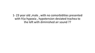
performed simulataneously to ensure the most rapid possible
treatment
11. • Each person should be familiar with basic trauma resuscitation
• 1-BASIC LIFE SUPPORT (BLS)
• 2-ADVANCED TRAUMA LIFE SUPPORT (ATLS)
12.
13. Role of team leader
• Organizes the group
• Monitors individual performance of team members
• Backs up team members
• Models excellent team behavior
• Facilitates understanding
• Focuses on comprehensive patient care
14. Effective resuscitation team dynamics
• Effective resuscitation team dynamics
• Effective resuscitation team dynamics
• Effective resuscitation team dynamics
• Effective resuscitation team dynamics
• Effective resuscitation team dynamics
• Effective resuscitation team dynamics
• Effective resuscitation team dynamics
16. Why ??
• Breakdowns in the care plan and medical mismanagement typically
occur due to one or more of four potential problems
●Communication breakdowns (eg, changes in the patient's physiologic
state or critical test results are not effectively communicated, overall
management plan or priority of tasks is not conveyed clearly by the
team leader)
●Failures in situational awareness (eg, failure to recognize shock, failure
to anticipate blood transfusion needs, failure to modify standard
management for higher risk patients).
17. ●Staffing or workload distribution problems (eg, insufficiently trained
staff conducting a procedure, inadequate staff for patient volume)
●Unresolved conflicts (eg, unresolved hostility about other team
members perceived to be performing inadequately, disagreement
about overall management plan, disagreement among senior clinicians
vying for team leadership)
18. • Principles of trauma patient management
• Treat the greatest threat to life first.
• Definitive diagnosis is not immediately important.
• Time matters (“golden hour” emphasizes urgency).
• Do no further harm.
• Assess, intervene, reassess
• Did the intervention work?
• Is the patient’s physiology returning to or staying normal
19. • While the most common causes of mortality from trauma are
hemorrhage, multiple organ dysfunction syndrome, and
cardiopulmonary arrest
• the most common preventable causes of morbidity are unintended
extubation , technical surgical failures, missed injuries, and
intravascular catheter-related complications .
24. • Triage:
• sorting and treating patients according to priority
identify, treat patients with life-threatening conditions first
• Priority may be determined by:
• Medical necessity
• Personnel skills
• Available equipment
25. Triage
• of trauma patients considers vital signs and prehospital clinical
course, mechanism of injury, patient age, and known or suspected
comorbid conditions.
• Findings that lead to an accelerated workup include multiple injuries,
extremes of age, evidence of severe neurologic injury, unstable vital
signs, and preexisting cardiac or pulmonary disease.
27. • Traumatic injuries can range from minor isolated wounds to complex
injuries involving multiple organ systems.
• All trauma patients require a systematic approach to management in
order to maximize outcomes and reduce the risk of undiscovered
injuries.
29. • In primary survey airway , breathing , circulation are assessed and
immediate life threatening problems are diagnosed and treated .
• A-airway
• B-breathing
• C –circulation
• D-disability
• E –exposure ,environment
30. ●Airway assessment and protection (maintain cervical spine
stabilization when appropriate)
●Breathing and ventilation assessment (maintain adequate
oxygenation)
●Circulation assessment (control hemorrhage and maintain adequate
end-organ perfusion)
●Disability assessment (perform basic neurologic evaluation)
●Exposure, with environmental control (undress patient and search
everywhere for possible injury, while preventing hypothermia)
31. • Primary survey usually takes no longer than few minutes , unless
procedures are required ,
• The primary survey must be repeated any time a patients status
changes , including changes in mental status , changes in vital signs
,or administration of new medications or treatment
32. • AIRWAY
• Always assess the airway
• • Talk to the patient – A patient speaking freely and clearly has an
open airway
• • Look and listen for signs of obstruction
• Snoring or gurgling
• Stridor or noisy breathing
• Foreign body or vomit in mouth
• if airway obstructed, open airway and clear obstruction
33. TECHNIQUES FOR OPENING THE AIRWAY
• No trauma
• Position patient on firm surface
• Tilt the head
• Lift the chin to open the airway
• Remove foreign body if visible
• Clear secretions
• Give oxygen 5 L/min
34. TECHNIQUES FOR OPENING THE AIRWAY
• In case of trauma •
• Stabilize cervical spine
• Do not lift head!
• Open airway using jaw thrust
• Remove foreign body if visible
• Clear secretions
• Give oxygen 5 L/min
35. AIRWAY DEVICES
• Oropharyngeal airway
• • Use if patient unconscious
• • Use correct size - measure from front of ear to corner of mouth
• • Slide airway over tongue
• If patient resists, gags or vomits, remove immediately!
36. • Nasopharyngeal airway
• • Better tolerated if patient is semi-conscious
• • Pass well lubricated into one nostril
• • Direct posteriorly, towards the throat
37. •In the unconscious patient, the airway
must be protected immediately once
any obstructions (eg, foreign body,
vomitus, displaced tongue) are removed
38. • Keep the following points in mind while performing the primary
survey:
• ●Airway obstruction is a major cause of death immediately following
trauma
• . The airway may be obstructed by the tongue, a foreign body,
aspirated material, tissue edema, or expanding hematoma.
• ●Once an airway has been established, it is important to secure it
well and to ensure it is not dislodged any time the patient is moved.
Unintended extubation is the most common preventable cause of
morbidity in trauma patients .(up to date)
39. TENSION PNEUMOTHORAX
• Air from lung puncture enters pleural space,
• cannot escape
• Progressive increase in intrathoracic
• pressure causes mediastinal shift and
• hypotension due to reduced venous return
• Patient becomes short of breath and hypoxic
• Diminished breath sounds on side of pneumothorax
• Requires urgent needle decompression,
• then chest drain as soon as possible
40. INDICATIONS FOR CHEST DECOMPRESSION
• Signs and Symptoms:
• Absent or diminished breath sounds on one side
• Evidence of chest trauma or rib fracture
• Open or "sucking" chest wound
• Diagnoses :
• Pneumothorax
• Tension pneumothorax
• Hemothorax
• Hemo-pnemothorax
41. Management :
• Give oxygen at 6-10 ml/min ,, via a non –rebreathing face mask , this
is indication for all patient suffering from polytrauma injuries
• Ventilate the patient with rescue breaths , a bag-valve device (bagging
the patient),or a ventilator but put it in mind that if the ventilation
problem is produced by a pneumothorax or tension
pneumothorax,intubation with vigorous bag-valve ventilation could
lead to further detoriartion of patient
• Treat open pneumothorax,tension pneumothorax,flail chest,massive
hemothorax( up to date)
42. • Insert a large bore needle over rib: – 2nd intercostal space – Over 3rd
rib at mid-clavicular line
• Listen for hissing sound of air escaping
• Insert chest drain
43.
44. BREATHING
• Assess ventilation - Is the patient in respiratory distress?
• Look – For cyanosis, wounds, deformities, ecchymosis, amplitude,
paradoxical movement
• Feel - Painful areas, abnormal movement
• Percuss - Dullness
• Listen - Reduced breath sounds
45.
46. • Airway patency alone doesn't assure adequate ventilation , adequate
gas exchange is required to maximize oxygenation and minimize
carbon dioxide accumulation ,
• Ventilation requires adequate function of the lungs ,chest wall ,
diaphragm , each component must be examined and evaluated
rapidly.
47. CIRCULATION: HAEMORRAGHIC SHOCK
• Assess the circulation
* Signs of hypoperfusion :
• Confusion, lethargy or agitation
• Pallor or cold extremities
• Weak or absent radial and femoral pulses
• Tachycardia
• Hypotension
Examine the abdomen for tenderness or guarding Carefully assess
pelvic stability
48.
49. •Hypotension generally does not
manifest until at least 30 percent of the
patient's blood volume has been lost
50. • Large volumes of blood may be hidden in thoracic, abdominal and
pelvic cavities, or from femoral shaft fractures
To decrease bleeding
Apply pressure to external wound
Apply splint to possible femur fracture
Apply pelvic binder to possible pelvic fracture
51. • Obtain two large bore IV catheters (14-16 gauge) using warmed fluids If
systolic BP <90 mmHg or pulse >110 bpm
• Give 500 ml bolus of Ringer’s Lactate or NS
• Keep patient warm
• Control hemorrhage by direct pressure over the wounds, tourniqutes
should be considered only in very limited conditions (traumatic
amputation).
• Perform CPR if needed
• Reassess vitals
• If still hypotensive after 2L of crystalloids, transfuse blood
52. INTRAVENOUS ACCESS
• Cannula should be placed in arm vein, not over joint, easy fixation.
Comfortable and convenient for drug administration and care
• Best veins in emergencies:
• Antecubital fossa
• Femoral
• External jugular
• Do not attempt subclavian vein due to high risk of pleural puncture
53. • Femoral vein
• If right handed, stand on patient’s right, palpate femoral artery
• Prep area carefully; site is contaminated
• Use a 14, 16 or 18 G (20 G in child) cannula mounted on 5 ml syringe
• Avoid injured extremities, if possible
54. • If patients is pregnant, she should not be on her back, put her on her
left side.
• Send blood for type and crossmatch
55. FLUIDS AND MEDICINES
• Avoid fluids containing dextrose during resuscitation
• Use Saline or Ringer's lactate
• For shocked patient: give fluids as fast as drip runs until blood
pressure responds
• May need a pressure infusion bag to push fluids
• Monitor response carefully; look at vital signs, urine output
Always give medicines intravenously during resuscitation
56. • Check the pulse using a pulse oximetre ,however ,remember that
pulse , oximetry can be unreliable in pateints with poor peripheral
perfusion after trauma..
57. • In trauma patients with significant hemorrhage, a lower score on the
Glasgow Coma Scale (GCS) and older age are both independently
associated with increased mortality, according to multivariable logistic
regression analysis of two large databases
58. Disability
• Checking for neurological damage: vital part of primary survey
Abbreviated neurological examination:
• – ALERT
• – VERBAL - responsive to verbal stimulus
• – PAIN - responsive to painful stimulus
• – UNRESPONSIVE
59. Glasgow coma scale
GCS is to be repeated
and recorded frequently.
It is the best way to
determine deterioration
60.
61. EXPOSURE
• Remove all patient's clothing
• Examine whole patient
• Front and back; log roll carefully
• Do not allow patient to get cold (especially children)
62.
63. • Cardiopulmonary resuscitation (CPR)
• (American heart association)
• >follow the C-A-B (compression –airway-breathing)
• >compression should be in a rate of 100/min moving the chest inward
at least 2 inches in adult and 13 of the chest diameter in children
• >compression ventilation ratio should be 30/2
• >minimize interruption to the chest compression .
66. • Start resuscitation at the same time as performing
primary survey Do not start secondary survey until
completing primary survey Constantly reassess
patient for response to treatment; if condition
deteriorates, reassess ABC.
67. Adjuncts to the primary survey
• Radiography :
• the “trauma triple”is portable cervical spine ,an anteroposterior
chest, an anteroposterior pelvis radiograph ,,, these provide the
maximum amount of information about potentially dangerous
conditions in a minimum amount of time
73. • Secondary survey
• Perform complete, thorough patient examination to ensure no other
injuries are missed
74. Secondary survey
• The secondary survey is performed only after the primary survey has
been finished and all immediate threats have been treated
• The secondary survey is a head to toe examination
• Dessigned to identify any injuries that might have been missed .
75. • Do not start definitive treatment until secondary survey is completed
unless required as life-saving measure
• When definitive treatment is not available, have a plan for safe
transfer of patient to another centre
76. Secondary survey
• Head Exam
• Scalp, eyes, ears
• Soft tissues
• Neck Exam
• Penetrating injuries
• Swelling or crepitus
• Neurological Exam
• Glasgow Coma Score
• Motor examination
• Sensory examination
• Reflexes
78. Patient history
• SAMPLE
• Symptoms –PAIN,shortness of breath ,other symptoms
• Allergies to medications
• Medications taken
• Past medical /surgical history
• Last meal –important to determine risk of aspiration
• Events Leading up to trauma
79. REASSESSMENT
• Always perform an ABCDE primary survey if patient deteriorates
• Signs of adequate resuscitation
• Slowing of tachycardia
• Urine output normalizes
• Blood pressure increases
80. MONITORING
• EKG monitoring if available
• Pulse oximeter
• Most widely used physiological monitoring device for heart rate,
oxygenation
• Especially useful in anaesthesia, ICU
• Simple to use
• Should be minimum standard of monitoring in every surgical theatre
• Blood pressure
• Manually or automated machine
81. Indication of referral to higher level
• 1- patient needs advanced care that is not available in the clinic
• 2- if there is a lack of skills , instruents , or equipment
• 3-lack of radiology services
82. Contraindication of referral to higher level
• 1-patients whose vital signs are unstable due to active bleeding ,
multiple fractures or suspicion of internal bleeding
• 2-patient with minor trauma not requiring any advanced medical
support.
83.
84. • Planning and preparation:
• Mode of transport
Accompanying personnel, including family
• Supplies needed for any possible treatment
• Identifying possible complications
• Communicate with all involved in transfer including receiving hospital
• Be prepared: if anything can go wrong, it will and at the worst
possible time
85. STABILIZATION AND TRANSFER
Resuscitation completed
Laboratory specimen sent
Controlled airway
Normalized circulation
Immobilized fractures
Appropriate analgesia
Functioning intravenous lines
Documentation completed
Transfer :
– Ward – Operating theatre – Higher level of care centre
86. • PATIENT SAFETY: Consent
• Informed consent means that patient and patient’s family understand
– What is to take place
• – Potential risks, complications of both proceeding and not
proceeding
• Have given permission for intervention
• Be attentive to legal, religious, cultural, linguistic, family norms and
differences Our job is not to judge, but to provide care to all without
regard to social status or any other considerations
87. • RECORD KEEPING
• • Essential that patients receive written note describing diagnosis,
procedure performed
• • All records should be clear, accurate, complete, signed
88.
89.
90. Elderly trauma
• Falls , motor vehicle accidents are the leading cause of trauma in
elderly .
• According to a systematic review of 18 studies, the probability of
falling at least once in any given year for individuals 65 years and
older is approximately 27 percent.(up to date ).
• Falls in the elderly most often occur from a standing position on a
level surface, with orthopedic injury (eg, hip or long bone fracture)
the most common significant complication.
91. • Elderly patients may be hypotensive relative to their baseline blood
pressure but still have blood pressure measurements in the "normal"
range. A single episode of hypotension substantially increases the
likelihood that a serious injury has occurred (up to date )
92. • Although there is little prospective data to guide triage decisions
about geriatric trauma patients, given the increased risk for severe
injury and death in this population, we suggest that trauma patients
over the age of 70 be evaluated at a trauma center with trauma team
activation whenever possible, regardless of the mechanism (ie, falls
from standing warrant such evaluation)(up to date )
93. Reference
• Who health organization (Emergency and Essential Surgical Care
(EESC) programme)
• Up to date
• American heart association (ATLS )