

Empfohlen
Weitere ähnliche Inhalte
Was ist angesagt?
Was ist angesagt? (20)
Andere mochten auch
Ähnlich wie Hplc presentation for class
Ähnlich wie Hplc presentation for class (20)
Mehr von Dr. Ravi Sankar
Mehr von Dr. Ravi Sankar (20)
Kürzlich hochgeladen
Kürzlich hochgeladen (20)
Hplc presentation for class
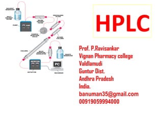
- 1. HPLC Prof. P.Ravisankar Vignan Pharmacy college Valdlamudi Guntur Dist. Andhra Pradesh India. banuman35@gmail.com 00919059994000
- 2. H P igh ressure L iquid C hromatography Since high pressure is used when compared to classical chromatography Separation of the components from a mixture is achieved by pumping mobile phase at High pressure using appropriate displacement pumps or gas pressure. (Due to the small particle size (3-5 um).
- 3. H P igh erformance L iquid Chromatography More ever it is improved performance when compared to other conventional column Chromatographic techniques.
- 5. • Michael Tswett (1906) -separation of plant pigments • Martin and Synge (1941) liquid-liquid partition chromatography • 1952 Nobel Prize • Other chromatography Techniques • Thin-layer chromatography (TLC) • Paper chromatography • Preparative column chromatography • Medium pressure chromatography • Gas chromatography • Ion-exchange chromatography • Size-exclusion chromatography (Gel Permeation Chromatography) • HPLC • VLC • Flash Chromatography • Affinity Chromatography • Chiral Chromatography • Super Critical Fluid Chromatography 5
- 6. What means chromatography ? AABC AC ABBC C C B C B A C B A B C CC C C C C BB B B B B A A A A A A Sample separated components Chromatography is the chemical / physical separation of components for qualitative and quantitative analysis PrinciplePrinciple 6
- 7. Different chromatography methods 7 Name Mechanism Stat. Phase Mobile phase Paper- partition liquid liquid Thin layer- adsorption solid liquid Gas- adsorption / partition solid or liquid gas Column- adsorption / partition Solid/liquid liquid
- 8. Liquid Chromatography (LC) • Two different phases are used to separate components of the mixture: stationary and mobile phase • The solute separates on the column via interactions based on physical mechanisms like retention 8
- 9. Partitioning • Separation is based on the analyte’s relative solubility between two liquid phases 9 Stationary PhaseMobile Phase Solvent Bonded Phase
- 10. 10
- 11. What is HPLC? • The most widely used analytical separations technique • Utilizes a liquid mobile phase & packed column to separate components of mixture • uses high pressure to push solvent through the column • Popularity: – sensitivity – ready adaptability to accurate quantitative determination – suitability for separating nonvolatile species or thermally fragile ones 11
- 12. • Popularity: – widespread applicability to substances that are of prime interest to industry, to many fields of science, and to the public • Ideally suited for separation and identification of amino acids, proteins, nucleic acids, hydrocarbons, carbohydrates, pharmaceuticals, pesticides, pigments, antibiotics, steroids, and a variety of other inorganic substances 12
- 13. History • Early LC carried out in glass columns – diameters: 1-5 cm – lengths: 50-500 cm • Size of solid stationary phase – diameters: 150-200 µm • Flow rates still low ! Separation times long! • Decrease particle size of packing causes increase in column efficiency! – diameters 3-10 µm • This technology required sophisticated instruments – new method called HPLC 13
- 14. Advantages of HPLC • Higher resolution and speed of analysis • Greater reproducibility due to close control of the parameters affecting the efficiency of separation • Easy automation of instrument operation and data analysis • Adaptability to large-scale, preparative procedures 14
- 15. Advantages to HPLC • Advantages of HPLC are result of 2 major advances: – stationary supports with very small particle sizes and large surface areas – appliance of high pressure to solvent flow 15
- 16. Chromatographic terms 16 Baseline Injection monitoring Eluent peak; dead volume Peak wide Peak width at half- height Baseline noise Peak fronting Tailing poor resolution Baseline drift
- 17. Parameters used in HPLC • Retention parameters • Column efficiency parameters • Retention : When a component in a sample interacts with the stationary phase in the column and a delay in elution occurs • Column efficiency : Goodness of a column 17
- 18. Column dead time, retention time 18 time signal t t t t 0 R 1 R 2 R 3 0 tR 1' tR 2' tR 3' t´R = tR - t0 t0 = column dead time = time an unretarded compound needs to pass the column tR = retention time
- 20. Capacity Factor • Capacity factor 20 k´ = tR - t0 t 0 t´R t 0 = •If the substance is not retained by the stationary phase, the capacity factor is k' = 0. •Small k' (k < 1) values show that the components are only retained slightly by the separation column. Their peaks are located close to the non-retained peak (k' = 0). •the optimum separation range to be k' values between 1 and 15. Values for k' > 5 mean long retention times with associated band broadening.
- 21. Resolution 21 If the peaks are separated almost down to the baseline, R » 1.5. Higher resolutions than R = 1.5 are not desirable because they significantly extend the analysis time but do not result in additional information. Generally the values of R » 1.0 are sufficient to achieve qualitative or quantitative results.Even values of R » 0.5 are sufficient to determine the number of components present. For quantitative analysis, however, the peak areas overlap too much
- 22. Condition for Good separation 22
- 23. Plate NumberPlate Number 23 The theoretical plate number or N is a quantitative measure for the column efficiency In formulas (1) or (2) the peak base width WB or the half width WH are compared with the retention time tR
- 24. Plate Number and Plate Height Since an efficient separation column delivers sharp peaks with narrow base widths, a better column has relatively high value of N The concept of the theoretical plates is a useful tool to describe the efficiency of a separation column The number of theoretical plates is proportional to the column length L. The longer a column, the more theoretical plates it has; however, the column back pressure increases To be able to compare separation columns of various lengths, the theoretical plate height, H is used 24 H = L/N
- 25. 25
- 26. Five modes in HPLC LC mode Packing materials Mobile phase Interaction Normal phase chromatography Silica gel n-Hexane/IPE Adsorption Reversed phase chromatography Silica-C18(ODS) MeOH/Water Hydrophobic Size exclusion chromatography Porous polymer THF Gel permeation Ion exchange chromatography Ion exchange gel Buffer sol. Ion exchange Affinity chromatography Packings with ligand Buffer sol. Affinity 26
- 27. 27 HPLC Basic Instrumentation Mobile phase Pump Solvent Delivery Injector Sample Injection Column Separation Detector Data Processor Waste
- 28. 28
- 29. Composition of HPLC System • Solvent • Solvent Delivery System (Pump) • Injector • Sample • Column • Detectors • Waste Collector • Recorder (Data Collection) 29
- 30. Mobile Phase Degassing • Dissolved gases in the mobile phase can come out of solution and form bubbles as the pressure changes from the column entrance to the exit – May block flow through the system • Sparging is used to remove any dissolved gas from the mobile phase – An inert and virtually insoluble gas, such as helium, is forced into the mobile phase solution and drives out any dissolved gas. • Degassing may also be achieved by filtering the mobile phase under a vacuum 30
- 31. Are used to store Mobile-Phase. The solvent reservoir must be made of inert material such as glass and must be smooth so as to avoid growth of microorganisms on its walls. It can be transparent or can be amber colored. A graduated bottle gives a rough estimate of mobile-phase volume in the bottle. Solvent reservoirs are placed above HPLC system (at higher level) in a tray. They should never be kept directly above the system as any spillage of solvent on the system may damage electronic parts of HPLC. 31 Solvent Reservoir
- 33. Mobile Phase Mixing • Solvent proportioning valve(3.) can be programmed to mix specific amounts of solvent from the various reservoirs to produce the desired mobile phase composition 33 3.
- 34. • Isocratic elution: Use of a constant-composition mobile phase in liquid chromatography • Gradient elution: – Vary the mobile phase composition with time – If there is a wide polarity range of components to be eluted. – Allows for faster runs. – Ex: mobile phase composition can be programmed to vary from 75% water: 25% acetonitrile at time zero to 25% water: 75% acetonitrile at the end of the run. • More polar components will tend to elute first. • More non-polar components will elute later in the gradient 34
- 35. Common Reverse Phase Solvents • Methanol 35 CH3OH • Acetonitrile CH3CN • Tetrahydrofuran • Water H2O
- 37. In order to reduce separation time and allow the use of smaller particle size packings (10 microns and below), we must force the liquid mobile phase through the column under pressure. This is the function of the pump (also called the "solvent delivery system") to maintain a constant flow of mobile phase through the HPLC regardless of the pressure (back pressure) caused by the flow resistance of the packed column. There are several types of pumps are available, Reciprocating Piston Pumps Syringe Type Pumps Constant Pressure Pumps . 37
- 38. 38 Reciprocating Piston Pumps Consist of a small motor driven piston which moves rapidly back and forth in a hydraulic chamber that may vary from 35- 400 µL in volume. On the back stroke, the separation column valve is closed, and the piston pulls in solvent from the mobile phase reservoir. On the forward stroke, the pump pushes solvent out to the column from the reservoir. A wide range of flow rates can be attained by altering the piston stroke volume during each cycle, or by altering the stroke frequency. Dual and triple head pumps consist of identical piston-chamber units which operate at 180 or 120 degrees out of phase
- 40. Syringe Type Pumps The syringe pump is a large, electrically operated simulation of a hypodermic syringe. Are most suitable for small bore columns because this pump delivers only a finite volume of mobile phase before it has to be refilled. These pumps have a volume between 250 to 500 mL. The pump operates by a motorized lead screw that delivers mobile phase to the column at a constant rate. The rate of solvent delivery is controlled by changing the voltage on the motor. 40
- 41. Constant Pressure Pumps The mobile phase is driven through the column with the use of pressure from a gas cylinder A low-pressure gas source is needed to generate high liquid pressures. The valving arrangement allows the rapid refill of the solvent chamber whose capacity is about 70 mL. This provides continuous mobile phase flow rates. 41
- 42. Tubing • Very small inner diameter • Consistent i.d. • Very strong • Easy to cut • Fittings available 42
- 43. Auto samplers Are fully automatic injection systems enabling greater productivity and the highest level of precision. The HPLC Autosampler incorporates an elegant swivel head that allows a manual injection and a special Rheodyne injection valve to give accurate full or partial loop fill injections. 43
- 44. Injection Port The sample introduction device such as injector to introduce the sample in a flow of mobile phase at high pressure. It is not possible to use direct syringe injection on column like GC as the inlet pressure in LC is too high. The valve injection through fixed or variable loop is a common way of introducing the sample. The Rheodyne valve is the mostly used devise. The loop can be partially or fully filled. There are both the types of injectors available. The advantage of partial filling is the possibility of using small amount of sample, when there is scarcity of sample. The precision of the injection is 1% RSD 44
- 45. Samples are injected into the HPLC via an injection port. The injection port of an HPLC commonly consists of an injection valve and the sample loop. The sample is typically dissolved in the mobile phase before injection into the sample loop. The sample is then drawn into a syringe and injected into the loop via the injection valve. A rotation of the valve rotor closes the valve and opens the loop in order to inject the sample into the stream of the mobile phase. Loop volumes can range between 10 µl to over 500 µl. In modern HPLC systems, the sample injection is typically automated 45
- 46. Column HPLC Column Hardware Stationary Phase 46
- 47. Columns • Solid Support - Backbone for bonded phases. – Usually 10µ, 5µ or 3µ silica or polymeric particles. • Bonded Phases - Functional groups firmly linked (chemically bound) to the solid support. – Extremely stable – Reproducible • Guard column - Protects the analytical column: – Prolongs the life of the analytical column 47 • Analytical column - Performs the separation.
- 48. HPLC Columns Particle size Column ID Sample Load Analytical 3 – 5 µ 0.3 − 4.6 mm ng – µg Semi-prep 10 µ 8 – 10 mm 1 – 100 mg Preparative 10 – 30 µ 5 – 200 mm gram scale 48 •An HPLC column consists of a stainless steel tube which is sealed with fittings on both ends. Steel frits in the end fittings keep the packing material in the column. •Analytical columns have inner diameters of 1 - 10 mm and lengths of 25 - 250 mm. They are operated at flow rates of 60 µl - 5.0 ml/min. •To protect the actual separation column from chemical contamination, a guard column with the same packing material as the separation column is installed.
- 49. Column Packing – Usually spherical silica particles of uniform diameter (2-10µm) • The smaller particles yield higher separation efficiencies. – The silica particles are very porous • Allows for greater surface area for interactions between the stationary phase and the analytes. – Other packing materials may also be used: • Zirconia (ZrO2) 49 http://hplc.chem.shu.edu/NEW/HP LC_Book/Adsorbents/ads_part.html
- 50. Particle Diameter • Has a greater effect on resolution than column length • Short columns with small particles ideal • 5µm is standard size • 3µm better, but restricted range of packings available • Downside is high back pressure and issues with retention of small particles inside the column, blockages 50
- 51. Cap nut with compression Guard column Split collets Housing with outer thread cartridge 51
- 52. Refers to the solid support contained within the column over which the mobile phase continuously flows. The sample solution is injected into the mobile phase through the injector port. As the sample solution flows with the mobile phase through the stationary phase, the components of that solution will migrate according to the non-covalent interactions of the compounds with the stationary phase. The chemical interactions of the stationary phase and the sample with the mobile phase, determines the degree of migration and separation of the components contained in the sample. For example, those samples which have stronger interactions with the stationary phase than with the mobile phase will elute from the column less quickly, and thus have a longer retention time, while the reverse is also true. Columns containing various types of stationary phases are commercially available. Stationary Phases 52
- 53. Monofunctional surface modification of SiO2 OH Si O Si O Si O SiO OH OH OH OH X Si CH3 CH3 CH2 CH2 CH2 CH2 CH2 CH2 CH2 CH2 CH2 CH2 CH2 CH2 C6H13 OH Si O Si O Si O SiO O OH O OH Si CH3 C18H37 CH3 Si CH3 C18H37 CH3 Si CH3 CH3 CH3 OH Si O Si O Si O SiO O O O O Si CH3 CH3 Si CH3 CH3 Si CH3 CH3 CH3 Endcapping monofunctional modification Endcapping Stationary Phases 53
- 54. Reversed Phase Chromatography • Bonded phases made by covalently bonding a molecule onto a solid stationary phase like silica • Typical stationary phases are nonpolar hydrocarbons, waxy liquids or bonded hydrocarbons (such as C18, C8, C4, etc.) • pH range 2.5 to 7.5 • 60-90% of all analytical LC separations are done on bonded phases in the reverse phase mode. 54
- 55. spherical irregular Structure of Packing Material monolithic 55
- 56. Synthesis of RP Packings 56
- 58. Reversed Phase ChromatographyReversed Phase Chromatography modification: RP-8, RP-select B, RP-18, -CN, -Diol, -NH2 base material: LiChrosorb® , LiChrospher® , Superspher® , Purospher® , Purospher® STAR, Chromolith® , Aluspher® , Polyspher® Normal Phase ChromatographyNormal Phase Chromatography modification: Si, -Diol, -CN, -NH2 base material: LiChrosorb® , LiChrospher® , Superspher® , Aluspher® ,Purospher® STAR , Chromolith® Ion Exchange ChromatographyIon Exchange Chromatography modification: -NR+ 3, -N+ HR2, NH2 for cations -SO3 - , -COO- for anions Base material: Polyspher® , LiChrosil® Size Exclusion ChromatographySize Exclusion Chromatography Sorbents: LiChrogel® PS for SEC in organic solvents Fractogel® for aqueous SEC 58
- 59. Detector 59 •Detect various compounds as they elute out from column. The detector gives response in terms of a milivolt signal that is then processed by the computer (integrator) to give a chromatogram. •Basically detector consists of a flow-cell through which the mobile phase and resolved sample moves optics shine through the detector cell and variation in optical properties are detected. •A Ultra violet or UV detector detects absorbance of UV light by chromophores in the analyte compound. •A refractive index detector will sense variation in refractive index of mobile phase stream passing through flow-cell • Similarly Fluorescence Detectors checks for Florescence.
- 60. Various Detetectors are Listed below • Ultraviolet (UV) Fixed wavelength detector Variable wavelength detector Diode Array • Fluorescence • Electrical Conductivity • Refractive Index • Electrochemical • Light scattering • IR Absorbance • Mass-Spectrometric 60
- 64. Picture of a Typical HPLC System 64
- 65. 65
- 67. 67
- 68. • 2004: Further advances in column technology and chromatography instrumentation – Utilized even smaller packing particle sizes (1.7µm) – Higher pressures (15000psi) – Allowed for significant increases in LC speed, reproducibility, and sensitivity. • New research utilizing particle sizes as small as 1µm and pressures up to 100,000psi! 68 What is Ultra Performance Liquid Chromatography?
- 70. Why UPLC more efficient • Peak capacity (P) is the number of peaks that can be resolved in a specific amount of time • P is proportional to the inverse of the square root of the Number of theoretical plates (N): N = L/H • Plate heights are correlated through the Van Deemter equation 70
- 71. Contrasting HPLC and UPLC • UPLC gives faster results with better resolution • UPLC uses less of valuable solvents like acetonitrile which lowers cost • The reduction of solvent use is more environmentally friendly • Increased productivity can increase you revenue in an industrial setting 71
- 73. Waters (USA) Agilent (USA) Thermo (USA) Varian (USA) Dionex (USA) Jasco (Germany) Knauer (Germany) Shimadzu (Japan) Manufacturers 73 Ningbo Yujie Optical Instruments Co., Ltd.
- 75. DETECTORS UV-Vis/PDA Refractive index Electrochemical Conductivity Fluorescence Radioactivity Evaporative light scattering Corona CAD PDA-MS PDA-NMR FTIR 75
- 76. OVERVIEW Detector Analyte /attributes sensitivity Nature UV-Vis Works with all molecules containing chromophores absorbing UV-Vis ng Specific PDA Works for all wavelengths in UV-Vis ng Fluorescence Compound with native fluorescence or fluorescence tag fg-pg Conductivity Anions, cations, organic acids, surfactants ng Radio activity Radioactive labeled compounds ng-pg 76
- 77. Detector Analyte /attributes sensitivity Nature Refractive Index Temperature sensitive; polymers, sugars, triglycerides, incompatible with gradients 0.1-10 µg Universal Evaporative light Scattering(ELSD) Uniform response; nonvolatile to semi volatile compounds; compatible to gradients ng Electrochemical Redox reactions pg Corona CAD Can detect non UV absorbing chromophores ng Mass Spectrometer Definite analyte identification fg-pg Both universal and specific IR Works with all molecules mg 77
- 78. Uv- visible detector Senstive Wide linear range Unaffected by changes in temp. And mobile phase composition Principle : • Lambert-Beer’s law: A= ε b c At constant cell thickness and constant wavelength a linear connection between the Absorbance A and the Concentration C is achieved.78
- 79. Types of uv-visible detectors A. Fixed wavelength detectors B. Variable wavelength detectors C. Photodiode array detectors 79
- 80. Fixed wavelength detectors Operates only at 1 wavelength (254 nm) Best overall precision is noted for peak area measurements as compared to variable wavelength detectors. Uses a discrete source: – Low pressure mercury lamp (253.6 nm) – Silicone photodiode light detector It can also be used at other wavelengths by filtering the emission source to give other lines or even using phosphor screens to give lines not available from mercury. 80
- 81. Variable wavelength detectors Uses continuous source of light • Deuterium or xenon lamp or tugston lamp • Desired wavelength is isolated by monochromator Improved sensitivity from operating at λmax of solute of interest Drawback: limited lamp lifetime as compared to mercury lamp (Deuterum lamp: 1000-2000 hrs) Dual wavelength detection feature: – Useful for simultaneous monitoring of two substances – Baseline noise is higher because this feature is achieved by toggling the monochromator between 2 wavelengths 81
- 82. 82
- 83. Photodiode array detectors Uses charge coupled DA with 512 to 1024 diodes capable of spectral resolution of 1 nm Earlier, they suffered from sensitivity problems, which has been solved by advanced flow cell design using fiber optics technology to extend path length without increasing noise or chromatographic band dispersion Performs a simultaneous measurement of absorption as a function of analysis time and over a chosen wavelength range, so we get UV spectrum for each eluted peak Used for method development where λmax of impurities in drug are unknown Every compound can be quantitated at its λmax , so useful for trace analysis Compounds not resolved chromatographically can sometimes be resolved spectrally Molecular absorption spectrum can be used for peak identification and peak tracking 83
- 84. Measures light intensity at each λ Measures light intensity at each λ 84
- 85. Indirect photometric detection Used when solute of interest does not possess chromophore Here, mobile phase possess a chromophore and absorbs light When analyte without a significant chromophore passes through the detector cell, the absorption of mobile phase is decreased and is recorded as negative peak 85
- 86. 86
- 87. 87
- 88. Features of modern uv-vis detectors Dual or multiple wavelength detection and stop flow scanning features Front panel access to self alligned sources and flow cells for easy maintanance Self validation features such as: – Power up diagnostics – Leak sensors – Time logs for lamps – Built in holmium oxide filters for wavelength calibration – Filter wheels for linearity verification 88
- 89. Refractive index detectors Measurement response is a function of the refraction index difference between pure mobile phase and the mobile phase with the dissolved separated components Due to the strong temperature dependence of the refraction index, good temperature control of the measurement cell must be ensured The detection limit is in the range of 10-6 - 10-8 g/ml To achieve a high sensitivity, in practice solvents are selected that have a very high or very low refraction index This ensures that the difference between the refractive indices of 89
- 90. 90
- 91. One half of the measurement cell is purged with the flowing mobile phase, the second half of the measurement cell (reference cell) is filled with mobile phase. The refraction index n is at first identical in both cells. If a sample component is added to the eluent, the refraction index n in the measurement cells changes. The light beam experiences a deflection on the path Operating principle 91
- 92. 92
- 93. Fluorescence detectors During fluorescence detection, the sample is irradiated with UV light of suitable wavelength and excited for emission of long wave light. The Xenon lamp continuously sends light of 325 - 410 nm. Provides LOD values 100 times lower than absorption detectors Highly selective 93
- 94. 94
- 95. 95
- 96. 96
- 97. 97
- 98. Electrochemical detectors Electron transfer processes offer highly selective and sensitive method Easily adaptable for use with microcolumns As background noise is dependent on mobile phase conditions, it is difficult to utilize these detectors with gradient elution separations 2 types: 1. Amperometric detection: fixed potential is applied to the electrode (glassy carbon) and a solute which will oxidize at that potential yields an output current 2. Coulometric detection: 100% of the solute species is converted, which offers advantages of no mobile phase flow dependence on signal and absolute quantitation through Faraday’s law 98
- 99. ` 99
- 100. Conductivity detectors Measurement of electrical conductance is a subset of ECD, although it is generally considered separately since it is non-Faradaic electrochemistry; i.e. no electron transfer reaction takes place The electrical conductivity of the mobile phase is used as characteristic for detection. Universal technique for ionic solutes and are used mainly for the separation of ions in water or polar eluents Since the conductivity can change approx. 2 % per °C, conductivity detector are often equipped with automatic temperature compensation 100
- 101. With the removal of electrolytes contained in the eluent system when using ion exchangers, a significant increase in the sensitivity can be achieved (otherwise it creates difficulty in measuring low concentration of an ionic solute in the presence of highly conducting mobile phase The resulting extremely low basic conductivity makes it possible, to generate good measurement signals from the smallest sample volumes. During conductivity detection the specific conductivity is measured continuously. The individual eluted components of the material sample are each displayed through a change in conductivity. 101 Conductivity detectors
- 102. Principle of 4 electrode method The two outer electrodes are used to transmit an electronically stable alternating current through the cell The electro-motor force (E), which is measured deenergized on the two inner electrodes, is a measure for the conductivity of the cell content according to the equation. χ = (K/E)*I χ = electric conductivity K = cell constant between the 2nd and 3rd electrode E = electro-motor force I = current strength The deenergized measurement of the electro-motor force at the two inner electrodes caused no passivity, hence the instrument operates stably for a long period of time. 102
- 103. 103
- 104. 104
- 105. ELSD can outperform traditional detectors when analysing non- chromophoric samples by HPLC Traditional HPLC detectors such as UV and RI have limited capabilities UV and RI are not compatible with a wide range of solvents RI detection is not gradient compatible Different analytes produce different UV responses depending on their extinction co-efficient ELSDs can detect anything that is less volatile than the mobile phase ELSD is universal and compatible with a wide range of solvents Evaporative Light Scattering Detector 105
- 106. Unique Method of Detection Three steps: • Nebulization • Mobile Phase Evaporation • Detection 106
- 107. Step 1: Nebulization Column effluent passes through nebulizer needle Mixes with nitrogen gas Forms dispersion of droplet 107
- 108. Step 2: Mobile Phase Evaporation Droplets pass through a heated zone Mobile phase evaporates from the sample particle Dried sample particles remain 108
- 109. Step 3: Detection Sample particles pass through an optical cell Sample particles interrupt laser beam and scatter light Photodiode detects the scattered light 109
- 110. ELSD – A Powerful Detector for HPLC Universal Sensitive Gradient Compatible AN ELSD IS AN EFFECTIVE REPLACEMENT OR A PERFECT COMPLEMENT TO EXISTING LC DETECTORS RI UV MS Fluorescence 110
- 111. Four Reasons to Replace an RI with an ELSD: 1. Better sensitivity 2. Gradient compatible 3. Stable baselines 4. No solvent front peaks 111
- 112. The ELSD Improves Baseline Stability and Detection Sensitivity Compared to RI 112
- 113. Reasons to operate an ELS detector in series with a UV detector: 1. Obtain a more accurate representation of sample mass than UV 2. See what may be missing from your UV chromatogram 113
- 114. The ELSD responds to all components in this antihistamine formulation 1. Diphenhydramine 2. PEG, Gelatin 3. Sorbitan 4. Glycerol 5. Sorbitol 114
- 115. Detect Difficult Samples like Triglycerides without Derivatizing 1. LLO 2. LLP 3. OOL 4. POL 5. PPL 6. OOO 7. OOP 8. PPO 9. OOS 115
- 116. Although the Mass Spectrometer is a Universal Detector, ELSDs offer many Benefits over MS: 1. Lower investment and operating costs 2. Less complicated operation 3. Less maintenance Since chromatographic requirements for ELSD and MS are similar, methods developed for ELSD are usually transferable to MS without Use ELSD in Parallel with MS to Obtain Maximum Structural and Concentration Information Use ELSD in Parallel with MS to Obtain Maximum Structural and Concentration Information 116
- 117. Replace Fluorescence Detectors with ELSD to Simply Methods by Eliminating Pre- or Post-column Derivatization 1. Glycine (Gly) 2. Serine / Asparagine (Ser/Asn) 3. Aspartic Acid (Asp) 4. Glutamine (Gln) 5. Alanine / Threonine (Ala/Thr) 6. Glutamic Acid (Glu) 7. Cysteine / Lysine (Cys/Lys) 8. Histidine (His) 9. Proline (Pro) 10. Arginine (Arg) 11. Valine (Val) 12. Methionine (Met) 13. Tyrosine (Tyr) 14. Isoleucine (Ile) 15. Leucine (Leu) 16. Phenylalanine (Phe) 17. Tryptophan (Trp) 117
- 118. Summary: Benefits of ELSD as a Replacement or Complement to Existing Detectors • See what you might be missing • More sensitive and stable than RI • Get a more accurate representation of sample mass than UV • Simplify methods by eliminating derivatization 118
- 119. 119
- 120. 120
- 121. 121
- 122. 122
- 123. 123
- 124. 124
- 125. 125
- 126. 126
- 127. 127 APPLICATIONS OF HPLC : Assay of cephalosporins Assay of theophylline Separation of barbiturates, phenothiazines, benzodiazepine derivatives, rauwolfia alkaloids etc. by C-18 reversed phases Quantitative analysis of several analgesics like aspirin, caffeine, paracetamol, phenacetin, etc. Analysis of urine and serum samples Separation of antipyrine and benzocaine in ear drops
- 130. 1. REFERENCES: 2. Instrumental methods of chemical analysis-B.K Sharma. 3. Pharmaceutical analysis by Ashutosh Kar 4. Fundamentalsof Analyttical chemistrySkoog,West,Holler 5. Instrumental analysis of chemical analysis- Gurudeep R Chatal 6. www.ctsholc.com/tools 7. 6.www.wikipedia.org/hplc 8. www.chemguide co.uk/analysis 9. www.hplcindia.com 130
- 131. 131
Hinweis der Redaktion
- It is common practice to make up these organic solvents as mixtures with water or, in a lot of cases, have each pure solvent mixed under instrument control and changed at a certain rate with time (gradient). Gradients can be simple or complex. Simple - as a linear gradient (ramp); Complex as steps (start and hold) and ramps together. You can have a solvent or several solvents being controlled at the same time with a changing modifier such as a pH buffer. Methanol - Most common solvent. Close to water in structure. Miscible in all proportions with H 2 O so that for less polar organics you can have the power of 90% Methanol with 10% H 2 O. Acetonitrile - Highly polar, very low UV absorbance. Also completely miscible with H 2 O but lacking in hydrogen bonding capability thus affording a different partitioning effect. Tetrahydrofuran - Molecule has high dipole moment. More soluble with non-polar compounds. Water - Also a very common solvent. Used to make up solvent modifiers to adjust pH (buffers) as well as ion-pairing reagents. Emphasize degassing (“bends”- bubble in detector) and for particle-free (dust could be up to 10X size of particles of solid support). 555- 25mM H 3 PO 4 :MeCN(90:10)->10:90 605 - 0.1M pH4.7 OAc: MeCN (1:1) isocratic. 610 - MeCN:H 2 O -> MeCN 8316 - 100% H 2 O 8330 - MeOH:H 2 O isocratic 8331 - MeOH:H 2 O(HOAc-C 10 SO 3 H) 8332- MeOH:H 2 O (3:2-CN), (1:1-C-18) 8325-A=MeCN:0.01M OAc (75:25), B= MeCN -> 60%MeCN total. λ= 190 for MeCN, λ= 205 for MeOH, λ= 190 for H 2 O, λ~ 290 for THF, λ~ 255 for 1% HOAc and λ~ 260 NH 4 OAc (1M)
- Here you can see the structure of the different packing materials of Merck. Starting from the irregular particles (e.g. LiChrosorb) there came the invention of spherical particles, used for the LiChrospher, Superspher, Purospher and Purospher STAR sorbents) and especially the latest innovation by Merck, the monolithic column technology in terms of the Chromolith column. On the different pictures you can compare the macropores (through-pores) and the mesopores of the different variations of these packing materials.