
Empfohlen
Weitere ähnliche Inhalte
Was ist angesagt?
Was ist angesagt? (20)
Ähnlich wie Glaucoma: Types, Causes, Symptoms and Treatment
Ähnlich wie Glaucoma: Types, Causes, Symptoms and Treatment (20)
Kürzlich hochgeladen
Kürzlich hochgeladen (20)
Glaucoma: Types, Causes, Symptoms and Treatment
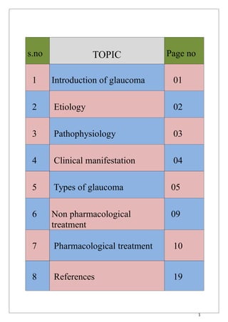
- 1. 1 s.no TOPIC Page no 1 Introduction of glaucoma 01 2 Etiology 02 3 Pathophysiology 03 4 Clinical manifestation 04 5 Types of glaucoma 05 6 Non pharmacological treatment 09 7 Pharmacological treatment 10 8 References 19
- 2. 2 GLAUCOMA Glaucoma is not a single disease process but a group of disorders characterized by a progressive optic neuropathy resulting in a characteristic appearance of optic disc & specific pattern of irreversible visual field defects that are associated frequently but not invariably with ↑IOP. The normal eye pressure is 12-22mm Hg. The word “glaucoma” is derived from Ancient Greek word glaukos which means grey, blue or green eyes. Glaucoma is the second leading cause of blindness globally after cataracts. The most common type is open-angle glaucoma with less common types including closed-angle glaucoma and normal-tension glaucoma. Open-angle glaucoma develops slowly over time and there is no pain. Peripheral vision may begin to decrease followed by central vision resulting in blindness if not treated. Closed-angle glaucoma can present gradually or suddenly. The sudden presentation may involve severe eye pain, blurred vision, mid-dilated pupil, redness of the eye, and nausea. Vision loss from glaucoma, once it has occurred, is permanent.
- 3. 3 ETIOLOGY All types of glaucoma – progressive optic neuropathy due to the death of retinal ganglion cells (RGCs). • RGCs death is initiated by: Block in transport of neurotrophins from brain to RGCs Damaging cascade activity Apoptosis of RGCs • RGCs death – loss of retinal fibers – optic neuropathy & visual field defects Mechanical theory: ↑IOP – mechanical stretch of lamina cribtosa – axonal deformation & altered capillary blood flow -- ↓neurotrophins to reach RGC`s • Pressure independent factors: 1 Failure of autoregulation 2. Vasospasm 3. Systemic hypotension
- 4. 4 PATHOPHYSIOLOGY The underlying cause of open-angle glaucoma remains unclear. Several theories exist on its exact etiology. “However, the major risk factor for most glaucomas and the focus of treatment is increased intraocular pressure.” Intraocular pressure is a function of production of liquid aqueous humor by the ciliary processes of the eye, and its drainage through the trabecular meshwork. Aqueous humor flows from the ciliary processes into the posterior chamber, bounded posteriorly by the lens and the zonules of Zinn, and anteriorly by the iris. It then flows through the pupil of the iris into the anterior chamber, bounded posteriorly by the iris and anteriorly by the cornea. From here, the trabecular meshwork drains aqueous humor via the scleral venous sinus (Schlemm's canal) into scleral plexuses and general blood circulation. In open/wide-angle glaucoma, flow is reduced through the trabecular meshwork, due to the degeneration and obstruction of the trabecular meshwork, Loss of aqueous humor absorption leads to increased resistance and thus a chronic, painless buildup of pressure in the eye. In close/narrow-angle, the iridocorneal angle is completely closed because of forward displacement of the final roll and root of the iris against the cornea, resulting in the inability of the aqueous fluid to flow from the posterior to the anterior chamber and then out of the trabecular network. This accumulation of aqueous humor causes an acute increase in pressure and pain.
- 5. 5 CLINICAL MANIFESTATION A- POAG: General POAG is usually bilateral with asymmetric disease progression 1. Loss of peripheral vision. 2. Presence of scotomata (blind spots) in vision field. 3. .Large cup-to-disc ratio. 4. .Notching of the optic nerve head rim. 5. .Splinter hemorrhages (using a slit-lamp biomicroscope) B- PACG: General (Medical emergency due to high risk of vision loss) Unilateral in presentation, the other eye is at risk Ocular pain & red eye Blurry vision. Nausea/vomiting. Headache / Diaphoresis. Cloudy cornea Pupil semidilated and fixed to light Eye will be harder on palpation.
- 6. 6 TYPES OF GLAUCOMA Primary open angle glaucoma: This is the most common type of glaucoma. It occurs when the trabecular meshwork becomes clogged. As the fluid builds up, it causes pressure to build within the eye. High pressure damages the optic nerve. Symptoms: Most patients do not have any symptoms in the early stages of the condition. However, if it is not treated early, gradual loss of sight starting with peripheral vision will occur – before finally resulting in permanent vision loss. In most cases of primary open angle glaucoma, medications can control the condition if it is diagnosed and treated in its early stage.
- 7. 7 Normal-tension glaucoma: Some patients develop glaucoma with eye pressure below 22 mmHg. Important risk factors include: o A family history of normal-tension glaucoma o Being of Japanese descent o Having a personal history of cardiovascular disease, hemodynamic crisis, low blood pressure at night, abnormal blood coagulation, migraines, or immune factors Angle-closure glaucoma: This type of glaucoma is less common than primary open angle glaucoma. It occurs when the iris bulges forward to block the drainage angle formed by the cornea and iris. As a result, fluid is unable to circulate through the eye and pressure increases. Symptoms: In angle-closure glaucoma, the symptoms vary: angle-closure glaucoma: This is a considered a Acute medical emergency due to the sudden extreme increase in eye pressure. Symptoms include severe headache and eye pain, eye redness,
- 8. 8 watery eyes, sensitivity to light, blurred vision, halos around lights, nausea and vomiting. Sub-acute angle-closure glaucoma: Symptoms are mild and intermittent. Patients may only have a headache. It is difficult to diagnose this form of glaucoma without an eye screening. Chronic acute angle-closure glaucoma: Patients usually have no symptoms in the early stage of this form of glaucoma, as it develops very slowly. Congenital glaucoma: This form of glaucoma is already present in babies and children, as it can be inherited. While it is rare, the symptoms are severe and hard to be controlled. Permanent vision loss occurs if it is not treated in its early stage. Secondary glaucoma: This type of glaucoma is caused by an eye disease or underlying medical condition, including an eye injury, eye inflammation, eye tumor, advanced cataract, diabetes, hyper tension and the use of certain medications such as steroids. Treatments vary, depending on the cause of the high eye pressure. Glaucoma suspect: In some cases, patients have an optic nerve or visual field that looks suspicious for glaucoma, requiring regular medical attention.
- 9. 9 NON-PHARMACOLOGICAL TREATMENT 1. Eat healthy diet. 2. Maintain good health. 3. Avoid stress. 4. Go for aromatic therapy. 5. Avoid cigarette, caffeine & alcohol. 6. Avoid allergic foods. 7. Eat fruits and leafy vegetables (especially citrus fruits).
- 10. 10 PHARMACOLOGICAL TREATMENT Ophthalmic Drugs for Glaucoma Lowering IOP by 1-REDUCING PRODUCTION OF AQUEOUS HUMOR: a) β- adrenergic Blockers. b) α2Adrenergic Agonists c) Carbonic Anhydrase Inhibitors. 2-DECREASING THE RESISTANCE TO OUTFLOW OF AQUEOUS HUMOR THROUGH TRABECULAR MESHWORK: Cholinergic and cholinesterase inhibitors. 3-IMPROVING THE OUTFLOW OF AQUEOUS HUMOR: a) Prostaglandins (uveoscleral outflow) b)sympathomimetics (trabecular meshwork and uveoscleral outflow).
- 11. 11 A. TOPICAL β-ADRENERGIC BLOCKING DRUGS Includes: 1. Non-selective: Timolol, Levobunolol, Metipranolol & Carteolol. 2. β1-Selective: Betaxolol. TIMOLOL -Mechanism of action: timolol blocks response to beta adrenergic stimulation to β1 and β2 receptors of ciliary muscle and decreases the production of aqueous muscle. -Dosing: 1 drop BID -Contraindicaions: 1- Bronchial asthma (COPD) 2- Heart block. 3- Sinus bradicardia. 4- 2nd or 3rd degree heart failure. -Drug interactions: 1- Hypotensive effect may be antagonised by NSAIDs 2- Additive effect with other antihypertensive drugs. -Side effects: 1- Bronchospasm. 2- Bradycardia. 3- Hypotension. 4- CHF exacerbation. 5- Mask hypoglycemia. 6- Tachyphylaxis (20% to 50% of pts) . -Nasolacrimal occlusion is a technique to decrease amount of drug absorbed systemically and decrease the incidence of side effects and improve medication effectiveness.
- 12. 12 B. α2-ADRENERGIC AGONISTS -Includes: Brimonidine(more selective) & Apraclonidine. -Brimonidine: (Effective long-term monotherapy / adjunctive therapy) - Apraclonidine (Short-term use only due to high rate of tachyphylaxis) Used for prevention & ttt of postsurgical IOP elevations. BRIMIDONE -Mechanism of action: It is a α2 adrenergic agonist through the activation of G protein coupled receptor inhibit activity of adenylate cyclise. This reduces cAMP and hence aqueous humour production by ciliary body. Vasoconstriction due to ↓ed in aqueous humor flow. The increased uveoscleral outflow from prolonged use may be explained by increased prostaglandin release due to α adrenergic stimulation. This may lead to relaxed ciliary muscle and increased uveoscleral outflow. -Dosing: 1 drop BID to TID. -Contraindication: 1. Hypersensitivity. 2. Patients receiving MOA inhibior therapy. -Drug interactions: 1. Additive effect with CNS depressants. 2. If used with cardiac tonic it increases effect of cardiac tonics. -Side effects: 1- Blepharoconjunctivitis. 2- Foreign body sensation. 3- Papillary mydrasis (Apraclonidine). 4- Eyelid retraction (Apraclonidine). 5- Mild systemic hypotension and lethargy. ( Brimonidine pass BBB ).
- 13. 13 C. CARBONIC ANHYDRASE INHIBITORS 1- Topical agents : -Includes: Dorzolamide and Brinzolamide. DORZOLAMIDE -Mechanism of action: The mechanism of diuresis involves the proximal tubule of the kidney. The enzyme carbonic anhydrase is found here, allowing the reabsorption of bicarbonate, sodium, and chloride. By inhibiting this enzyme, these ions are excreted, along with excess water, lowering blood pressure, intracranial pressure, and intraocular pressure. By excreting bicarbonate. -Dosing: 1 drop BID with beta blockers or TID alone. Combination of Timolol+Dorzolamide is commonly used -Contraindications: 1. Patients with history of hypersensitivity to sulphonamides. 2. It reduces phenytoin excretion, hence increasing the potential for toxicity. 2. It reduces plasma levels of primidone. Hence reducing anticonvulsant effect. -Drug interactions: 1-May increase adverse/toxic effects of other carbonic anhydrase inhibitors. 2-May increase serum concentration of α/β agonist(indirect acting). -Side effects: 1-Burning. 2-Stinging. 3-Itching. 4-Dry eyes. 5-Conjunctivitis
- 14. 14 2- Orally-administered CAIs: -Includes: Acetazolamide , Dichlorphenamide, and Methazolamide. ACETAZOLAMIDE -Mechanism of action: It is a highly specific carbonic anhydrase II (CA II) inhibitor, which is the main CA isoenzyme involved in aqueous humor secretion. Inhibition of CA-II in ciliary processes of the eye decreses aqueous humor secretions, presumably by slowing the formation of bicarbonate ion with subsequent reduction in sodium and fluid transport. -Dosing: 250-1000mg daily , given in divided doses for amount over 250mg. -Contraindications: 1-Hypokalemia. 2-Hyponatremia. -Drug interactions: 1-Dorzolamide may increase the hypotensive activities of Acebutolol. 2-Atenolol may increase the hypotensive activities of Dorzolamide. -Side effects: 1-Paresthesia of hands and feet. 2-Hypokalemia and hyponatremia. 3-Nephrolethiasis and renal failure. 4-Hepatic insufficiency. 5-Blooddyscrasias from bone marrow suppression
- 15. 15 D. CHOLINERGICS AND CHOLINESTERASE INHIBITORS 1- Cholinergics: -Includes: Pilocarpine and Carbachol. PILOCARPINE. Mechanism of action: Pilocarpine is a cholinergic parasympathomimetic agent. It increase secretion by the exocrine glands, and produces contraction of the iris sphincter muscle and ciliary muscle (when given topically to the eyes) by mainly stimulating muscarinic receptors. -Dosing: 1–2 drops TID or QID. -Used with caution for closed angle -Contraindications: 1. Severe myopia to avoid detachment. 2. pregnancy 3. Severe myopia to avoid detachment -Drug interactions: 1. concurrent use with β blockers may lead to toxicity. 2. concomitant admin. of 2 miotics may ↑es risk of toxic reactions. -Side effects:(due to miosis) 1-Brow ache and headache. 2-Affect night vision. 3-Bradycardia at high conc. 4-Retinal detachment.
- 16. 16 2- Cholinesterase inhibitors: -Includes: Echothiophate iodide &Demecarium bromide. -Irreversible AchE inhibitors e’ long durations of action. -Stop at least 1 week before general surgical procedure. ECHOTHIOPHATE IODIDE -Mechanism of action: Echothiophate Iodide is a long-acting cholinesterase inhibitor for topical use which enhances the effect of endogenously liberated acetylcholine in iris, ciliary muscle, and other parasympathetically innervated structures of the eye. Echothiophate iodide binds irreversibly to cholinesterase, and is long acting due to the slow rate of hydrolysis by cholinesterase. It causes miosis, increase in facility of outflow of aqueous humor, fall in intraocular pressure, and potentiation of accommodation. - Dosing: 1 drop BID. -Contraindications: Parkinson Symptoms Detachment of the Retina of the Eye high blood pressure heart attack within the last 30 days asthma seizures -Drug interactions: 1. The therapeutic efficacy of Echothiophate can be decreased when used in combination with Dipyridamole 2. Echothiophate may increase the neuromuscular blocking activities of Succinylcholine.. -Side effects: 1- Depletion of systemic cholinesterase and pseudocholinesterase. 2- Cataracts: occur in 30–50% of elderly patients using these drugs for at least 6 months
- 17. 17 E. PROSTAGLANDINS Includes: Latanoprost, Bimatoprost, Travoprost. -First-line alternatives to topical β-blockers. -Patients required to lower IOP by greater than 25%. -Lower IOP by 25% to 35% (Lower nocturnal IOP). LATANOPROST Mechanism of action: latanoprost is an ester prodrug that is activated to the free acid in the cornea. Also like the related drugs, latanoprost acid is an analogue of prostaglandin F2α that acts as a selective agonist at the prostaglandin F receptor. Prostaglandins increase the sclera's permeability to aqueous fluid. So, an increase in prostaglandin activity increases outflow of aqueous fluid thus lowering intraocular pressure. Dosing: 1 drop once a day at bedtime Containdications 1-pregnancy 2-close angle glaucoma 3-ocular trauma or surgery 4-Hepatic or renal failure Drug interaction: The concomitant use of latanoprost and bimatoprost or other prostaglandins may result in increased intraocular pressure. Non-steroidal anti-inflammatory drugs (NSAIDs) can reduce or increase the effect of latanoprost.[9][10] Side effects: 1- Conjunctival hyperemia. 2- Stinging on instillation 3- Inc. in iris pigmentation. 4- Hypertrichosis. 5- Eyelashes darkening.
- 18. 18 F. SYMPATHOMIMETICS Includes: dipivefrin hcl , epinephrine. -IOP is reduced by 20–25%. -Last line agents due to their systemic S.E. Profile. EPINEPHRINE Mechanism of action: It is a direct acting sympathomimetic amine and it acts by decreasing aqueous humor formation in the early phase presumably due to its α-adrenergic effect. It also increases trabecular outflow probably by stimulating β2 -adrenergic receptors in the trabecular meshwork. Dosing:1 drop BID. Intolerance to ocular adverse effects leads to discontinuation of epinephrine in 80% of patients. Contraindications: 1-In narrow angle glaucoma 2 – may worsening of symptoms in patient with parkinsons 3- patients with heart problems Drug interactions: 1 –Epinephrine and dopamine both decreses sedation. 2 – doxepin increases effect of epinephrine. Side effects: 1 -Burning, tearing. 2-Reactive conjunctival hyperemia. 3-Allergic blepharoconjunctivitis. 4-Mydriasis → Blurring of vision.
- 19. 19 REFERENCES Clinical pharmacy and therapeutics – Roger and walker. Pharmacotherapy: A pathological approach – Joseph T. Dipiro et al. Appleton & Lange. Pathologic basis of disease – Robins SL. Internet and wikipedia.