Chronic Superficial Venous insufficiency
•Als PPTX, PDF herunterladen•
30 gefällt mir•5,533 views

a slide on chronic venous insufficiency of the lower limb superficial veins, highlighting the varocose veins

Empfohlen
Weitere ähnliche Inhalte
Was ist angesagt?
Was ist angesagt? (20)
Ähnlich wie Chronic Superficial Venous insufficiency
Ähnlich wie Chronic Superficial Venous insufficiency (20)
Kürzlich hochgeladen
Kürzlich hochgeladen (20)
Chronic Superficial Venous insufficiency
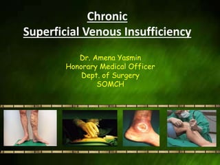
- 1. Chronic Superficial Venous Insufficiency Dr. Amena Yasmin Honorary Medical Officer Dept. of Surgery SOMCH
- 2. CHRONIC SUPERFICIAL VENOUS INSUFFICIENCY (CSVI) • occurs when the venous wall and/or valves in the superficial leg veins are not working effectively, making it difficult for blood to return to the heart from the legs.
- 3. Clinical hallmarks: Distal venous hypertension, which follows the development of valvular incompetence, reflux, and/or venous obstruction.
- 4. VENOUS SYSTEM Superficial venous system 1. Saphenous veins 2. Lateral venous complex Deep venous system Perforating veins Deep femoral v. Femoral v. Popliteal v. Small saphenous v. Great saphenous v. Perforating v. Perforating v. Image source: Fundamentals of Phlebology: Venous Disease for Clinicians. Illustration by Linda S. Nye. American College of Phlebology 2004. Deep femoral v. Perforating v.
- 5. PERFORATING VEINS AND REFLUX Maintain one-way flow from superficial to deep veins Perforator valve failure causes: • Higher venous pressure and GSV/branch dilation • Increasing pressure results in GSV valve failure • Additional vein branches become varicose • Further GSV incompetence and dilation
- 7. RISK FACTORS • Advancing age • Family history of venous disease • Prolonged standing • Increased body mass index • Smoking • Sedentary lifestyle
- 8. RISK FACTORS CONTD. • Lower extremity trauma • Prior venous thrombosis • Arterio-venous shunt • High estrogen states • Pregnancy • Ligamentous laxity ( hernia, flat fleet)
- 9. CEAP classification – an international consensus conference initiated the Clinical- Etiology-Anatomy-Pathophysiology classification. C: Clinical E: Etiology A: Anatomy P: Pathophysiology
- 10. CEAP classification cont. • C 0 – no evidence of venous disease. • C 1 – telangiectasias/reticular veins. • C 2 – varicose veins. • C 3 – edema associated with vein disease. • C 4a – pigmentation or eczema. • C 4b – lipodermatosclerosis. • C 5 – healed venous ulcer. • C 6 – active venous ulcer.
- 11. CEAP classification cont. • E c – congenital • E p – primary venous disease. • E s – secondary venous disorder. • E n – not specified.
- 12. CEAP classification cont. • A s – superficial veins. • A d – deep veins. • A p – perforating veins. • A n – not specified. • P r – venous reflux. • P o – venous obstruction. • P n – not specified.
- 13. 20+ million 2 to 6 million Skin Ulcers 500,000 Manifestations of Venous Insufficiency Superficial venous reflux is progressive and if left untreated, may worsen over time. Below are manifestations of the disease.5 Photos courtesy of Rajabrata Sarkar, MD, PhD. Swollen Legs Skin ChangesVaricose Veins
- 14. Systemic Reflux in Venous Ulceration Incompetent perforators found in 63% of venous ulcer patients Comprehensive care treats all sources of refluxPhotos courtesy of Steven A. Kaufman, MD. Sources of Reflux in Venous Ulcer Patients8 Superficial Perforating Deep 79% 63% 50%
- 15. SKIN CHANGES AT CSVI Gravitational dermatitis Hyperpigmentation Lipodermatosclerosis
- 16. LIPODERMATOSCLEROSIS There is a proliferation of the dermal capillaries and fibrosis on subcutaneous tissue It is a combination of: • induration • pigmentation • inflammation
- 17. VENOUS ULCER Clinical Findings: Inner aspect of the distal third of the leg (commonly the pressure areas) Shape - rounded, elongated or very large like a cuff (so-called gaiter ulcer) Base - flat, covered with fibrous slough Margins - sharp or rolled
- 19. Diagnostic Evaluation Level 1 : history and clinical examination. Level 2 : non-invasive vascular laboratory testing which now routinely include Duplex color scanning. Level 3 : invasive investigations or more complex imaging studies including ascending and descending venography, Varicography, venous pressure measurements, magnetic resonance imaging.
- 22. Management of Venous Stasis Ulcers • Dressings -Occlusive dressings -Low adherent gauze dressings • Surgical debridement used to remove devitalized tissue. • Enzymatic agents used to break down necrotic tissue (Santyl).
- 23. MANAGEMENT contd. • Growth factors synthesized by many cell types such as platelets, neutrophils, and epithelial cells (e.g. Regranex). • Bioengineered tissue used for a variety of non-healing ulcers (e.g. Apligraf, Dermagraft). • Skin grafting an option for non-healing ulcers.
- 24. MANAGEMENT OF CHRONIC SUPERFICIAL VENOUS INSUFFICIENCY A. Conservative treatment B. Vein ablation treatments C. Surgical procedure
- 25. 1. Conservative Treatment • Avoiding long periods of standing • While sitting, legs should be above the thigh • Avoiding crossing legs • Ideal body weight • Walking programme • Compression therapy • Micronised purified flavonoid fraction (diosmin+ hesperidin)
- 26. Compression therapy - elastic compression bandages - compression stockings - Pneumatic compression therapy
- 27. - Sclerotherapy - Foam sclerotherapy (USG guided) 2. Vein ablation treatments
- 28. Alternative techniques Radiofrequency ablation Endovenous laser ablation therapy. Indications Persistent signs/symptoms of venous disease after a minimum of 3 months of medical therapy Documented reflux (e.g. >0.5 seconds of reflux GSV).
- 29. Radiofrequency ablation • Radiofrequency devices generate a high frequency alternating current for which the energy heats the adjacent vein walls to the probe which alters the protein structure of the vein effecting its closure.
- 30. Endovenous laser ablation therapy • Lasers emit a single, coherent wavelength of light. Laser therapy of venous structures is based upon the concept of photothermolysis. Vein wall injury is mediated directly by absorption of photon energy by the vein wall and indirectly by thermal convection from heated blood.
- 31. ENDOVENOUS LASER ABLATION THERAPY
- 33. 3. SURGICAL OPTIONS 1. Sapheno-femoral/ sapheno-popliteal flush ligation 2. Venous stripping 3. Multiple phelebectomies 4. Ligation of the perforators
- 34. SAPHENO-FEMORALFLUSH LIGATION VENOUS STRIPPING • SFJ is identified after giving a groin incision lateral to pubic tubercle. • LSV tributaries are ligated and divided • A flush SFJ ligation is then performed • LSV retrogradely stripped to the knee • Tributaries of varicocities then avulsed through small incisions
- 35. • The sapheno-femoral junction, where all the tributaries have been ligated. • Sutured small groin incision
- 36. Stripper passed from groin to upper leg.
- 37. • Graduated compression stockings or bandages are worn day & night for 7-10days; thereafter they are worn only during day for one month • Patient should sit with his feet elevated • Patient should return to work and driving within 10days of surgery • Swimming and cycling are allowed after dressing have been removed Post-operative care
- 38. Venous Ulcer Patient Outcomes • Treating the underlying cause of venous ulceration results in improved clinical outcomes • Treating both the superficial and perforator hypertension results in: • Faster ulcer healing time • Lower ulcer recurrence rate than with compression therapy alone9,10
- 39. Complication • Eczema • Ulcers • Bleeding • Thrombophlebitis • DVT
- 40. Prevention Weight control Adequate physical exercise Avoidance of smoking Avoidance of sedentary activities Control of hypertension Modification of profession
- 41. REFERENCES 1. American Heart Association, SIR, Brand et al. “The Epidemiology of Varicose Veins: The Framingham Study” 2. US Markets for Varicose Vein Treatment Devices 2006, Millennium Research Group 2005. 3. Coon WW, Willis PW, Keller JB: Venous thromboembolism and other venous disease in the Tecumseh Community Health Study Circulation 1973; 48:839-846. 4. Barron HC, Ross BA. Varicose Veins: A guide to prevention and treatment. NY, NY: Facts on File, Inc. [An Infobase Holdings Company]; 1995;vii. 5. White JV, Ryjewski C. Chronic venous insufficiency. Perspect Vasc Surg Endovasc Ther 2005;17:319- 27 6. Dietzek A, Two-Year Follow-Up Data From A Prospective, Multicenter Study Of The Efficacy Of The ClosureFAST Catheter, 35th Annual Veith Symposium. November 19, 2008. New York. 7. Alameida JI. Lessons Learned After 2000 Endovenous Ablations. 34th Veith Symposium. Nov 14-18, 2007. New York 8. Hanrahn L. et al. Distribution of valvular incompetence in patients with venous stasis ulceration. JVS 13,6, 805-812 June 1991 9. Jamie R Barwell, Colin E Davies, Comparison of surgery and compression with compression alone in chronic venous ulceration (ESCHAR study): randomized controlled trial,THE LANCET, Vol 363, June 04 10. Nelzen O. Fransson I. True long-term healing and recurrence of venous leg ulcers following SEPS combined with superficial venous surgery: a prospective study. Eur J Vasc Endovasc Surg 34, 605- 612 (2007)
- 42. Thank you