SlideShare ist ein Scribd-Unternehmen logo
1 von 4
Downloaden Sie, um offline zu lesen
ORIGINAL ARTICLE

            Porcelain Fused to Metal (PFM) Crowns and Caries in
                               Adjacent Teeth
                                         Durr-e-Sadaf and Muhammad Zubair Ahmad


  ABSTRACT
  Objective: To assess the discrepancies in marginal integrity and contact points of Porcelain Fused to Metal (PFM) crowns
  and its association with caries in adjacent teeth.
  Study Design: Cross-sectional study.
  Place and Duration of Study: Dental Section of the Aga Khan University Hospital, Karachi, from January to August 2009.
  Methodology: Ninety six (96) patients with PFM crowns on maxillary and mandibular premolars and molars were included
  in this study. Single unit PFM crowns that were part of fixed bridge with at least one natural adjacent tooth were studied.
  PFM crown with no adjacent tooth or badly broken down teeth were excluded. Marginal integrity, contact points of PFM
  crowns and caries in adjacent teeth were assessed clinically and radiographically. The data was collected on a structured
  Proforma. Fisher's exact test was used for statistical analysis.
  Results: Marginal overhang and marginal gaps on the mesial surfaces of PFM crowns were observed in 17.7% and 13.5%
  respectively. Tight contact points and open contacts of PFM crowns with adjacent teeth were 15.6% and 17.8%
  respectively on mesial surfaces. Caries were present in 33.3% and 20% of teeth present mesial and distal to PFM crowns
  respectively. The association of faulty contact points of PFM crowns with caries in adjacent teeth was found significant
  (p < 0.001). The association of marginal discrepancy with caries in adjacent teeth was found significant (p = 0.002).
  Conclusion: A significant association of faulty contact points and margins of PFM crowns with caries in adjacent teeth
  was found.

  Key words:     Porcelain fused to metal (PFM) crowns. Marginal integrity. Contact points. Caries.


                    INTRODUCTION                                     extremely difficult to keep clean an area of crown with
In a traditional porcelain fused to metal (PFM) crown,               faulty margins and contact points which may result in
the strength is provided by the metal substructure, and              caries in adjacent teeth (Figure 1B). Therefore, the
a porcelain veneer provides esthetics.1 Porcelain fused              reproduction of contact points of proper size and
to metal (PFM) crowns are used to restore badly broken               location is essential for the success of the restoration.6
down teeth to protect remaining tooth structure. It may              Acceptable contact points in full coverage crowns are
also be responsible for maintaining occlusion and                    those that allow the floss to pass through with the same
providing aesthetics.2 Margins are unacceptable, if the              amount of resistance offered by the other contacts in
gap between margins of the crown and tooth structure is              natural dentition.3
greater than 50 um allowing insertion of the tip of the
explorer inside.3 Marginal gap between the crown and
the tooth may lead to microleakage and secondary
caries (Figure 1A). Secondary caries beneath crown
margins is considered the most frequent reason for
failure of crowns and fixed prosthodontic treatment.4,5
Defective cast crowns may also have adverse effects on
adjacent teeth. Correct formation, location and size of
contact points is essential. Any discrepancy in contact                 A                                      B
points leads to food impaction. Patients may find it                   Figure 1 (A and B): (A). Caries beneath open margin of PFM crown on
                                                                       mandibular first molar. Note that distal margin of this crown is open.
  Dental Section, Department of Surgery, The Aga Khan University       (B). Caries in mandibular second premolar adjacent to PFM crown on
  Hospital, Karachi.                                                   mandibular first molar.

  Correspondence: Dr. Durr-e-Sadaf, Associate Professor,             To our knowledge no research study has done so far to
  Department of Operative Dentistry, Margalla Institute of Health    observe the effects of marginal discrepancy and faulty
  Sciences, Quaid-e-Azam Avenue, Gulrez-III, Rawalpindi.             contact points of PFM crowns on adjacent natural teeth.
  E-mail: drdurresadaf@hotmail.com                                   Marginal integrity and contact points of PFM crowns
  Received October 15, 2009; accepted February 14, 2011.             were assessed clinically and radiographically. Diagnosis


134                                                           Journal of the College of Physicians and Surgeons Pakistan 2011, Vol. 21 (3): 134-137
Porcelain fused to metal (PFM) crowns and caries in adjacent teeth


of caries beneath crown margins with the help of radio-
graphs is well documented.5
The objective of this study was to assess discrepancies
in contact points and marginal integrity of PFM crowns
and its association with caries in adjacent natural teeth.

                        METHODOLOGY
It was a cross-sectional study conducted from January
to August 2009. Ninety six (96) patients visiting dental
clinics at the Aga Khan University Hospital, Karachi,
Pakistan with PFM crowns on maxillary and mandibular
molars and premolar teeth were selected. The crowns
were prepared by dental practitioners. Patients who
gave informed consent were included in the study. The                              Figure 2: Distribution of teeth with PFM crowns.
study was done after the approval of ethical review
committee of the institution. PFM crowns as a single unit                        22.9% (Figure 2). Single crowns were 82 (85.4%) and
or part of bridge with at least one natural tooth adjacent                       14 (14.6%) were abutment and part of fixed denture.
to crown were included. PFM crowns with no adjacent                              Crowns with root treated teeth were 65 (67.7%)
tooth or badly broken down tooth were excluded. Both                             and         31 (32.3%) were without any history of
root-treated and vital teeth with PFM crowns were                                endodontic treatment.
included. Clinical and radiographic assessment of
contact points and marginal integrity of teeth with PFM                          Marginal overhang and marginal gaps on the mesial
                                                                                 surface of crowns were 17 (17.7%) and 13 (13.5%)
crowns was done. Caries in teeth adjacent to PFM
                                                                                 respectively. Marginal integrity was satisfactory in 65
crowns was also recorded. Clinical assessment of
                                                                                 (67.7%) of mesial surfaces (Table I).
contact points of PFM crowns was assessed with the
help of dental floss. Contact points were categorized as                         Distal surfaces of crowns presented 10 (10.4%) marginal
acceptable, open contact points, tight contact points and                        overhang and 17 (17.7%) marginal gaps. Marginal
lost contacts due to caries in adjacent teeth. Acceptable                        integrity on distal surface was satisfactory in 56 (58.3%)
contact points were considered if dental floss could be                          of surfaces (Table I). Caries was observed in 18 (20%)
passed with little résistance. Open contact points were                          of teeth mesial to crowns and in 23 (33.3%) of teeth
those, which allowed the dental floss to pass without                            distal to PFM crowns (Table I).
resistance. If dental floss shredded or could not be                             The association of discrepancy in mesial margins of
passed, it was categorized as tight contact points.                              crowns with presence of caries in mesial adjacent teeth
Marginal integrity of PFM crowns was assessed with                               was found significant (p = 0.003). The association of
bitewing and periapical views of digital radiographs.                            discrepancy in distal margins of crowns with presence of
Marginal integrity was categorized as overhanged                                 caries in distal adjacent teeth was also found significant
margins, intact margins, space beneath margins, and                              (p = 0.021) (Table I).
caries beneath margins. The data was collected on a                              Tight contact points were observed in 14 (15.6%) on
structured proforma. It was analyzed using SPSS 17.0                             mesial surfaces and 20 (29%) on the distal surfaces of
and Fisher's exact test was used for statistical analysis                        crowns. Open contacts were observed on distal
to determine the association between distal crown                                surfaces in 20 (29%) and 16 (17.8%) were on mesial
margins and presence of caries in distal adjacent teeth,                         surfaces. Acceptable contact points were present on
between distal contact points and presence of caries in                          mesial surfaces in 51 (56.7%) and 24 (34.8%) were on
distal adjacent teeth, between mesial crown margins                              distal surfaces of the crowns (Table I).
and presence of caries in mesial adjacent teeth,                                 A significant association of faulty contact points with
between mesial contact points and presence of caries in                          presence of caries in adjacent mesial teeth was found
mesial adjacent teeth. P-value of less than 0.05 was                             (p < 0.001). The association of faulty contact points with
considered statistically significant at 95% confidence                           presence of caries in distal adjacent teeth was also
interval.                                                                        significant (p < 0.001) (Table I).
                                                                                 Caries beneath margins of crowns were seen in 1 (1%)
                               RESULTS                                           on mesial surfaces and 13 (13.5%) on distal surfaces.
There were 50 (52.1%) male patients and 46 (47.9%)                               Contact points were lost on mesial surfaces due to
female patients. Mandibular first molars with crowns                             caries in 9 (10%) of cases and on distal surfaces in
were 33.3% and maxillary first molars with crowns were                           5 (7.2%) of cases.


Journal of the College of Physicians and Surgeons Pakistan 2011, Vol. 21 (3): 134-137                                                  135
Durr-e-Sadaf and Muhammad Zubair Ahmad


Table I: Association of status of margins and contact points of PFM crowns with the condition of adjacent natural teeth. (Fisher's exact test is
         used at 95% Confidence Interval, α = 5%).
Crown margins                       Mesial             Distal                Contact points                     Mesial                  Distal
Intact margins                      65 (67.7%)         56 (58.3%)            Acceptable contact points          51 (56.7%)              24 (34.8%)
Overhang margins                    17 (17.7%)         10 (10.4%)            Tight contact points               14 (15.6%)              20 (29%)
Space beneath margins               13 (13.5%)         17 (17.7%)            Loose/ Open contact points         16 (17.8%)              20 (29%)
Caries beneath margins              1 (1%)             13 (13.5%)            Lost contact points                9 (10%)                 5 (7.2%)
                                                                             Missing                            6 (6.25%)               27 (28.12%)
Status of adjacent natural teeth                                             Status of adjacent natural teeth
Healthy                             62 (68.9%)         34 (49.3%)            Healthy                            62 (68.9%)              34 (49.3%)
Restored                            10 (11.1%)         12 (17.4%)            Restored                           10 (11.1%)              12 (17.4%)
Carious                             18 (20%)           23 (33.3%)            Carious                            18 (20%)                23 (33.3%)
Missing                             6 (6.25%)          27 (28.12%)           Missing                            6 (6.25%)               27 (28.12%)
P-value (Fisher's exact test)       0.002              0.017                 P-value (Fisher's exact test)      < 0.001                 < 0.001


                                DISCUSSION                                  points. One reason of tight contact points may be due to
The data obtained in this study showed marginal and                         over contoured crown on proximal surfaces. It also
                                                                            reduces embrasure space. Reduced embrasure space
contact points discrepancies in PFM crowns and caries
                                                                            results in broadening of the Col area, causing pressure
in adjacent teeth. Discrepancies in contact points,
                                                                            and irritation on the papilla. Over-contoured crown
marginal integrity and anatomic contour of the crowns
                                                                            decreases gingival embrasure leading to gingival
may have adverse effects on surrounding tissues.
                                                                            inflammation and inhibit effective oral hygiene.12,13
Marginal integrity is one of the most important critical
                                                                            Gordon suggested that the axial reduction of tooth
factors in success and failure of fixed restoration.6
                                                                            structure should follow the original contour of the tooth
Marginal defects were present in the form of overhang                       so that final restoration is more close to the natural
margins, space beneath margins and caries beneath                           anatomy of that tooth.14 Frequently, dentists prepare the
margins in this study. Space beneath margins was                            axial surfaces to be flat, forcing technicians to make over
observed to be the most common among all defects on                         contoured crown with wide occlusal tables. Many times
distal surfaces and overhang margins were most                              it may not be possible for even good technicians to
common among all defects on mesial surfaces of PFM                          overcome the discrepancies of preparation.
crowns.
                                                                            Tight contact points make the interdental area to floss
Crown margins should blend with the tooth structure                         extremely difficult for patients. It also makes the area
without overextention, marginal gaps and under                              highly susceptible for caries. In this study, faulty (tight,
extension.7 A study on clinical evaluation of all-ceramic                   open or lost) contact points were associated with caries
crowns showed visible marginal discrepancy in 30% of                        in adjacent teeth significantly. Contact points within
all-ceramic crowns and 3% caries contiguous with the                        normal limits were associated with less number of
margin.7 The gap between the crown margins and the                          carious lesions in adjacent natural teeth. Tight contact
prepared tooth can dissolve dental cement leading to                        points had greater association with presence of caries in
microleakage and caries development.9                                       adjacent natural teeth than open contact. Open contact
Marginal discrepancies are related to irregular or absent                   also leads to food impaction which is a favorable
                                                                            environment for cariogenic bacteria and results in dental
tooth preparation margins, impression defects or casting
                                                                            caries and gingival inflammation. Although open contact
shrinkage.10 Marginal discrepancy is seen more on
                                                                            points make the area easily accessible for oral hygiene
distal margins of the crown than on the mesial margins
                                                                            but it is not desirable because this condition may lead to
in this study. It may be related to difficulty in access to
                                                                            several problems including drifting/tilting of adjacent
distal surface while preparing teeth and difficult to
                                                                            tooth. In this study presence of carious lesion was
establish well defined and smooth margins. Practitioners                    observed less in teeth adjacent to crown with open
may face such problem in preparing distal surface of                        contact points than those with tight contact points.
maxillary and mandibular first and second molars.
Prevalence of marginal discrepancy in PFM crowns was                        Greater discrepancies in contact points between crown
observed to be up to 49% in a study.8                                       and distal teeth were observed than that between a
                                                                            crown and mesial tooth.
Porcelain fused to metal crowns with porcelain margins
showed less fracture resistance than that with metal                        Crown contours should facilitate plaque removal.12
margins which should be considered by operator in                           Ramfjord recommended placement of contact areas as
treatment planning phase particularly in posterior teeth                    far occlusally as possible to facilitate access for
where heavy occlusal forces can cause fracture of                           interproximal plaque control.15
restoration.11 Discrepancies in contact points were                         Interproximal space slightly larger than normal may be
observed in the form of tight contacts and open contact                     desirable since it provides adequate room for the gingival


136                                                                  Journal of the College of Physicians and Surgeons Pakistan 2011, Vol. 21 (3): 134-137
Porcelain fused to metal (PFM) crowns and caries in adjacent teeth


papilla and more accessible to clean.16,17 There are                                 2.          Ikai H, Kanno T, Kimura K. [A review of clinical follow-up studies
concerns regarding lateral impaction of food with open                                           focusing on pretreatment conditions of abutment and clinical
embrasures.16 However, another study reported that                                               examination parameters]. Nihon Hotetsu Shika Gakkai Zasshi 2006;
even with grossly undercontoured, open embrasure                                                 50:245-55. Japanese.
space, lateral food impaction rarely occurs as long as                               3.          Jalalian E, Jannati H, Mirzaei M. Evaluating the effect of a
interproximal contacts are properly maintained.18                                                sloping shoulder and a shoulder level on the marginal integrity of
                                                                                                 porcelain-fused-to-metal (PFM) veneer crowns. J Contemp Dent
                                                                                                 Pract 2008; 9:17-24.
Another study on most common complication associated
with fixed prosthesis showed caries incidence to be
0-2.7% in crowns and 0.7-26% in fixed partial dentures.8                             4.          Rosenstiel SF, Land MF, Fujimoto J, editors. Contemporary fixed
Prevalence of secondary caries in crowned teeth is                                               prosthodontics, 4th ed. St. Louis: Mosby; 2006.
reported up to 11.2% when examined clinically and up                                 5.          Zoellner A, Heuermann M, Weber HP, Gaengler P. Secondary
to 8.3% when examined radiographically.5 Crowned                                                 caries in crowned teeth: correlation of clinical and radiographic
tooth should be examined both clinically and                                                     findings. J Prosthet Dent 2002; 88:314-9.
radiographically. In a five year clinical study of posterior
Cercon FPD's by Sailer et al., secondary caries was
                                                                                     6.          Becker CM, Kaldahl WB. Current theories of crown contour,
                                                                                                 margin placement, and pontic design. 1981. J Prosthet Dent 2005;
found in 21.7% in crowned teeth.19 Presence of caries                                            93:107-15.
beneath margins was observed in 1% of mesial margins                                 7.          Goodacre CJ, Bernal G, Rungcharassaeng K, Kan JY. Clinical
and 13.5% of distal margins in this study. Visible                                               complications in fixed prosthodontics. J Prosthet Dent 2003; 90:
marginal discrepancy was found about 14% in all-                                                 31-41.
ceramic crowns in a study.17 All ceramic crowns showed
                                                                                     8.          Sjogren G, Lantto R, Tillberg A. Clinical evaluation of all-ceramic
                                                                                                 crowns (Dicor) in general practice. J Prosthet Dent 1999; 81:
changes in the surface texture in areas of occlusal
contact which may result in crack propagation leading to
                                                                                                 277-84.
porcelain fracture, an occlusal splint should be
considered for patients with heavy occlusal forces /                                 9.          Felton DA, Kanoy BE, Bayne SC, Wirthman GP. Effect of in vivo
parafunctional habits to prevent this situation.1 This                                           crown margin discrepancies on periodontal health. J Prosthet Dent
                                                                                                 1991; 65:357-64.
study showed marginal gaps in 13.5% of mesial
surfaces and 17.7% of the distal surfaces of crowns.                                 10. Bishop K, Briggs P, Kelleher M. Margin design for porcelain
                                                                                         fused to metal restorations which extend onto the root. Br Dent J
It is recommended on the basis of results of this study
                                                                                         1996; 180:177-84.
that crown should be evaluated both clinically and
radiographically before final cementation. At try in stage                           11. Michalakis KX, Stratos A, Hirayama H, Kang K, Touloumi F,
the margins and contact points of definitive crown                                       Oishi Y. Fracture resistance of metal ceramic restorations with
                                                                                         two different margin designs after exposure to masticatory
                                                                                         simulation. J Prosthet Dent 2009; 102:172-8.
should be assessed for any discrepancy. If any fault is
detected it should be adjusted by the dental laboratory.
After final cementation of the crown another bitewing                                12. Reitemeier B, Hansel K, Walter MH, Kastner C, Toutenburg H.
radiograph should be taken to check the excess cement                                    Effect of posterior crown margin placement on gingival health.
which if present should be immediately removed.                                          J Prosthet Dent 2002; 87:167-72.
                                                                                     13. Reeves WG. Restorative margin placement and periodontal
                           CONCLUSION                                                    health. J Prosthet Dent 1991; 66:733-6.
Faulty contact points of PFM crowns are found to be                                  14. Christensen GJ. Frequently encountered errors in tooth
associated with presence of carious lesions in adjacent                                  preparations for crowns. J Am Dent Assoc 2007; 138:1373-5.
natural teeth significantly. Discrepancies in crown                                  15. Ramfjord SP. Periodontal aspects of restorative dentistry. J Oral
margins are associated with presence of caries in                                        Rehabil 1974; 1:107-26.
adjacent teeth significantly. Caries beneath crown
                                                                                     16. Orkin DA, Reddy J, Bradshow D. The relationship of the position
                                                                                         of crown margins to gingival health. J Prosthet Dent 1987; 57:
margins are also found frequently in such cases.
Marginal discrepancies and defective contact points are                                  421-4.
seen more commonly on the distal surfaces of crowns
than on the mesial surfaces. Presence of carious lesions                             17. Smukler H, Chaibi M. Periodontal and dental considerations in
                                                                                         clinical crown extension: a rational basis for treatment. Int J
                                                                                         Periodontics Restorative Dent 1997; 17:464-77.
is seen more commonly on the teeth distal to PFM
crowns than those on the mesial to the PFM crowns.
                                                                                     18. Linkow LI. Contact areas in natural dentitions and fixed
                            REFERENCES                                                   prosthodontics. J Prosthet Dent 1962; 12:132-7.

1.   Etman MK, Woolford MJ. Three-year clinical evaluation of two                    19. Sailer I, Faher A, Filser F, Gauckler LJ, Luthy H, Hammerle CH.
     ceramic crown systems: a preliminary study. J Prosthet Dent 2010;                   Five year clinical results of zirconia frameworks for posterior
     103:80-90.                                                                          fixed partial denture. Int J Prosthodont 2007; 20:383-8.


                                                                         G G G G G   *
                                                                                     G G G G G


Journal of the College of Physicians and Surgeons Pakistan 2011, Vol. 21 (3): 134-137                                                                            137

Weitere ähnliche Inhalte

Was ist angesagt?

Managing the vertical dimension refined begg mechanics - d
Managing the vertical dimension   refined begg mechanics - dManaging the vertical dimension   refined begg mechanics - d
Managing the vertical dimension refined begg mechanics - dIndian dental academy
 
prosthodontic management of maxillectomy/obturators part 2 final copy
prosthodontic management of maxillectomy/obturators part 2 final copyprosthodontic management of maxillectomy/obturators part 2 final copy
prosthodontic management of maxillectomy/obturators part 2 final copyNAMITHA ANAND
 
Immediate placement and provisionalization of maxillary anterior single impla...
Immediate placement and provisionalization of maxillary anterior single impla...Immediate placement and provisionalization of maxillary anterior single impla...
Immediate placement and provisionalization of maxillary anterior single impla...Nguyễn Thị Minh Hiền
 
Implant treatment plan for completely edentulous patient
Implant treatment plan for completely edentulous patientImplant treatment plan for completely edentulous patient
Implant treatment plan for completely edentulous patientDr. Shashi Kiran
 
obturators / prosthodontic management of maxillectomy - part 1
obturators / prosthodontic management of maxillectomy - part 1obturators / prosthodontic management of maxillectomy - part 1
obturators / prosthodontic management of maxillectomy - part 1NAMITHA ANAND
 
JOURNAL CLUB PRESENTATION IN PROSTHODONTICS on ANDREW'S BRIDGE SYSTEM
JOURNAL CLUB PRESENTATION IN PROSTHODONTICS on ANDREW'S BRIDGE SYSTEMJOURNAL CLUB PRESENTATION IN PROSTHODONTICS on ANDREW'S BRIDGE SYSTEM
JOURNAL CLUB PRESENTATION IN PROSTHODONTICS on ANDREW'S BRIDGE SYSTEMNAMITHA ANAND
 
A Clinical Classification System for the Treatment of Postextraction SitesArt...
A Clinical Classification System for the Treatment of Postextraction SitesArt...A Clinical Classification System for the Treatment of Postextraction SitesArt...
A Clinical Classification System for the Treatment of Postextraction SitesArt...Nguyễn Thị Minh Hiền
 
Implant occlusion and failures1/ dental crown & bridge courses
Implant occlusion and failures1/ dental crown & bridge coursesImplant occlusion and failures1/ dental crown & bridge courses
Implant occlusion and failures1/ dental crown & bridge coursesIndian dental academy
 
Esthetic Management of Congenitally Missing Lateral Incisors With Single Toot...
Esthetic Management of Congenitally Missing Lateral Incisors With Single Toot...Esthetic Management of Congenitally Missing Lateral Incisors With Single Toot...
Esthetic Management of Congenitally Missing Lateral Incisors With Single Toot...Abu-Hussein Muhamad
 
The abo discrepancy index (di)
The abo discrepancy index (di)The abo discrepancy index (di)
The abo discrepancy index (di)Ali Waqar Hasan
 
Journal club presentation on tooth supported overdentures
Journal club presentation on tooth supported overdentures Journal club presentation on tooth supported overdentures
Journal club presentation on tooth supported overdentures NAMITHA ANAND
 
JOURNAL CLUB PRESENTATION IN PROSTHODONTICS ON PLATELET RICH FACTOR
JOURNAL CLUB PRESENTATION IN PROSTHODONTICS ON PLATELET RICH FACTORJOURNAL CLUB PRESENTATION IN PROSTHODONTICS ON PLATELET RICH FACTOR
JOURNAL CLUB PRESENTATION IN PROSTHODONTICS ON PLATELET RICH FACTORNAMITHA ANAND
 
Prosthetic options in implant dentistry
Prosthetic options in implant dentistryProsthetic options in implant dentistry
Prosthetic options in implant dentistryNAMITHA ANAND
 
Trt plan in implant/prosthodontic courses
Trt plan in implant/prosthodontic coursesTrt plan in implant/prosthodontic courses
Trt plan in implant/prosthodontic coursesIndian dental academy
 
Functional and Esthetic Restoration of the Worn Dentition
Functional and Esthetic Restoration of the Worn DentitionFunctional and Esthetic Restoration of the Worn Dentition
Functional and Esthetic Restoration of the Worn DentitionThe Dawson Academy
 

Was ist angesagt? (20)

Managing the vertical dimension refined begg mechanics - d
Managing the vertical dimension   refined begg mechanics - dManaging the vertical dimension   refined begg mechanics - d
Managing the vertical dimension refined begg mechanics - d
 
prosthodontic management of maxillectomy/obturators part 2 final copy
prosthodontic management of maxillectomy/obturators part 2 final copyprosthodontic management of maxillectomy/obturators part 2 final copy
prosthodontic management of maxillectomy/obturators part 2 final copy
 
Immediate placement and provisionalization of maxillary anterior single impla...
Immediate placement and provisionalization of maxillary anterior single impla...Immediate placement and provisionalization of maxillary anterior single impla...
Immediate placement and provisionalization of maxillary anterior single impla...
 
Implant treatment plan for completely edentulous patient
Implant treatment plan for completely edentulous patientImplant treatment plan for completely edentulous patient
Implant treatment plan for completely edentulous patient
 
obturators / prosthodontic management of maxillectomy - part 1
obturators / prosthodontic management of maxillectomy - part 1obturators / prosthodontic management of maxillectomy - part 1
obturators / prosthodontic management of maxillectomy - part 1
 
Presentation
PresentationPresentation
Presentation
 
JOURNAL CLUB PRESENTATION IN PROSTHODONTICS on ANDREW'S BRIDGE SYSTEM
JOURNAL CLUB PRESENTATION IN PROSTHODONTICS on ANDREW'S BRIDGE SYSTEMJOURNAL CLUB PRESENTATION IN PROSTHODONTICS on ANDREW'S BRIDGE SYSTEM
JOURNAL CLUB PRESENTATION IN PROSTHODONTICS on ANDREW'S BRIDGE SYSTEM
 
A Clinical Classification System for the Treatment of Postextraction SitesArt...
A Clinical Classification System for the Treatment of Postextraction SitesArt...A Clinical Classification System for the Treatment of Postextraction SitesArt...
A Clinical Classification System for the Treatment of Postextraction SitesArt...
 
Jc ppt
Jc pptJc ppt
Jc ppt
 
Implant occlusion and failures1/ dental crown & bridge courses
Implant occlusion and failures1/ dental crown & bridge coursesImplant occlusion and failures1/ dental crown & bridge courses
Implant occlusion and failures1/ dental crown & bridge courses
 
Cracks in Endodontics
Cracks in EndodonticsCracks in Endodontics
Cracks in Endodontics
 
Periimplantitis
PeriimplantitisPeriimplantitis
Periimplantitis
 
Esthetic Management of Congenitally Missing Lateral Incisors With Single Toot...
Esthetic Management of Congenitally Missing Lateral Incisors With Single Toot...Esthetic Management of Congenitally Missing Lateral Incisors With Single Toot...
Esthetic Management of Congenitally Missing Lateral Incisors With Single Toot...
 
The abo discrepancy index (di)
The abo discrepancy index (di)The abo discrepancy index (di)
The abo discrepancy index (di)
 
Journal club presentation on tooth supported overdentures
Journal club presentation on tooth supported overdentures Journal club presentation on tooth supported overdentures
Journal club presentation on tooth supported overdentures
 
JOURNAL CLUB PRESENTATION IN PROSTHODONTICS ON PLATELET RICH FACTOR
JOURNAL CLUB PRESENTATION IN PROSTHODONTICS ON PLATELET RICH FACTORJOURNAL CLUB PRESENTATION IN PROSTHODONTICS ON PLATELET RICH FACTOR
JOURNAL CLUB PRESENTATION IN PROSTHODONTICS ON PLATELET RICH FACTOR
 
Part 7 fact and fantasy about orthodontics
Part 7 fact and fantasy about orthodonticsPart 7 fact and fantasy about orthodontics
Part 7 fact and fantasy about orthodontics
 
Prosthetic options in implant dentistry
Prosthetic options in implant dentistryProsthetic options in implant dentistry
Prosthetic options in implant dentistry
 
Trt plan in implant/prosthodontic courses
Trt plan in implant/prosthodontic coursesTrt plan in implant/prosthodontic courses
Trt plan in implant/prosthodontic courses
 
Functional and Esthetic Restoration of the Worn Dentition
Functional and Esthetic Restoration of the Worn DentitionFunctional and Esthetic Restoration of the Worn Dentition
Functional and Esthetic Restoration of the Worn Dentition
 

Andere mochten auch

Alloys for pfm crown &bridg /certified fixed orthodontic courses by Indian de...
Alloys for pfm crown &bridg /certified fixed orthodontic courses by Indian de...Alloys for pfm crown &bridg /certified fixed orthodontic courses by Indian de...
Alloys for pfm crown &bridg /certified fixed orthodontic courses by Indian de...Indian dental academy
 
Repairing fractured pfm
Repairing fractured pfmRepairing fractured pfm
Repairing fractured pfmjhe0869
 
The metal ceramic crown preparation
The metal ceramic crown preparationThe metal ceramic crown preparation
The metal ceramic crown preparationguest33a456f1
 

Andere mochten auch (6)

Alloys for pfm crown &bridg /certified fixed orthodontic courses by Indian de...
Alloys for pfm crown &bridg /certified fixed orthodontic courses by Indian de...Alloys for pfm crown &bridg /certified fixed orthodontic courses by Indian de...
Alloys for pfm crown &bridg /certified fixed orthodontic courses by Indian de...
 
PFM
PFMPFM
PFM
 
Repairing fractured pfm
Repairing fractured pfmRepairing fractured pfm
Repairing fractured pfm
 
Base metal alloys
Base metal alloysBase metal alloys
Base metal alloys
 
Fixed prosthodontics lesson 3
Fixed prosthodontics lesson 3Fixed prosthodontics lesson 3
Fixed prosthodontics lesson 3
 
The metal ceramic crown preparation
The metal ceramic crown preparationThe metal ceramic crown preparation
The metal ceramic crown preparation
 

Ähnlich wie 03

Teeth in The Line of Mandibular Fractures
Teeth in The Line of Mandibular FracturesTeeth in The Line of Mandibular Fractures
Teeth in The Line of Mandibular FracturesAhmed Adawy
 
furcation involvmentt dental disease.pptx
furcation involvmentt dental disease.pptxfurcation involvmentt dental disease.pptx
furcation involvmentt dental disease.pptxMohamedYElZahar
 
Managing congenitally missing lateral incisors with single tooth implants
Managing congenitally missing lateral incisors with single tooth implants Managing congenitally missing lateral incisors with single tooth implants
Managing congenitally missing lateral incisors with single tooth implants Abu-Hussein Muhamad
 
Abutment evaluation / cosmetic dentistry training
Abutment evaluation  / cosmetic dentistry trainingAbutment evaluation  / cosmetic dentistry training
Abutment evaluation / cosmetic dentistry trainingIndian dental academy
 
Supernumerary teeth(hyperdontia)
Supernumerary teeth(hyperdontia)Supernumerary teeth(hyperdontia)
Supernumerary teeth(hyperdontia)Abu-Hussein Muhamad
 
Do pre existing micro cracks play a role in the fracture resistance of roots ...
Do pre existing micro cracks play a role in the fracture resistance of roots ...Do pre existing micro cracks play a role in the fracture resistance of roots ...
Do pre existing micro cracks play a role in the fracture resistance of roots ...Nadeem Aashiq
 
Extraction teeth for gaining space in orthodontics
Extraction teeth for gaining space in orthodontics Extraction teeth for gaining space in orthodontics
Extraction teeth for gaining space in orthodontics ameen qulah
 
The uses of orthodontic study models in DIAGNOSIS AND TREATMENT PLANNING
The uses of orthodontic study models in DIAGNOSIS AND TREATMENT PLANNINGThe uses of orthodontic study models in DIAGNOSIS AND TREATMENT PLANNING
The uses of orthodontic study models in DIAGNOSIS AND TREATMENT PLANNINGMaher Fouda
 
Jc 3 mother article
Jc 3 mother articleJc 3 mother article
Jc 3 mother articleAnju Bansal
 
Selection and choice of abutments/ dental implant courses
Selection and choice of abutments/ dental implant coursesSelection and choice of abutments/ dental implant courses
Selection and choice of abutments/ dental implant coursesIndian dental academy
 
Selection and choice of abutments / orthodontics training courses
Selection and choice of abutments / orthodontics training coursesSelection and choice of abutments / orthodontics training courses
Selection and choice of abutments / orthodontics training coursesIndian dental academy
 
Esthetic Management of Congenitally Missing Lateral Incisors With Single Toot...
Esthetic Management of Congenitally Missing Lateral Incisors With Single Toot...Esthetic Management of Congenitally Missing Lateral Incisors With Single Toot...
Esthetic Management of Congenitally Missing Lateral Incisors With Single Toot...Abu-Hussein Muhamad
 
prosthodontic concept of crown to root ratio.pptx
prosthodontic concept of crown to root ratio.pptxprosthodontic concept of crown to root ratio.pptx
prosthodontic concept of crown to root ratio.pptxDr vaishali shrivastava
 
Pre prosthetic orthodontic implant for management of congenitally unerupted l...
Pre prosthetic orthodontic implant for management of congenitally unerupted l...Pre prosthetic orthodontic implant for management of congenitally unerupted l...
Pre prosthetic orthodontic implant for management of congenitally unerupted l...Abu-Hussein Muhamad
 
Perio-ortho
Perio-orthoPerio-ortho
Perio-orthoshufei
 
Determination Of Dental Midline in Camouflage Orthodontic Treatment Of Facial...
Determination Of Dental Midline in Camouflage Orthodontic Treatment Of Facial...Determination Of Dental Midline in Camouflage Orthodontic Treatment Of Facial...
Determination Of Dental Midline in Camouflage Orthodontic Treatment Of Facial...Rahul Roy
 

Ähnlich wie 03 (20)

Teeth in The Line of Mandibular Fractures
Teeth in The Line of Mandibular FracturesTeeth in The Line of Mandibular Fractures
Teeth in The Line of Mandibular Fractures
 
furcation involvmentt dental disease.pptx
furcation involvmentt dental disease.pptxfurcation involvmentt dental disease.pptx
furcation involvmentt dental disease.pptx
 
Gingival esthetics
Gingival estheticsGingival esthetics
Gingival esthetics
 
Managing congenitally missing lateral incisors with single tooth implants
Managing congenitally missing lateral incisors with single tooth implants Managing congenitally missing lateral incisors with single tooth implants
Managing congenitally missing lateral incisors with single tooth implants
 
Abutment evaluation / cosmetic dentistry training
Abutment evaluation  / cosmetic dentistry trainingAbutment evaluation  / cosmetic dentistry training
Abutment evaluation / cosmetic dentistry training
 
Supernumerary teeth(hyperdontia)
Supernumerary teeth(hyperdontia)Supernumerary teeth(hyperdontia)
Supernumerary teeth(hyperdontia)
 
Do pre existing micro cracks play a role in the fracture resistance of roots ...
Do pre existing micro cracks play a role in the fracture resistance of roots ...Do pre existing micro cracks play a role in the fracture resistance of roots ...
Do pre existing micro cracks play a role in the fracture resistance of roots ...
 
186th publication jamdsr- 5th name
186th publication  jamdsr-  5th name186th publication  jamdsr-  5th name
186th publication jamdsr- 5th name
 
Extraction teeth for gaining space in orthodontics
Extraction teeth for gaining space in orthodontics Extraction teeth for gaining space in orthodontics
Extraction teeth for gaining space in orthodontics
 
The uses of orthodontic study models in DIAGNOSIS AND TREATMENT PLANNING
The uses of orthodontic study models in DIAGNOSIS AND TREATMENT PLANNINGThe uses of orthodontic study models in DIAGNOSIS AND TREATMENT PLANNING
The uses of orthodontic study models in DIAGNOSIS AND TREATMENT PLANNING
 
Jc 3 mother article
Jc 3 mother articleJc 3 mother article
Jc 3 mother article
 
Selection and choice of abutments/ dental implant courses
Selection and choice of abutments/ dental implant coursesSelection and choice of abutments/ dental implant courses
Selection and choice of abutments/ dental implant courses
 
Selection and choice of abutments / orthodontics training courses
Selection and choice of abutments / orthodontics training coursesSelection and choice of abutments / orthodontics training courses
Selection and choice of abutments / orthodontics training courses
 
Esthetic Management of Congenitally Missing Lateral Incisors With Single Toot...
Esthetic Management of Congenitally Missing Lateral Incisors With Single Toot...Esthetic Management of Congenitally Missing Lateral Incisors With Single Toot...
Esthetic Management of Congenitally Missing Lateral Incisors With Single Toot...
 
Abutment selection in fpd
Abutment selection in fpdAbutment selection in fpd
Abutment selection in fpd
 
prosthodontic concept of crown to root ratio.pptx
prosthodontic concept of crown to root ratio.pptxprosthodontic concept of crown to root ratio.pptx
prosthodontic concept of crown to root ratio.pptx
 
Pre prosthetic orthodontic implant for management of congenitally unerupted l...
Pre prosthetic orthodontic implant for management of congenitally unerupted l...Pre prosthetic orthodontic implant for management of congenitally unerupted l...
Pre prosthetic orthodontic implant for management of congenitally unerupted l...
 
Perio-ortho
Perio-orthoPerio-ortho
Perio-ortho
 
Single tooth implants
Single tooth implantsSingle tooth implants
Single tooth implants
 
Determination Of Dental Midline in Camouflage Orthodontic Treatment Of Facial...
Determination Of Dental Midline in Camouflage Orthodontic Treatment Of Facial...Determination Of Dental Midline in Camouflage Orthodontic Treatment Of Facial...
Determination Of Dental Midline in Camouflage Orthodontic Treatment Of Facial...
 

03

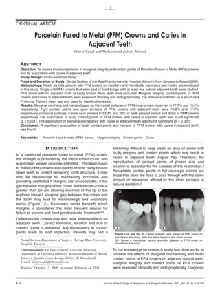
  • 1. ORIGINAL ARTICLE Porcelain Fused to Metal (PFM) Crowns and Caries in Adjacent Teeth Durr-e-Sadaf and Muhammad Zubair Ahmad ABSTRACT Objective: To assess the discrepancies in marginal integrity and contact points of Porcelain Fused to Metal (PFM) crowns and its association with caries in adjacent teeth. Study Design: Cross-sectional study. Place and Duration of Study: Dental Section of the Aga Khan University Hospital, Karachi, from January to August 2009. Methodology: Ninety six (96) patients with PFM crowns on maxillary and mandibular premolars and molars were included in this study. Single unit PFM crowns that were part of fixed bridge with at least one natural adjacent tooth were studied. PFM crown with no adjacent tooth or badly broken down teeth were excluded. Marginal integrity, contact points of PFM crowns and caries in adjacent teeth were assessed clinically and radiographically. The data was collected on a structured Proforma. Fisher's exact test was used for statistical analysis. Results: Marginal overhang and marginal gaps on the mesial surfaces of PFM crowns were observed in 17.7% and 13.5% respectively. Tight contact points and open contacts of PFM crowns with adjacent teeth were 15.6% and 17.8% respectively on mesial surfaces. Caries were present in 33.3% and 20% of teeth present mesial and distal to PFM crowns respectively. The association of faulty contact points of PFM crowns with caries in adjacent teeth was found significant (p < 0.001). The association of marginal discrepancy with caries in adjacent teeth was found significant (p = 0.002). Conclusion: A significant association of faulty contact points and margins of PFM crowns with caries in adjacent teeth was found. Key words: Porcelain fused to metal (PFM) crowns. Marginal integrity. Contact points. Caries. INTRODUCTION extremely difficult to keep clean an area of crown with In a traditional porcelain fused to metal (PFM) crown, faulty margins and contact points which may result in the strength is provided by the metal substructure, and caries in adjacent teeth (Figure 1B). Therefore, the a porcelain veneer provides esthetics.1 Porcelain fused reproduction of contact points of proper size and to metal (PFM) crowns are used to restore badly broken location is essential for the success of the restoration.6 down teeth to protect remaining tooth structure. It may Acceptable contact points in full coverage crowns are also be responsible for maintaining occlusion and those that allow the floss to pass through with the same providing aesthetics.2 Margins are unacceptable, if the amount of resistance offered by the other contacts in gap between margins of the crown and tooth structure is natural dentition.3 greater than 50 um allowing insertion of the tip of the explorer inside.3 Marginal gap between the crown and the tooth may lead to microleakage and secondary caries (Figure 1A). Secondary caries beneath crown margins is considered the most frequent reason for failure of crowns and fixed prosthodontic treatment.4,5 Defective cast crowns may also have adverse effects on adjacent teeth. Correct formation, location and size of contact points is essential. Any discrepancy in contact A B points leads to food impaction. Patients may find it Figure 1 (A and B): (A). Caries beneath open margin of PFM crown on mandibular first molar. Note that distal margin of this crown is open. Dental Section, Department of Surgery, The Aga Khan University (B). Caries in mandibular second premolar adjacent to PFM crown on Hospital, Karachi. mandibular first molar. Correspondence: Dr. Durr-e-Sadaf, Associate Professor, To our knowledge no research study has done so far to Department of Operative Dentistry, Margalla Institute of Health observe the effects of marginal discrepancy and faulty Sciences, Quaid-e-Azam Avenue, Gulrez-III, Rawalpindi. contact points of PFM crowns on adjacent natural teeth. E-mail: drdurresadaf@hotmail.com Marginal integrity and contact points of PFM crowns Received October 15, 2009; accepted February 14, 2011. were assessed clinically and radiographically. Diagnosis 134 Journal of the College of Physicians and Surgeons Pakistan 2011, Vol. 21 (3): 134-137
  • 2. Porcelain fused to metal (PFM) crowns and caries in adjacent teeth of caries beneath crown margins with the help of radio- graphs is well documented.5 The objective of this study was to assess discrepancies in contact points and marginal integrity of PFM crowns and its association with caries in adjacent natural teeth. METHODOLOGY It was a cross-sectional study conducted from January to August 2009. Ninety six (96) patients visiting dental clinics at the Aga Khan University Hospital, Karachi, Pakistan with PFM crowns on maxillary and mandibular molars and premolar teeth were selected. The crowns were prepared by dental practitioners. Patients who gave informed consent were included in the study. The Figure 2: Distribution of teeth with PFM crowns. study was done after the approval of ethical review committee of the institution. PFM crowns as a single unit 22.9% (Figure 2). Single crowns were 82 (85.4%) and or part of bridge with at least one natural tooth adjacent 14 (14.6%) were abutment and part of fixed denture. to crown were included. PFM crowns with no adjacent Crowns with root treated teeth were 65 (67.7%) tooth or badly broken down tooth were excluded. Both and 31 (32.3%) were without any history of root-treated and vital teeth with PFM crowns were endodontic treatment. included. Clinical and radiographic assessment of contact points and marginal integrity of teeth with PFM Marginal overhang and marginal gaps on the mesial surface of crowns were 17 (17.7%) and 13 (13.5%) crowns was done. Caries in teeth adjacent to PFM respectively. Marginal integrity was satisfactory in 65 crowns was also recorded. Clinical assessment of (67.7%) of mesial surfaces (Table I). contact points of PFM crowns was assessed with the help of dental floss. Contact points were categorized as Distal surfaces of crowns presented 10 (10.4%) marginal acceptable, open contact points, tight contact points and overhang and 17 (17.7%) marginal gaps. Marginal lost contacts due to caries in adjacent teeth. Acceptable integrity on distal surface was satisfactory in 56 (58.3%) contact points were considered if dental floss could be of surfaces (Table I). Caries was observed in 18 (20%) passed with little résistance. Open contact points were of teeth mesial to crowns and in 23 (33.3%) of teeth those, which allowed the dental floss to pass without distal to PFM crowns (Table I). resistance. If dental floss shredded or could not be The association of discrepancy in mesial margins of passed, it was categorized as tight contact points. crowns with presence of caries in mesial adjacent teeth Marginal integrity of PFM crowns was assessed with was found significant (p = 0.003). The association of bitewing and periapical views of digital radiographs. discrepancy in distal margins of crowns with presence of Marginal integrity was categorized as overhanged caries in distal adjacent teeth was also found significant margins, intact margins, space beneath margins, and (p = 0.021) (Table I). caries beneath margins. The data was collected on a Tight contact points were observed in 14 (15.6%) on structured proforma. It was analyzed using SPSS 17.0 mesial surfaces and 20 (29%) on the distal surfaces of and Fisher's exact test was used for statistical analysis crowns. Open contacts were observed on distal to determine the association between distal crown surfaces in 20 (29%) and 16 (17.8%) were on mesial margins and presence of caries in distal adjacent teeth, surfaces. Acceptable contact points were present on between distal contact points and presence of caries in mesial surfaces in 51 (56.7%) and 24 (34.8%) were on distal adjacent teeth, between mesial crown margins distal surfaces of the crowns (Table I). and presence of caries in mesial adjacent teeth, A significant association of faulty contact points with between mesial contact points and presence of caries in presence of caries in adjacent mesial teeth was found mesial adjacent teeth. P-value of less than 0.05 was (p < 0.001). The association of faulty contact points with considered statistically significant at 95% confidence presence of caries in distal adjacent teeth was also interval. significant (p < 0.001) (Table I). Caries beneath margins of crowns were seen in 1 (1%) RESULTS on mesial surfaces and 13 (13.5%) on distal surfaces. There were 50 (52.1%) male patients and 46 (47.9%) Contact points were lost on mesial surfaces due to female patients. Mandibular first molars with crowns caries in 9 (10%) of cases and on distal surfaces in were 33.3% and maxillary first molars with crowns were 5 (7.2%) of cases. Journal of the College of Physicians and Surgeons Pakistan 2011, Vol. 21 (3): 134-137 135
  • 3. Durr-e-Sadaf and Muhammad Zubair Ahmad Table I: Association of status of margins and contact points of PFM crowns with the condition of adjacent natural teeth. (Fisher's exact test is used at 95% Confidence Interval, α = 5%). Crown margins Mesial Distal Contact points Mesial Distal Intact margins 65 (67.7%) 56 (58.3%) Acceptable contact points 51 (56.7%) 24 (34.8%) Overhang margins 17 (17.7%) 10 (10.4%) Tight contact points 14 (15.6%) 20 (29%) Space beneath margins 13 (13.5%) 17 (17.7%) Loose/ Open contact points 16 (17.8%) 20 (29%) Caries beneath margins 1 (1%) 13 (13.5%) Lost contact points 9 (10%) 5 (7.2%) Missing 6 (6.25%) 27 (28.12%) Status of adjacent natural teeth Status of adjacent natural teeth Healthy 62 (68.9%) 34 (49.3%) Healthy 62 (68.9%) 34 (49.3%) Restored 10 (11.1%) 12 (17.4%) Restored 10 (11.1%) 12 (17.4%) Carious 18 (20%) 23 (33.3%) Carious 18 (20%) 23 (33.3%) Missing 6 (6.25%) 27 (28.12%) Missing 6 (6.25%) 27 (28.12%) P-value (Fisher's exact test) 0.002 0.017 P-value (Fisher's exact test) < 0.001 < 0.001 DISCUSSION points. One reason of tight contact points may be due to The data obtained in this study showed marginal and over contoured crown on proximal surfaces. It also reduces embrasure space. Reduced embrasure space contact points discrepancies in PFM crowns and caries results in broadening of the Col area, causing pressure in adjacent teeth. Discrepancies in contact points, and irritation on the papilla. Over-contoured crown marginal integrity and anatomic contour of the crowns decreases gingival embrasure leading to gingival may have adverse effects on surrounding tissues. inflammation and inhibit effective oral hygiene.12,13 Marginal integrity is one of the most important critical Gordon suggested that the axial reduction of tooth factors in success and failure of fixed restoration.6 structure should follow the original contour of the tooth Marginal defects were present in the form of overhang so that final restoration is more close to the natural margins, space beneath margins and caries beneath anatomy of that tooth.14 Frequently, dentists prepare the margins in this study. Space beneath margins was axial surfaces to be flat, forcing technicians to make over observed to be the most common among all defects on contoured crown with wide occlusal tables. Many times distal surfaces and overhang margins were most it may not be possible for even good technicians to common among all defects on mesial surfaces of PFM overcome the discrepancies of preparation. crowns. Tight contact points make the interdental area to floss Crown margins should blend with the tooth structure extremely difficult for patients. It also makes the area without overextention, marginal gaps and under highly susceptible for caries. In this study, faulty (tight, extension.7 A study on clinical evaluation of all-ceramic open or lost) contact points were associated with caries crowns showed visible marginal discrepancy in 30% of in adjacent teeth significantly. Contact points within all-ceramic crowns and 3% caries contiguous with the normal limits were associated with less number of margin.7 The gap between the crown margins and the carious lesions in adjacent natural teeth. Tight contact prepared tooth can dissolve dental cement leading to points had greater association with presence of caries in microleakage and caries development.9 adjacent natural teeth than open contact. Open contact Marginal discrepancies are related to irregular or absent also leads to food impaction which is a favorable environment for cariogenic bacteria and results in dental tooth preparation margins, impression defects or casting caries and gingival inflammation. Although open contact shrinkage.10 Marginal discrepancy is seen more on points make the area easily accessible for oral hygiene distal margins of the crown than on the mesial margins but it is not desirable because this condition may lead to in this study. It may be related to difficulty in access to several problems including drifting/tilting of adjacent distal surface while preparing teeth and difficult to tooth. In this study presence of carious lesion was establish well defined and smooth margins. Practitioners observed less in teeth adjacent to crown with open may face such problem in preparing distal surface of contact points than those with tight contact points. maxillary and mandibular first and second molars. Prevalence of marginal discrepancy in PFM crowns was Greater discrepancies in contact points between crown observed to be up to 49% in a study.8 and distal teeth were observed than that between a crown and mesial tooth. Porcelain fused to metal crowns with porcelain margins showed less fracture resistance than that with metal Crown contours should facilitate plaque removal.12 margins which should be considered by operator in Ramfjord recommended placement of contact areas as treatment planning phase particularly in posterior teeth far occlusally as possible to facilitate access for where heavy occlusal forces can cause fracture of interproximal plaque control.15 restoration.11 Discrepancies in contact points were Interproximal space slightly larger than normal may be observed in the form of tight contacts and open contact desirable since it provides adequate room for the gingival 136 Journal of the College of Physicians and Surgeons Pakistan 2011, Vol. 21 (3): 134-137
  • 4. Porcelain fused to metal (PFM) crowns and caries in adjacent teeth papilla and more accessible to clean.16,17 There are 2. Ikai H, Kanno T, Kimura K. [A review of clinical follow-up studies concerns regarding lateral impaction of food with open focusing on pretreatment conditions of abutment and clinical embrasures.16 However, another study reported that examination parameters]. Nihon Hotetsu Shika Gakkai Zasshi 2006; even with grossly undercontoured, open embrasure 50:245-55. Japanese. space, lateral food impaction rarely occurs as long as 3. Jalalian E, Jannati H, Mirzaei M. Evaluating the effect of a interproximal contacts are properly maintained.18 sloping shoulder and a shoulder level on the marginal integrity of porcelain-fused-to-metal (PFM) veneer crowns. J Contemp Dent Pract 2008; 9:17-24. Another study on most common complication associated with fixed prosthesis showed caries incidence to be 0-2.7% in crowns and 0.7-26% in fixed partial dentures.8 4. Rosenstiel SF, Land MF, Fujimoto J, editors. Contemporary fixed Prevalence of secondary caries in crowned teeth is prosthodontics, 4th ed. St. Louis: Mosby; 2006. reported up to 11.2% when examined clinically and up 5. Zoellner A, Heuermann M, Weber HP, Gaengler P. Secondary to 8.3% when examined radiographically.5 Crowned caries in crowned teeth: correlation of clinical and radiographic tooth should be examined both clinically and findings. J Prosthet Dent 2002; 88:314-9. radiographically. In a five year clinical study of posterior Cercon FPD's by Sailer et al., secondary caries was 6. Becker CM, Kaldahl WB. Current theories of crown contour, margin placement, and pontic design. 1981. J Prosthet Dent 2005; found in 21.7% in crowned teeth.19 Presence of caries 93:107-15. beneath margins was observed in 1% of mesial margins 7. Goodacre CJ, Bernal G, Rungcharassaeng K, Kan JY. Clinical and 13.5% of distal margins in this study. Visible complications in fixed prosthodontics. J Prosthet Dent 2003; 90: marginal discrepancy was found about 14% in all- 31-41. ceramic crowns in a study.17 All ceramic crowns showed 8. Sjogren G, Lantto R, Tillberg A. Clinical evaluation of all-ceramic crowns (Dicor) in general practice. J Prosthet Dent 1999; 81: changes in the surface texture in areas of occlusal contact which may result in crack propagation leading to 277-84. porcelain fracture, an occlusal splint should be considered for patients with heavy occlusal forces / 9. Felton DA, Kanoy BE, Bayne SC, Wirthman GP. Effect of in vivo parafunctional habits to prevent this situation.1 This crown margin discrepancies on periodontal health. J Prosthet Dent 1991; 65:357-64. study showed marginal gaps in 13.5% of mesial surfaces and 17.7% of the distal surfaces of crowns. 10. Bishop K, Briggs P, Kelleher M. Margin design for porcelain fused to metal restorations which extend onto the root. Br Dent J It is recommended on the basis of results of this study 1996; 180:177-84. that crown should be evaluated both clinically and radiographically before final cementation. At try in stage 11. Michalakis KX, Stratos A, Hirayama H, Kang K, Touloumi F, the margins and contact points of definitive crown Oishi Y. Fracture resistance of metal ceramic restorations with two different margin designs after exposure to masticatory simulation. J Prosthet Dent 2009; 102:172-8. should be assessed for any discrepancy. If any fault is detected it should be adjusted by the dental laboratory. After final cementation of the crown another bitewing 12. Reitemeier B, Hansel K, Walter MH, Kastner C, Toutenburg H. radiograph should be taken to check the excess cement Effect of posterior crown margin placement on gingival health. which if present should be immediately removed. J Prosthet Dent 2002; 87:167-72. 13. Reeves WG. Restorative margin placement and periodontal CONCLUSION health. J Prosthet Dent 1991; 66:733-6. Faulty contact points of PFM crowns are found to be 14. Christensen GJ. Frequently encountered errors in tooth associated with presence of carious lesions in adjacent preparations for crowns. J Am Dent Assoc 2007; 138:1373-5. natural teeth significantly. Discrepancies in crown 15. Ramfjord SP. Periodontal aspects of restorative dentistry. J Oral margins are associated with presence of caries in Rehabil 1974; 1:107-26. adjacent teeth significantly. Caries beneath crown 16. Orkin DA, Reddy J, Bradshow D. The relationship of the position of crown margins to gingival health. J Prosthet Dent 1987; 57: margins are also found frequently in such cases. Marginal discrepancies and defective contact points are 421-4. seen more commonly on the distal surfaces of crowns than on the mesial surfaces. Presence of carious lesions 17. Smukler H, Chaibi M. Periodontal and dental considerations in clinical crown extension: a rational basis for treatment. Int J Periodontics Restorative Dent 1997; 17:464-77. is seen more commonly on the teeth distal to PFM crowns than those on the mesial to the PFM crowns. 18. Linkow LI. Contact areas in natural dentitions and fixed REFERENCES prosthodontics. J Prosthet Dent 1962; 12:132-7. 1. Etman MK, Woolford MJ. Three-year clinical evaluation of two 19. Sailer I, Faher A, Filser F, Gauckler LJ, Luthy H, Hammerle CH. ceramic crown systems: a preliminary study. J Prosthet Dent 2010; Five year clinical results of zirconia frameworks for posterior 103:80-90. fixed partial denture. Int J Prosthodont 2007; 20:383-8. G G G G G * G G G G G Journal of the College of Physicians and Surgeons Pakistan 2011, Vol. 21 (3): 134-137 137