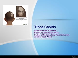
Tinea Capitis
•Als PPT, PDF herunterladen•
47 gefällt mir•17,987 views

Interactive Seminar, Dermatology Week Block 4.1 , College of Medicine, King Faisal University Al-Ahsa, Saudi Arabia

Empfohlen
Weitere ähnliche Inhalte
Was ist angesagt?
Was ist angesagt? (20)
Ähnlich wie Tinea Capitis
Ähnlich wie Tinea Capitis (20)
Mehr von Abdullatif Al-Rashed
Mehr von Abdullatif Al-Rashed (20)
Kürzlich hochgeladen
Kürzlich hochgeladen (20)
Tinea Capitis
- 1. Tinea Capitis Abdullatiff Sami Al-Rashed Block 4.1 (Dermatology Week) College of Medicine, King Faisal University Al-Ahsa, Saudi Arabia
- 2. Case A 3-year-old boy presents with a 3-week history of a circular scalp area of hair loss and flaky skin. He attends daycare and is provided with a sleep mat for an afternoon nap, which is not exclusively for his use. The scalp lesion is not itchy, but has not gone away with an anti-dandruff shampoo. There are no other skin lesions present.
- 4. Definition • Tinea capitis is a fungal infection of the scalp that most often presents with pruritic, scaling areas of hair loss. • Several synonyms are used, including ringworm of the scalp and tinea tonsurans.
- 5. Epidemiology • Most common among toddlers and school age children. • Much more common in blacks than in whites.
- 6. Etiology • Tinea capitis is a dermatophyte infection. • Dermatophytes are filamentous fungi in the genera Trichophyton, Microsporum, and Epidermophyton that infect keratinized tissue of skin, hair, or nails.
- 7. Etiology • Organisms in the These genera causes Tinea Capitis:
- 8. Etiology Etiology varies from country to country and from region to region:
- 9. Transmission Person-to-person, animal-to-person, via fomites. Spores are present on asymptomatic carriers, animals, or inanimate objects.
- 10. Clinical presentation Non- inflammatory infection Partial alopecia, often circular in shape, showing numerous broken-off hairs, dull gray from their coating of arthrospores. Fine scaling with fairly sharp margin. Infammatory response minimal, but massive scaling.
- 11. Clinical presentation Black dot Broken off hairs near the scalp give appearance of “dots”. Tends to be diffuse and poorly circumscribed. Low-grade folliculitis may be present.
- 12. Clinical presentation kerion Inflammatory mass in which remaining hairs are loose. Characterized by boggy, purulent, inflamed nodules, and plaques Usually painful; drains pus from multiple openings, like honeycomb. thick crusting with matting of adjacent hairs. Frequently, associated with lymphadenopathy.
- 13. Clinical presentation Favus Early cases show perifollicular erythema and matting of hair. Later, thick yellow adherent crusts (scutula)composed of skin debris and hyphae that are pierced by remaining hair shafts. Fetid odor. Shows little tendency to clear spontaneously. Often results in scarring alopecia
- 14. History
- 15. History
- 17. Physical Exam and Investigations Examination of the affected area with a Wood's light can help identify tinea capitis in patients with some ectothrix infections and favus. Ectothrix infections secondary to M. canis often exhibit green- yellow fluorescence. T. tonsurans does not fluoresce. Wood’s light
- 18. Diagnosis skin scales contain hyphae and arthrospores. Ectothrix: arthrospores can be seen surrounding the hair shaft. Endothrix: spores within hair shaft. Favus: loose chains of arthrospores and airspaces in hair shaft Direct Microscopy ”potassium hydroxide” Growth of dermatophytes usually seen in 10-14 days. Rule out bacterial infection, usually S. aureus or GAS. Fungal Culture Bacterial Culture
- 20. Treatment
- 21. Treatment Adjunctive interventions: • Antifungal shampoo : Selenium sulfide 5-10 ml on wet scalp, 2 applications each week for 2 weeks will provide control.
- 22. Prognosis • The prognosis of tinea capitis is excellent, with complete clearance occurring in most patients after a course of treatment. • Complete hair regrowth occurs in most children with hair loss. • Patients with chronic or severe infections (eg, kerion, favus) have the greatest risk for permanent scarring alopecia.
- 23. Reference
Hinweis der Redaktion
- Partial alopecia, often circular in shape, showing numerous broken-off hairs, dull gray from their coating of arthrospores. Fine scaling with fairly sharp margin. Hair shaft becomes brittle, breaking off at or slightly above scalp. Small patches coalesce, forming larger patches. Infammatory response minimal, but massive scaling.
- Broken-offhairs near the scalp give appearance of “dots” (Fig. 26-44) (swollen hair shafts) in dark-haired patients. Dots occur as affected hair breaks at surface of scalp. Tends to be diffuse and poorly circum- scribed. Low-grade folliculitis may be pres- ent.
- latin for honeycomb. Early cases show perifollicular erythema and matting of hair. Lat- er, thick yellow adherent crusts (scutula) com- posed of skin debris and hyphae that are pierced by remaining hair shafts (Fig. 26-46). Fetid odor. Shows little tendency to clear spontaneously. Often results in scarring alopecia The scutula contain fungi, neutrophils, dried serum, and epidermal cells. Scutula eventually coalesce to form adherent masses above areas of severe alopecia.