
Empfohlen
Weitere ähnliche Inhalte
Was ist angesagt?
Was ist angesagt? (20)
Ähnlich wie Doctor's case history of fever and lymphadenopathy
Ähnlich wie Doctor's case history of fever and lymphadenopathy (20)
Kürzlich hochgeladen
Kürzlich hochgeladen (20)
Doctor's case history of fever and lymphadenopathy
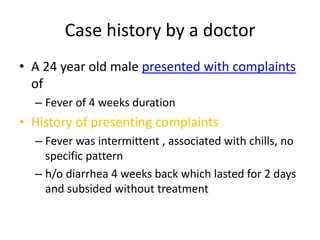
- 1. Case history by a doctor A 24 year old male presented with complaints of Fever of 4 weeks duration History of presenting complaints Fever was intermittent , associated with chills, no specific pattern h/o diarrhea 4 weeks back which lasted for 2 days and subsided without treatment
- 2. h/o loss of appetite for the past 1 month h/o loss of weight around 3-4 kilos over the last month Past history Not a known diabetic No h/o previous Anti Tuberculous Treatment No h/o any drug intake
- 3. Personal history Known alcoholic - occasional 3-4 drinks twice a week Not a smoker No h/o any other addictions Married 1 ½ years back, no kids, no pets Employed at a vehicle repair shop Family history No family h/o malignancy or premature death
- 4. Medical questions Treatment history Took antibiotics, anti-malarialsand antipyretics for fever from a general practitioner Got admitted outside for the above complaints and was evaluated. But even after investigating for a week no cause could be attributed for the symptoms. Incidentally ultra sonogram of the abdomen picked up peripancreatic lymphadenopathy. Patient was then referred to our institution for further evaluation.
- 5. Clinical findings General examination Well built, moderately nourished Febrile No pallor Not icteric No clubbing No edema of extremities Skin warm, dry, free of rashes Cervical and inguinal nodes were palpable: 3-4 in number, bilateral, around 1.0- 1.5 cm in size, discrete, mobile, firm, non tender.
- 6. Vitals Pulse 92 per minute Blood pressure 126/80 mm hg Respiratory rate 16/min Temperature 101.8F
- 7. Cardiovascular system: Heart sounds S1, S2 heard, no murmurs Respiratory system: Normal vesicular breath sounds heard, no added sounds Abdomen: Soft, no organomegaly Nervous system: No neurological deficits
- 8. Diagnosis Fever of unknown origin (FUO) ? Infectious ? Malignancy
- 9. Investigations ECG – sinus rhythm, WNL CXR – normal Hemogram WBC 3,000 46% polymorphs, 48% lymphocytes, 6% eosinophils Hemoglobin 10.5 g/l Hematocrit 36 Platelet 220 000 ESR 20mm/hr
- 10. Random blood sugar 90mg/dl Renal function tests: Urea 24 mg/dl Serum creatinine 0.9 mg/dl Electrolytes : Na+ 136 K+ 4.0 Cl- 95 HCO3- 25
- 11. Liver function tests: Total bilirubin 1.2 mg/dl Direct bilirubin 1.0 mg/dl AST 45 ALT 46 GGT 67 SAP 149 Total proteins 7.6mg/dl Albumin 4.2mg/dl Globulin 3.4mg/dl
- 12. Blood group A+ Coagulation profile: PT 14 sec INR 1.0 aPTT 26 sec Urine analysis pH 6.5 Specific gravity 1.025 Deposits 3-4/HPF HbsAg - neg Anti HCV - neg
- 13. Fever profile Smear for malarial parasite – neg Dengue IgM – neg WIDAL neg 1 in 25 dilution MSAT 2+ Blood culture - no growth Urine culture - no growth HIV – negative VDRL – non reactive
- 14. TB screening: Sputum AFB Sample A – neg Sample B – neg Mantoux – neg Chest x ray - normal Ultrasonogram abdomen: Liver 15 cm – mild hepatomegaly Peripancreaticadenopathy Paraaortic areas free No free fluid
- 15. Treatment IVF for hydration Tepid sponging Antipyretics Antibiotics - 2 weeks 3rd generation cephalosporin IV Doxycycline oral Antimalarial: Quinine – 5 day course
- 16. With the above treatment for two weeks the fever did not subside…… Common tropical infections (dengue, leptospirosis, malaria, filariasis, typhoid) being ruled out..
- 17. How to proceed? What are the differential diagnosis considered in this patient?
- 18. Approach to a patient with FUO
- 19. Hematologist opinion To rule out CTD (connective tissue disorder); Post viral adenopathy LPD (lympho proliferative disorder) – Hodgkin’s Peripheral smear, CBC at hematology lab
- 20. Report: CBC TC 7,000 DC P68, L30, M2 Hb 10 g/dl Platelets 180 000 ESR 25/55 Peripheral smear Normal study Final opinion: Review with AFB culture, lymph node biopsy.
- 21. Rheumatologist opinion: Fever with lymphadenopathy Suggested ANA, RF To r/o connective tissue disorder ANA, RF turned out to be negative
- 22. Evaluation of lymphadenopathy Lymphadenopathy was an important clue, efforts were then directed towards evaluating the cause for lymph node enlargement CT chest: Mediastinaladenopathy, Supraclavicular adenopathy with one large node having intrathoracic extension.
- 23. CT abdomen with contrast: Liver normal in size Spleen 10.3 x 4.3 cm - normal Para aortic region – multiple small rounded and soft tissue density lesions with low attenuated center with peripheral enhancement on I.V. contrast encasing paraaortic region and aortocaval region suggestive of lymphadenopathy
- 24. Incision biopsy of lymph nodeunder IV anesthesia: 0.5 x 0.3 cm lymph node architecture showing capsule with underlying lymphoid follicle composed of prominent germinal center, few follicles show polymorphous population of cells composed of lymphocytes , neutrophils and macrophages .
- 25. Comment on the lymph node biopsy…
- 26. Lymph node biopsy report: Non – specific lymphadenitis
- 27. Fever for more than 3 weeks, evaluated for more than 1 week -FUO CBC, differentials, peripheral smear, ESR, urine analysis, LFT, RFT, Electrolytes, HIV, VDRL, serology- (dengue, lepto, typhoid), ANA, RF. Blood culture, urine culture Potential diagnostic clue- lymphadenopathy Further evaluation directed towards it
- 28. CT chest and abdomen with contrast Multiple nodes in neck, mediastinum, abdomen. Incision biopsy done Biopsy inconclusive ? Empirical therapy
- 29. ? Empirical ATT Empirical ATT was an attractive option but since the initial TB screen was negative we proceeded with serology Anti TB IgM – 0.41 < 0.8 negative Anti TB IgA – 183 < 200 negative Anti TB IgG – 505 > 225 positive
- 30. Since the serology was not unambiguous we sought the opinion of the chest physician for empirical ATT CT guided needle biopsy of the mediastinal node was advised Bone marrow aspiration for AFB studies