
Empfohlen
Weitere ähnliche Inhalte
Was ist angesagt?
Was ist angesagt? (20)
Andere mochten auch
Andere mochten auch (16)
Ähnlich wie Fractures of the Distal Humerus
Ähnlich wie Fractures of the Distal Humerus (20)
Mehr von Arun Shanbhag
Mehr von Arun Shanbhag (11)
Kürzlich hochgeladen
Kürzlich hochgeladen (20)
Fractures of the Distal Humerus
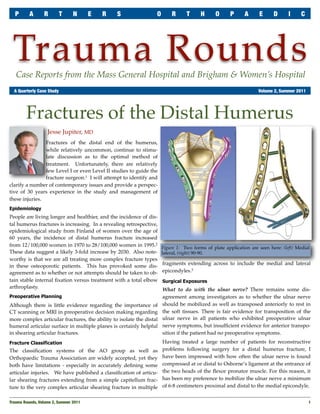
- 1. P A R T N E R S O R T H O P A E D I C Trauma Rounds Case Reports from the Mass General Hospital and Brigham & Women’s Hospital A Quarterly Case Study Volume 2, Summer 2011 Fractures of the Distal Humerus Jesse Jupiter, MD Fractures of the distal end of the humerus, while relatively uncommon, continue to stimu- late discussion as to the optimal method of treatment. Unfortunately, there are relatively few Level I or even Level II studies to guide the fracture surgeon.1 I will attempt to identify and clarify a number of contemporary issues and provide a perspec- tive of 30 years experience in the study and management of these injuries. Epidemiology People are living longer and healthier, and the incidence of dis- tal humerus fractures is increasing. In a revealing retrospective, epidemiological study from Finland of women over the age of 60 years, the incidence of distal humerus fracture increased from 12/100,000 women in 1970 to 28/100,000 women in 1995.2 Figure 1: Two forms of plate application are seen here: (left) Medial These data suggest a likely 3-fold increase by 2030. Also note- lateral, (right) 90-90. worthy is that we are all treating more complex fracture types fragments extending across to include the medial and lateral in these osteoporotic patients. This has provoked some dis- agreement as to whether or not attempts should be taken to ob- epicondyles.3 tain stable internal fixation versus treatment with a total elbow Surgical Exposures arthroplasty. What to do with the ulnar nerve? There remains some dis- Preoperative Planning agreement among investigators as to whether the ulnar nerve Although there is little evidence regarding the importance of should be mobilized as well as transposed anteriorly to rest in CT scanning or MRI in preoperative decision making regarding the soft tissues. There is fair evidence for transposition of the more complex articular fractures, the ability to isolate the distal ulnar nerve in all patients who exhibited preoperative ulnar humeral articular surface in multiple planes is certainly helpful nerve symptoms, but insufficient evidence for anterior transpo- in shearing articular fractures. sition if the patient had no preoperative symptoms. Fracture Classification Having treated a large number of patients for reconstructive The classification systems of the AO group as well as problems following surgery for a distal humerus fracture, I Orthopaedic Trauma Association are widely accepted, yet they have been impressed with how often the ulnar nerve is found both have limitations - especially in accurately defining some compressed at or distal to Osborne’s ligament at the entrance of articular injuries. We have published a classification of articu- the two heads of the flexor pronator muscle. For this reason, it lar shearing fractures extending from a simple capitellum frac- has been my preference to mobilize the ulnar nerve a minimum ture to the very complex articular shearing fracture in multiple of 6-8 centimeters proximal and distal to the medial epicondyle. Trauma Rounds, Volume 2, Summer 2011 1
- 2. P A R T N E R S O R T H O P A E D I C T R A U M A R O U N D S Approach to the Fracture Enthusiasm has been tempered for an olecranon osteotomy ap- proach to the distal humerus due to reported complications of nonunion at the osteotomy site, yet it still provides excellent exposure for those complex very distal articular fractures. I still favor this approach, creating a chevron shaped osteotomy with the apex pointing distally and secured with standard tension wire fixation modified only by two tension wires. Alternative exposures include a midline split of the triceps, tri- ceps elevation with a small fragment of the proximal olecranon, or the extended lateral approach, which is especially useful for anterior shearing articular fractures. The literature suggests that a triceps-splitting approach will lead to functional outcomes similar to that of the olecranon os- teotomy, without associated complications. Fracture Stabilization Some current discussion focuses on whether to apply plates and Figure 2: Radiograph of a patient who underwent total elbow screws parallel to each other, or placing the implants orthogonal arthroplasty for a distal humerus fracture. to one other (Figure 1). Biomechanical studies have not demon- strated a major advantage for either method, but there is some seen renewed interest following the publication of the Canadian evidence to support the use of parallel plate applications for Multi-center Trial.4 Patients over 65 years were prospectively comminuted or osteoporotic fractures. randomized to total elbow arthroplasty or open reduction and The development of anatomically shaped implants with angu- internal fixation. The results suggested that those patients who lar stable locking screw fixation may offer some advantages in had the arthroplasty functioned better on both objective and providing stable internal fixation in the osteoporotic patient. patient rated scores (Figure 2). These data combined with addi- There is, however, little evidence in the literature to support this tional studies would support the indications for total elbow concept. One should always realize the ability to anatomically arthroplasty for patients over the age of 65 years who have sus- shape standard implants to meet the unique requirements of tained a displaced comminuted fracture NOT amenable to sta- complex fractures. ble internal fixation.1 For stable fixation of complex, articular shearing fractures, I References 1. Nauth A et al, Current Concepts Review. Distal Humeral Fractures in Adults: J find that headless screws placed from anterior to posterior di- Bone Joint Surg 2011; 93:686-700. rection through an extended lateral approach are predictable for 2. Palvaren M et al, Secular trends in distal humeral fractures of elderly women: most fracture patterns. Bone 2010; 46: 1355-8. Indications for Total Elbow Arthroplasty 3. Mckee M, Jupiter J, Bamberger HB, Coronal shear fractures of the distal end of the humerus: J Bone Joint Surg 1996; 78: 49-54. Arthroplasty for complex fractures in the older aged patient, 4. McKee M et al, A multicenter prospective, randomized, controlled trial of open especially those with complex multi-fragmented fractures has reduction-internal fixation versus total elbow arthroplasty for displaced intra- articular distal humerus fractures in elderly patients: J Shoulder Elbow Surg AchesAndJoints.org/Trauma 2009; 18: 3-12. Please share your comments online, or by email: Trauma Faculty Michael Weaver, MD — 617-525-8088 Mark Vrahas, MD / mvrahas@partners.org BWH Orthopedic Trauma Mark Vrahas, MD — 617-726-2943 Yawkey Center for Outpatient Care, Suite 3C mjweaver@partners.org Partners Chief of Orthopaedic Trauma 55 Fruit Street, Boston, MA 02114 mvrahas@partners.org Jesse Jupiter, MD — 617-726-5100 MGH Hand & Upper Extremity Service Editor in Chief Mitchel B Harris, MD — 617-732-5385 jjupiter@partners.org Mark Vrahas, MD Chief, BWH Orthopedic Trauma mbharris@partners.org David Ring, MD — 617-724-3953 MGH Hand & Upper Extremity Service Program Director R Malcolm Smith, MD, FRCS — 617-726-2794 dring@partners.org Suzanne Morrison, MPH Chief, MGH Orthopaedic Trauma (617) 525-8876 Brandon E Earp, MD — 617-732-8064 smmorrison@partners.org rmsmith1@partners.org BWH Hand & Upper Extremity Service David Lhowe, MD — 617-724-2800 bearp@partners.org Editor, Publisher MGH Orthopaedic Trauma George Dyer, MD — 617-732-6607 Arun Shanbhag, PhD, MBA dlhowe@partners.org BWH Hand & Upper Extremity Service www.MassGeneral.org/ortho gdyer@partners.org www.BrighamAndWomens.org/orthopedics 2 Trauma Rounds, Volume 2, Summer 2011