Poster atrial signals
•
0 gefällt mir•121 views

ADAS-AF: LGE-MRI Analysis of Fibrosis in the Atrial Wall, Rosa Maria Figueras et al. Poster at Atrial Signals Conference, Valencia (Spain), 2017

Empfohlen
Weitere ähnliche Inhalte
Ähnlich wie Poster atrial signals
Ähnlich wie Poster atrial signals (20)
Kürzlich hochgeladen
Kürzlich hochgeladen (20)
Poster atrial signals
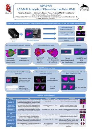
- 1. Follow-up and revisionIntra-intervention EP guide ADAS-AF: LGE-MRI Analysis of Fibrosis in the Atrial Wall Rosa M. Figueras i Ventura1, Xavier Planes1, Lluís Mont2, Luis Serra1 1 Galgo Medical S.L. (Barcelona, Catalonia, Spain) 2 UFA (Unitat de Fibril·lació Auricular de l’Hospital Clínic), Hospital Clínic, Universitat de Barcelona & IDIBAPS, Barcelona, Catalonia, ADAS-AF: software tool designed to bring atrial fibrosis information extracted from LGE-MRI into current clinical practice Late Gadolinium Enhancement 3D MRI Characterize substrate structure “Non-Invasive Mapping™” Pre-interventional treatment planning Intra-intervention EP guide Follow-up and procedure revision Pre-interventional treatment planning Isolation of fibrotic patches Closure of gaps in re-do ablation procedures Sphericity and volume computation Fibrosis quantification Fibrosis quantification and gap visualization Joint EAM and MRI visualization ADAS 3D ADAS-AF features and tools Side by side comparison of MRI and EAM or MRI1 and MRI2 Export 3D atrial model to main navigation systems Semi-automatic 3D atrial fibrosis map generation Semi-automatic atrial wall delineation and 3D model creation Automatic quantification of anatomical features CARTO® (Biosense Webster) EnsiteTM (Abbott) MRI-EAM MRI pre – MRI post ADAS-AF in clinical papers Authors Title Publication year Achievement Benito, E.M.; Andreu, D.: Mont, L.; Berruezo, A. Correlation between functional electrical gaps identified by ultrahigh-density mapping and by late gadolinium enhancement cardiac magnetic resonance in repeat atrial fibrillation procedure Heart Rhythm Case Reports 3(5): 282-285 2017 Anatomical gap localization performed with ADAS-AF. Match with electrical gap. Benito, E.M.; Carlosena-Remirez, A.; Guasch, E.; et al. Left atrial fibrosis quantification by late gadolinium- enhanced magnetic resonance: a new method to standardize the thresholds for reproducibility EP Europace 19(8): 1272- 1279 2017 Analysis of 10 healthy patients to derive thresholds for atrial fibrosis in LGE-MRI. Pontecorboli, G.; Figueras i Ventura, R.M.; Carlosena, A.; et al. Use of delayed-enhancement magnetic resonance imaging for fibrosis detection in the atria: a review EP Europace 19(2): 180-189 2017 Review of several methods to analyze atrial fibrosis. Andreu, D.; Gomez- Pulido, F.; Calvo, M.; et al. Contact force threshold for permanent lesion formation in atrial fibrillation ablation: A cardiac magnetic resonance–based study to detect ablation gaps Heart Rhythm 13(1): 37-45 2016 Study to derive minimum force required to achieve anatomical isolation of the veins. Bisbal, F.; Andreu, D.; Berruezo, A. Simplified mapping and ablation of a scar-related atrial tachycardia using magnetic resonance imaging tissue characterization EP Europace 17(2): 186 2015 Case report showing that fibrosis information obtained thanks to ADAS-AF helps to ablate atrial tachycardia. Bisbal, F.; Guiu, E.; Cabanas-Grandío, P.; et al. CMR-Guided Approach to Localize and Ablate Gaps in Repeat AF Ablation Procedure JACC: Cardiovascular Imaging 7(7): 653-663 2014 Study that shows that LGE-MRI can provide useful information for gap ablation in repeat AF procedures.