
Empfohlen
Weitere ähnliche Inhalte
Was ist angesagt?
Was ist angesagt? (18)
Andere mochten auch
Andere mochten auch (20)
Ähnlich wie 12 1
Ähnlich wie 12 1 (20)
Mehr von arislantern
Kürzlich hochgeladen
Kürzlich hochgeladen (20)
12 1
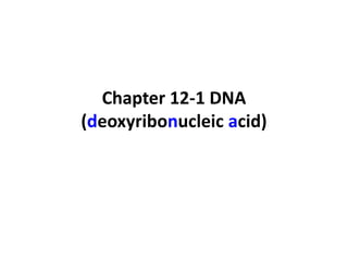
- 1. Chapter 12-1 DNA (deoxyribonucleic acid)
- 2. DNA function • Nucleic acid • Store and transmit hereditary information
- 3. Experimental Proof of DNA as Genetic Material...
- 4. 1) Griffith’s Transformation Experiments
- 5. 1) Griffith’s Transformation Experiments • What two strains of bacteria did Griffith use? – S (smooth) strain –cause pneumonia – R (rough) strain –harmless • What happened in the fourth experiment? – The mice died. There were live S strains found in the mice body. • What can be concluded from the experiment? – Cell components in the dead S strains transformed the live R strains to live S strains
- 6. 2) Oswald Avery experiment • The transforming factor is DNA
- 8. 3) The Hershey-Chase’s bacteriophages experiment • Bacteriophages – viruses that infect bacteria – consist of protein coat and a DNA or RNA core – inject their hereditary material into bacteria
- 9. 3) The Hershey-Chase’s bacteriophages experiment
- 10. virus particle virus particle labeled with 35S labeled with 32P • Radioactive Markers – Radioactive bacterial cell (cutaway view) isotope – 32P could only mark DNA – 35S could only mark protein coat label outside cell label inside cell
- 15. DNA • Made by nucleotide – 5-carbon sugar (deoxyribose) 5 – phosphate group 4 1 – nitrogenous bases 3 2 • purine (2 rings): adenine , guanine; • pyrimidine (1 ring): cytosine , thymine
- 16. 5 4 1 3 2
- 17. Purines Pyrimidines Adenine Guanine Cytosine Thymine 1 1 1 1 2 2 2 2 3 3 3 3 5 4 5 4 5 4 5 4 Phosphate group Deoxyribose
- 18. DNA Structure • Chargaff’s Rule : – Amount of adenine = amount of thymine, amount of guanine = amount of cytosine – A=T and G=C
- 19. DNA Structure • Rosalind Franklin – Used x-ray diffraction to examine DNA fibers – Concluded that DNA was helix Rosalind Franklin's X-ray diffraction photograph of DNA, 1953 Photo: courtesy HarperCollins
- 20. DNA Structure • Watson and Crick’s model – DNA have two strands run in opposite directions – Strands are held together by hydrogen bonds – A binds with T and C with G – Molecule is a double helix Hydrogen bond
- 21. DNA Structure
- 22. Nucleotide Hydrogen bonds Sugar-phosphate backbone Key Adenine (A) Thymine (T) Cytosine (C) Guanine (G)
- 23. James D. Watson Francis H. C. Crick Wilkins Rosalind Franklin
- 28. Questions: • 20% A in DNA strands, how many C ? • A=T=20%, • A+T+G+C=100% • G=C, • C=30%
- 29. The drawing below shows a short section of a DNA molecule. What is labelled by I, II and III? I II III I II III A. 3′ end purine hydrogen bond B. 5′ end pyrimidine covalent bond C. 3′ end pyrimidine hydrogen bond D. 5′ end purine covalent bond