1. Carbohydrate metabolism
The metabolism of the sugars found in our food is discussed in all textbooks and I
will not take up all of the details here. The points I do wish to discuss are
concerned with maintenance of blood sugar levels under differing physiological
conditions. How do we start up storage of glucose after a meal? How do we
preserve blood glucose levels between meals? What are the differences in
metabolism of common sugars in various organs?
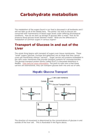
Transport of Glucose in and out of the
Liver
The whole thing begins with transport of sugars over tissue membranes. These
"small" sugars (glucose, fructose and galactose) are so large that they cannot
cross cell membranes without "carriers". Sugar carriers are proteins embedded in
the cell's outer membrane that provide transport systems for monosaccharides.
The glucose transport protein family (called GLUT) is discussed elsewhere in
MedBio. Click here for more information. The point to note now is that these
carriers are bidirectional; they can transport glucose both into and out of cells.
The direction of movement is determined by the concentrations of glucose in and
outside of the liver cell. This is illustrated in the figure above.
1
2. Drawing "1" shows the situation when the portal blood and the liver cell have
equal concentrations of glucose; sugar moves in both directions simultaneously.
This may seem to be wasteful, but gears the system to react to small changes in
glucose concentration.
The second drawing shows what happens when blood glucose tends to fall.
Glucose production in the liver accelerates and the net flow of glucose is outward,
stabilizing the blood sugar level. This is extremely important. The total amount
of sugar present in the blood can support resting activity for about 40 minutes.
Just walking increases glucose use to a point where the entire blood content is
used up in about 15 minutes. Since mental activity is completely dependent
upon stable blood glucose levels, there must be a way of evening out blood
glucose levels. This is one of the major duties of the liver. On a short-term
basis, this is the only organ capable of replacing blood sugar used by other
organs. Click here for the details.
Following a meal, the portal blood sugar level increases. This is shown in the
third drawing where we see that the liver rapidly takes up glucose from the
blood. Once again, the liver stabilizes blood sugar. In principle, this two-way
flow of glucose can forego in most tissues. However, only the liver and kidneys
are sugar producers and export of glucose occurs only in these tissues. Most of
our organs are sugar-burners, taking up glucose from the blood and using it for
energy production.
What determines this limit on release of glucose from most of the body? Why
cannot skeletal muscles release glucose from their large glycogen stores? The
secret is that uptake of sugars to our organs involves immediate phosphorylation
at either carbon 1 or 6. The phosphorylated sugar derivatives cannot "leak" out
of the cell. There is no mechanism for their cross-membrane transport.
Once sugars are phosphorylated they stay put!
What is the key to production of glucose in the liver and kidneys? These organs
produce a specific enzyme, glucose-6-phosphatase, that cleaves the glucose-
phosphate bond. Regulation of the balance between phosphorylating and
dephosphorylating enzymes is crucial and determines the net direction of uptake
and release of glucose in these organs.
2
3. Regulation of sugar phosphorylation
As stated above, monosaccharides enter metabolism through phosphorylation.
That is, they react with ATP and a kinase to yield phosphorylated derivatives.
Those small structural differences we noted between the three monosaccharides
found in our food determine which enzyme initiates sugar metabolism. Glucose
can be phosphorylated by either hexokinase or glucokinase. The former is
involved in the cell's energy metabolism. Glucokinase is implicated in either
energy storage or in glucose-signaling systems. Fructose metabolism is initiated
through fructokinase while galactokinase is the first enzyme involved in galactose
metabolism.
3
4. All organs exhibit hexokinase activity. As the name implies, this enzyme is
relatively nonspecific and can react with most 6-carbon sugars. However, its
affinity for these sugars varies with their structure. Hexokinase reacts strongly
with glucose at levels found in plasma and tissues. While it in principle can
catalyze phosphorylation of fructose and galactose, its affinity for these is quite
low. This excludes active handling of fructose and galactose by hexokinase at the
concentrations of these found in our bodies. Hexokinase is product-inhibited.
That is, if the glucose-6-phosphate formed by the enzyme is not rapidly removed,
hexokinase activity promptly falls. Hexokinase is well-adapted as the first
enzyme in tissues utilizing glucose as an energy source, but not as the initiator of
energy storage in the liver. We find, therefore, specialized kinases in the liver to
handle all three of the sugars found in food. The following table summarizes the
distribution and substrate specificity of the kinases involved in the initiation of
sugar metabolism. Note that glucokinase is found in tissues that require a
"glucose sensing system". I'll come back to this under a discussion of insulin
secretion.
Note also that it is the liver that has fructokinase and galactokinase activity. The
liver is the only organ that actively metabolizes these sugars. I will take up
replacing table sugar with fructose later but you can see now that fructose is a
substrate for hepatic metabolism alone. Aside from sperm, no other organ has
an active fructokinase. No other organ is perfused with concentrations of fructose
large enough to allow reaction with hexokinase. The liver very effectively
removes absorbed fructose from the portal blood. In fact, this is necessary for
uptake of fructose in the gut. Remember that transport there is passive, relying
on a steep concentration gradient to drive fructose uptake. Fructose is not a
direct energy source for muscles and the brain as many of its producers claim.
These tissues rely on the hexokinase catalyzed phosphorylation of glucose for
4
5. energy metabolism. Sorry, but you do not become stronger and smarter by
eating fructose.
Glucose metabolism and glucokinase
Let us take up glucokinase first. This enzyme is specific for glucose and is not
inhibited by its product, glucose-6-phosphate. While glucokinase has a high Km
(low affinity) for its substrate, it reacts strongly with glucose at the
concentrations found in portal blood after a carbohydrate meal. The Km of the
liver enzyme, around 10-12 mmolar, lies above fasting blood glucose levels. This
means that glucokinase activity is "turned on " by the glucose in portal blood
following a meal (10-30 mmolar), and is "turned off" after glucose from the meal
is absorbed. Remember, a major function of the liver is to release glucose when
blood sugar levels begin to fall. Liver has an enzyme (glucose-6-phosphatase)
that cleaves phosphate from glucose-6-phosphate, yielding free glucose. The
balance between glucokinase and glucose-6-phosphatase slides back and forth,
increasing uptake to the liver when the level of blood glucose is high, and
releasing glucose when blood glucose falls. It was commonly believed that this
Km-steered regulation of glucokinase was the key to understanding glucose
uptake and release in the liver. However, we now know that a much more
refined mechanism controls glucokinase activity.
Translocation of glucokinase between
cytosol and nucleus
Hepatic glucokinase has a high Km and this will serve as a "switch" to activate and
inactivate the enzyme. But, think about the situation which evolves when blood
glucose tends to fall. Glucose is synthesized from stored glycogen and through
gluconeogenesis. The cytoplasmic concentration of glucose must rise if the liver
shall export sugar and this would "turn on" glucokinase, drive forward
phosphorylation and spoil the system. And that at a time when glucokinase must
be inactivated to allow glucose dephosphorylation and export. So, how do we
stop glucokinase activity?
5
6. One of the best ways to "turn off" an enzyme is to put it away. Just move the
protein to a compartment where it is not needed and inactivate it by binding to a
parking place! Several publications during the past few years have shown that
glucokinase is translocated to the liver cell's nucleus when plasma glucose
concentrations approach fasting levels (around 5mmoles/l). It is bound there to
a glucokinase regulatory protein called GKRP. Release and translocation back to
the cytosol is stimulated by increases in plasma glucose, trace amounts of
fructose, and insulin. This translocation system may well be dominant in
directing glucose flow in and out of the hepatocyte. The following figure is taken
from "Regulation of Hepatic Glucose Metabolism by Translocation of Glucokinase
between the Nucleus and the Cytoplasm in Hepatocytes", Y. Toyoda et. al., Horm
Metab Res 33, 329-336 (2001). The bright areas in the pictures are
immunofluoresent areas in hepatocytes in culture. The fluorescence comes from
glucokinase. Clearly, the enzyme moves from the nuclei to the cytosol when
glucose levels in the surrounding medium is increased from 5 to 25 mmol/liter.
Small amounts of fructose greatly promote this transfer.
While the total GK activity (cytoplasmic plus nuclear activity) was not altered
after incubation with glucose, the enzyme migrated from the nuclei to the cytosol
in these cells. Inclusion of fructose at very low levels led to increased
cytoplasmic glucokinase at 5 mM, and appeared to give a total transfer to the
6
7. cytosol in the presence of 25 mM glucose. The GK-GKRP complex was previously
shown to disassociate in the presence of fructose-1-phosphate. I believe that
these are the first pictures showing a simultaneous translocation from nuclei to
the cytosol.
The time course of this migration has been recently investigated by Chu et al.
Look up "Rapid Translocation of Hepatic Glucokinase in Response to
Intraduodenal Glucose infusion and Changes in Plasma Glucose and Insulin in
Conscious Rats, Am J Physiology (in press 01.04) for details. The next figure,
taken from that work with permission from Dr. Shiota, clearly shows that
migration from the liver cell's nucleus occurs rapidly and within the time interval
required to participate in the observed increases in hepatic glucokinase seen after
a meal. While GKRP remained in the nucleus, the GK moved to the cytoplasm
following perfusion of fasted rats with glucose. Significant increases were noted
after only 10 minutes. This corresponds well with the time course of glucokinase
activation in these animals.
In summary, glucose , insulin and fructose control the activity of glucokinase
through translocation in liver cells. Storage of active enzymes and carriers as a
method of metabolic control has been well-documented previously with respect to
the insulin-sensitive glucose carrier GLUT4.
7
8. Fructose Metabolism and its Influence
on Glucose Metabolism
One of the strange things here is the role of fructose. Why is fructose such a
strong signal for release of glucokinase. Remember, glucokinase is "not
interested" in reacting with fructose. It is specific for glucose. The enzyme
required to initiate fructose metabolism, fructokinase, is only found in quantity in
the liver (and sperm cells). Furthermore, it is not under metabolic control. If
fructose comes to the liver, it will be taken up and very quickly metabolized!
Carbohydrate metabolism starting from glucose or galactose proceeds under
strict control; fructose is in a special class! We are "constructed" to conserve
energy. The rapid entry of fructose into glycolysis leads to fatty acid synthesis in
the liver. Because fructose metabolism "fills" glycolysis with substrate at a very
high rate, frequent use of sucrose (remember sucrose is a dimer of fructose and
glucose) or fructose promotes fat production. Plasma triglyceride levels are
increased by the ingestion of large amounts of sugar. There is a correlation
between sugar consumption, high plasma lipid levels and atherosclerosis.
Starch, which yields glucose during digestion, has been a main energy source for
mankind since the agricultural revolution 8000-10000 years ago. Fructose is
found in small quantities in many fruits and honey. The amount of fructose in our
diets was far lower than starch-derived glucose found in the food we have eaten
for thousands of years. Combine an apple (5-7% fructose) and some wheat,
potatoes or corn, and you get translocation of glucokinase and an active glucose
metabolizing system (with a little fructose taken along for the ride). Fructose
seems to have acted as a signal substance, used to activate glucose metabolism.
Fructose in our diet
Commercial production of fructose began in Finland in1969. Since then it has
become "modern" to exchange fructose for sugar to cut down on the caloric
content of sweetened food. Fructose is sweeter than
sucrose. But, it is the 5-ring form of fructose that is sweet,
the 6-ring form tastes about the same as usual table sugar.
Unfortunately, warming fructose leads to formation of the 6-
ring form. Sweetening coffee and tea and baking cakes with
fructose requires just about as much of this sugar as sucrose
to get the same taste. Baking is also difficult as one needs
to use about the same amount of fructose as table sugar and
it burns at lower temperatures.
By the way, shifting out sucrose with less fructose may
reduce total calorie intake, but it increases fructose
consumption. Statistics from the USA suggest that people
8
9. do not cut back sugar use when they use fructose. They seem to choose sweeter
food! Remember, half of table sugar is fructose, all of fructose is fructose! That
excess fructose will be largely converted to fat!
The new sweetener, high fructose corn
syrup (HFCS)
While consumption of sucrose has actually fallen in the USA, the use of high-
fructose corn syrup (HFCS) has increased markedly during the past 30 years.
Conservative estimates suggest that 16 % of the energy intake of average
Americans is fructose. HFCS contains either 42 % or 55 % fructose. This is
produced from corn through hydrolysis of starch. The resulting glucose syrup is
then isomerized to the sweeter high fructose syrup enzymatically. High fructose
corn syrup is widely used today in commercial production of soft drinks, breakfast
cereals, baked goods and condiments. As we can see from the following figure
from Elliott et.al., total sugar consumption has increased in the USA during the
past 30 years despite a real fall in sucrose consumption. The decreased use of
sucrose is more than balanced by substituting fructose for sugar in commercial
food production. The global distribution of soft drinks, breakfast cereals and fast
food is leading to similar increases in sugar consumption in many areas.
Fructose does not cause insulin release from beta cells, as these lack
fructokinase. One of the results of this is that fructose consumption does not
dampen appetite. This may lead to increased caloric intake with obesity and the
metabolic syndrome as a result. You can read more about fructose, weight gain
and insulin resistance in an excellent article by Elliott et al, Am J Clin Nutr 76,
911-922 (2002). Just click here if you have access to a library.
9
10. In the next 3 figures I will try to clarify this fructose business. First, remember
that sugars do not just diffuse through the hepatocytes plasma membrane.
There must be a carrier there or they “cannot come in”. That carrier is GLUT2.
This glucose transport protein reacts with both fructose and glucose.
Let us take up interaction with glucose
first.
Once again, recall that glucose transporters carry sugar down a concentration
gradient. If glucose concentration is high in portal blood, the liver will take up
glucose. This will be stored as glycogen which can later be used to replace blood
sugar. Part of the glucose load is oxidized completely to CO2 and drives ATP
production. Excess carbohydrates are converted to fatty acids and exported as
triglycerides. The trace of fructose found in food gives F-1-P and this serves as
part of the glucokinase activating system. Thus, low levels of fructose, judged by
today's urban standards, activate uptake and metabolism of glucose.
10
11. Falling levels of glucose in the blood flowing through the liver favor glucose
export, shown in the next figure. Hormone activation of hepatic gluconeogenesis
and glycogenolysis lead to increased hepatic glucose concentrations and export of
glucose. The direction is determined through allosteric and hormonal control of
the enzymes involved. The final actors in this picture are glucokinase and
glucose-6-phosphatase. Recall that release of glucose from the liver demands
stopping glucokinase activity. As I hope I have explained above, glucokinase
activity is controlled to a large degree by "parking" the enzyme in the
hepatocyte's nucleus (or nuclei; they are often binuclear). The absence of
fructose in the portal blood assures minimal F-1-P concentrations: Glucokinase
can be "parked" in the cell's nucleus.
11
12. Sugar and Fructose metabolism
So, what happens if we consume large quantities of table sugar (sucrose) or high
fructose corn syrup? The body responds to the glucose content of the meal in the
usual manner; insulin secretion increases and glycogen and lipid metabolism
proceed as would be expected. However, fructose uptake and phosphorylation
occurs at the same time, providing additional substrate for these anabolic
processes. Remember that fructose does not stimulate insulin secretion. The
hormonal steering of metabolism applies only to that induced by glucose. The
excess substrate from fructose is poured into fat synthesis which is stimulated by
the insulin released in response to simultaneously consumed glucose.
The liver has a huge capacity for the uptake and phosphorylation of fructose. The
phosphorylation capability is about twice that of the glucose phosphorylation
system. We find large increases in uptake and metabolism of fructose with
increased sugar levels in portal blood. In individuals with fructose intolerance we
find an increase in fructose-1-phosphate due to the fact that fructokinase activity
can exceed that of aldolase B. This special aldolase is required to cleave F-1-P to
dihydroxyacetone phosphate and glyceraldehyde. The fructose taken up cannot
be stored as such. It is converted to intermediates in glycolysis WHICH COME
AFTER THAT ALL-IMPORTANT CONTROL POINT, PHOSPHOFRUCTOKINASE 1. So
what? Well, some of that fructose winds up as glycogen and glucose. That is ok;
we need glucose in the blood. The rest of the fructose rushes through pyruvate,
is transferred to mitochondria, and finally converted to fatty acids and exported
from the liver as triglycerides.
That fat formation is a major problem. People that consume large amounts of
sugar seem to demonstrate increased plasma triglyceride levels. This is quite
parallel to eating large quantities of saturated fats. While the mechanisms here
12
13. are not completely clarified, there is good reason to believe that increased plasma
triglyceride are a moment in development of atherosclerosis and CVD.
So, why do we have such an active fructose metabolism in the liver? Speculation
again, but an interesting hypothesis is that traces of fructose in the blood
perfusing the liver indicate consumption of carbohydrates and a need for
glucokinase release and activation. As is the case in many other physiological
systems, it may well be that a combination of hormonal and allosteric factors
steer the body's reactions. Fructose may have initially been a signal stuff. The
"new" today is that it has become a major energy source in the diet.
To summarize and Speculate a little:
Control of the ratio of glucokinase and glucose-6-phosphatase activity is essential
for regulation of the liver's uptake and release of glucose. Control of the blood
glucose level is vital for mental processes. In fact, a 50% cut in blood glucose
levels leads to dizziness, nausea and eventually loss of consciousness.
Our diet through thousands of years included starches as the major carbohydrate
source and fructose as a sweet extra in fruits and honey. Remember, progression
to the “hunter stadium” came long after development of the first humanoids. You
can click here to find more information about this. A very active conversion of
that limited fructose to fructose-1-phosphate (not 6-phosphate) appears to have
been elemental in regulation of hepatic glucokinase and glucose metabolism. The
epidemic growth of overweight, diabetes type 2, hypertension and CVD in urban
societies around the globe today may well have our exceedingly high sugar
consumption as a major underlying factor. We were simply "not made" to eat
25% of our food in the form of sugar. Changing to fructose will not help!
Table sugar and fructose used as a "spice" also provide excess calories and
resulting obesity. Perhaps the most important thing to remember here is that
excessive chronic sugar intake can lead to increased plasma fatty acid
concentrations with the danger of developing atherosclerosis. In susceptible
individuals this may lead to metabolic syndrome and associated diabetes type 2,
hypertension, and damage the kidneys. For a deeper discussion of the metabolic
syndrome click here.
13
14. Hereditary fructose intolerance disease
As stated above, hepatic fructose metabolism is quite rapid. That is, the initial
step, phosphorylation by fructokinase is rapid. Further metabolism of fructose is
dependent upon aldolase B. Normally, fructose consumption leads to a rapid flux
into glycolysis at the triose phosphate level, enhancing gluconeogenesis,
glycolysis and triglyceride synthesis. However, individuals who have reduced
levels of aldolase B exhibit so-called fructose intolerance. They build up
excessively high hepatic fructose-1-phosphate levels, trapping inorganic
phosphate and reducing ATP synthesis accordingly. In these people, fructose is
not a good substrate for glycolysis or gluconeogenesis.
While the statistics on this are not clear, it appears that somewhere between 1 in
10,000 to 1 in 50,000 persons exhibit fructose intolerance. Declining ATP levels
interfere with many of the liver's functions, among these are ureogenesis and
gluconeogenesis. You can get more information about fructose intolerance
syndrome by clicking here.
14
15. To summarize, reducing calorie intake by using fructose instead of sucrose does
not give big winnings. And there is that fat synthesis business. And, not least
important, metabolism of fructose happens in the liver. One does not get extra
and long-lasting energy from fructose as suggested on some fructose packages!
Muscles and the brain get energy from fructose only after the liver has converted
fructose to glucose or fat.
Galactose metabolism
Carbohydrates in food give us only three monosaccharides that are taken up from
the intestine and metabolized further. Go back to the chapter on the structure of
sugars to review this (it's just a click away). Remember, sucrose (table sugar)
gives us equal amounts of glucose and fructose, lactose (milk sugar) gives equal
amounts of galactose and glucose.
Galactose is almost identical to glucose; remember it is only the position of the
hydroxy group on carbon four that differs here. That "little" difference is enough
to markedly reduce binding of galactose to hexokinase. A specific enzyme,
galactokinase, is essential to initiate galactose metabolism. Galactokinase reacts
with galactose and gives us galactose-1-phosphate. There is no direct oxidative
pathway for galactose-1-P. It has to be converted to an intermediate in glucose
metabolism to come further. We have an activated form of glucose, uridine
diphosphoglucose (UDPG) that reacts with gal-1-P. UDPG has a glucose-1-P
"tail". Using UDPG-gal-1-P transferase, we simply exchange that "tail" with gal-
1-P and convert the Gal-1-P to glucose-1-P. Another enzyme, UDPGal-UDPG
isomerase "wins" back that UDPG for us and the process can start up again.
15
16. Unlike fructose metabolism, glucose and galactose metabolism are subject to
precise allosteric and hormonal control. Metabolism of these sugars goes
through phosphofructokinase and FDPase, two enzymes under exacting hormonal
and allosteric control.
The metabolism of galactose is usually non-problematic for those of us who have
lactase (the enzyme which splits lactose). All of the others (about 90% of the
world's adult population) are lactose intolerant and cannot consume milk and milk
products unless these are fermented first. Recall that lactase is a very special
enzyme, the only one we have that can split the ß-sugar bond. No lactase, no
hydrolysis of milk sugar. That means that lactose will be sent to the large
intestine giving "explosive diarrhea".
Galactose intolerance does occur in children lacking either galactokinase or the
converting enzyme galactose-1-phopshate-UDPG transferase. In the latter case,
phosphorylated galactose levels build up in the liver, trapping inorganic
phosphate and preventing ATP production. This will ”knockout” liver functions
and lead to low blood sugar levels and brain damage all too quickly. All newborn
babies are controlled for galactosemia during the first few days of life. They are
switched from a milk diet to one based on sucrose if they have excessive plasma
galactose.
16
17. There is something strange about
lactate...
We usually think of pyruvic and lactic acids as normal end products of glycolysis.
Release of lactate follows increased energy utilization, especially in skeletal
muscles. Anaerobic glycolysis is something that extra work brings forth. What
we often neglect is that some tissues serve as "lactate producers" with the
intention of nourishing others.
Perhaps the easiest to understand here are erythrocytes. After all, they do not
have mitochondria and cannot oxidize glucose to CO2. Furthermore, glycolysis in
these cells is largely inefficient. Erythrocytes produce (and require) 2-3
bisphosphoglycerate in amounts about equal to their hemoglobin content. While
this gives control over oxygen-binding to hemoglobin it precludes a net
production of ATP. Erythrocytes can take up and metabolize quantities of glucose
without the inhibitory effects of high ATP levels. What happens to all that
lactate? It just so happens that our kidneys thrive on lactate and devour as
much as the red cells produce. Check the following diagram taken from the
chapter about insulin. Almost 10% of the carbohydrate content of a normal meal
wings up as lactate which is used as an energy substrate in the kidneys. About
2-3 g per hour are used here and most goes to aerobic metabolism and ATP
production. This is a rather simple case and is perhaps not so exciting. Let's look
at brain metabolism for some action!
17
18. Lactate metabolism in the brain
We have all learned that the brain (and spinal cord and retina) require a steady
supply of glucose. These tissues have little or no glycogen; lack of glucose is just
about as damaging as an oxygen cut in the CNS. Even under starvation, the
brain must cover about 50% of its energy needs from blood sugar. ß-OH
butyrate and acetoacetate can supply only half of the substrate required. Why?
The blood-brain-barrier protects the brain from most substances in plasma. Even
fatty acids are excluded here. And, in a way, glucose is among the excluded
goodies. That is, most of the glucose that crosses the blood-brain-barrier never
comes over the barrier (or glia cell). The next figure, taken from a publication by
Tsacopoulou and Magistretti (click here if you are connected to a library) explains
this phenomenon.
The blood-brain-barrier is comprised of glia cells, primarily astrocytes. A small
fraction of the glucose released from capillaries wanders directly to nerves and
synapses. However, most is "trapped" in astrocytes and oxidized to lactate by
these cells. Lactate goes further into the brain and nourishes the brain's
neurons. This seems to be especially important for glutamatergic neurons which
comprise much of the brain. The astrocytes also efficiently pick up released
glutamate, convert this to glutamine, and send the product back to glutamatergic
neurons where it continues to cycle as a neurotransmitter. The axons do not
contain glycogen and are, therefore, completely dependent upon the lactate sent
18
19. from astrocytes to maintain ATP levels. Astrocytes and other glia cells appear to
have some glycogen which can serve as a very short-term source of lactate.
So the answer to the preceding question is that much of the brain is dependent
upon lactate from glia cells to provide substrate for aerobic energy production;
ketone bodies cannot cover their substrate requirements. One can reduce
glucose consumption and use ketone bodies during starvation. However, some
neurons must have lactate and the brain must continue to use blood sugar.
Testicles too...
We find the same sort of work division in the testicles. Here, the sertoli cells
enclose the seminiferous tubules, effectively shielding them from the circulation
and direct uptake of glucose. This resembles the brain and the blood-brain-
barrier. Substrates for energy metabolism in the tubules are delivered by the
sertoli cells. These take up glucose, convert it to lactate, and send this further to
the tubules. The lactate serves as the substrate of choice for spermatogenesis.
19