Myocardial Perfusion SPECT coregistered Coronary CTA
•
8 gefällt mir•1,384 views

My Slide Presentation in Journal Club Thanks Author for Good Medical Research

Empfohlen
Weitere ähnliche Inhalte
Was ist angesagt?
Was ist angesagt? (20)
Ähnlich wie Myocardial Perfusion SPECT coregistered Coronary CTA
Ähnlich wie Myocardial Perfusion SPECT coregistered Coronary CTA (20)
Mehr von Apichaya Claimon
Mehr von Apichaya Claimon (9)
Kürzlich hochgeladen
Kürzlich hochgeladen (20)
Myocardial Perfusion SPECT coregistered Coronary CTA
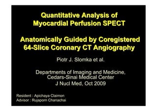
- 1. Quantitative Analysis of Myocardial Perfusion SPECT Anatomically Guided by Coregistered 64-Slice Coronary CT Angiography Piotr J. Slomka et al. Departments of Imaging and Medicine, Cedars-Sinai Medical Center J Nucl Med, Oct 2009 Resident : Apichaya Claimon Advisor : Rujaporn Chanachai
- 2. Coronary CT Myocardial perfusion angiography (CTA) SPECT (MPS) • precise localization • detect and estimate and classification of severity of ischemia coronary artery plaques + depiction of coronary anatomy • Inconclusive results obtained by 1 of the tests -> sequential testing by both modalities • Visual analysis of fused MPS and coronary CTA images -> improve the diagnostic value • Manual tools for the purpose of combined visual analysis have been developed
- 3. Previous studies • Need interactive alignment • Complicate protocol • Reduce clinical usability
- 4. Aim of the study • To develop tool for rapid automatic – Coregistration – Visualization – Combined quantification Between coronary CTA and MPS ; obtained from stand- alone scanners in different scanning sessions • To showed that coregistered MPS–CTA data can be used to improve quantitative MPS analysis
- 6. Patient Selection • Between October 2005 and May 2007 • Retrospectively 40 consecutive patients – who underwent myocardial MPS, CTA, and invasive coronary angiography (ICA) within a 90-d period • 2 patients excluded ; the relevant imaging data could not be retrieved • 22 patients ; evaluation of symptoms – either chest pain or dyspnea; 8 had prior MI • 16 patients ; asymptomatic
- 7. The imaging indications • post–myocardial infarction (3 cases) • post–percutaneous coronary intervention risk stratification (3 cases) • risk stratification without prior event (10 cases) • 3 patients excluded ; because of CABG surgery The remaining 35 patients • 5 cases, CTA and MPS were performed on the same day • 20 cases, CTA was performed after MPS – (range, 1–49 d; median, 9 d) • 10 cases, MPS was performed after CTA – (range, 1–73 d; median, 13 d)
- 9. CT Image Acquisition Unenhanced CT scan CT coronary calcium scores Coronary CTA Electrocardiogram (ECG)-gated during a 9- to 12-s breath hold ECG-based dose modulation 40%-80% of the cardiac cycle ; to limit radiation dose
- 10. Coronary CTA Image Reconstruction Raw CTA data → retrospectively gated reconstruction performed at 40%-80% of the R-R interval Extract coronary arterial trees using vendors’ software Transferred to a Windows workstation for MPS–CTA fusion
- 11. CTA Image Evaluation • A coronary CTA reader – with experienced >300 coronary CTA interpretations – Unaware of MPS and ICA results • Evaluate coronary segments > 1.5 mm in diameter • Evaluate for the presence and degree of stenosis. • Any stenosis narrowing the luminal diameter by > 50% or > 70% was recorded. • If a segment could not be assessed because of artifacts, no stenosis was recorded.
- 12. ICA Image Acquisition and Evaluation • Standard technique of intensive coronary angiography • Evaluate by interventional cardiologist, unaware of coronary CTA and MPS results • By visual inspection • Whether luminal diameter narrowing > 50% or > 70% was present
- 13. • Left main stenosis > 50% was considered as significant for the LAD and LCX territories • If present the ramus intermedius -> assigned to the LCX territory.
- 14. Angiographic Characteristics of Data (n = 35)
- 15. MPS Protocol • Standard protocol – 1- or 2-d protocols – dual-isotope (thallium–technetium) protocol • MPS acquisitions ; 64 projections, 45o RAO to 45o LPO • Stress scan ; exercise, adenosine injection, or adenosine–walk protocol • No attenuation or scatter correction • Reconstruct gated images to original transverse orientation – with filtered backprojection and a Butterworth filter
- 18. MPS image processing static MPS images in end-diastolic (ED) phase to match the diastolic cardiac phase of coronary CTA
- 20. MPS image processing Validation of Automatic Registration (error analysis) manual alignment parameters (3 translations and 3 rotations) Visual alignment was performed without knowledge of the automatic results
- 22. MPS image processing 2-dimensional/3D textures Segmented CTA voxel maps → rendered in 3D within QPS and within the same coordinates as the epicardial 3D surface display with overlaid MPS function and perfusion
- 24. CTA-Guided MPS Contour and Territory Adjustment • Fused coronary CTA and MPS images were evaluated with overlaid contours in multiplanar orientations • If discrepancies between the MPS and CTA valve plane position -> manually adjust the contour • Overlaid the default vascular territory boundaries with the 3D LV MPS surfaces – with color-coded perfusion information – and with a coregistered volume-rendered segmented 3D coronary tree
- 25. • Adjust vascular territories segment by segment – based on a 17-segment American Heart Association model – using anatomic information provided by the coronary CTA • If adjusted MPS contours or vascular territories -> repeat QMPS analysis
- 27. MPS image processing •perfusion-defect performed individually for each vessel, 17 segments vascular territory •total perfusion deficit (TPD) of territory -> automated quantification in each vessel •threshold of 2% •record QMPS before and after adjustments
- 28. QMPS
- 29. RESULTS
- 30. • Of 35 cases with all 3 scans (CTA, MPS, and ICA) available – 20 patients underwent CTA after MPS – 15 underwent MPS after CTA • In cases performed CTA after MPS – 11 had equivocal reversible defects on visual evaluation of MPS – 9 done CTA because MPS were discordant with clinical or suspected multivessel disease
- 31. • In cases underwent MPS after CTA – 7 had at least 1 nondiagnostic major coronary segment on CTA – 4 had maximal luminal stenosis in the LAD estimated at 50% and considered of borderline significance – 4 patients done for assess hypoperfusion
- 32. • unenhanced CT calcium score ; average was 942 + 1,530 (range, 0–7,781) – Heavy calcification (score > 500) in 15/33
- 33. • 10 cases ; motion artifacts on CTA • Interpretation difficulties ; 9 cases • Significant CT disease ; 27/35, with – 6 LCX lesions – 11 RCA lesions – 21 LAD lesions – 2 left main lesions • MPS ejection fractions – 57.4% + 14% (range, 32%-83%) on stress – 57.2% + 14 (range, 25%-83%) on rest
- 34. • TID ; 1.15 + 0.14 (range, 0.96–1.4) MPS findings • Visually ; – normal in 3 cases – probably normal in 3 cases – borderline in 6 cases – probably abnormal in 1 case – abnormal in 22 cases • Quantitatively; total perfusion deficit (TPD) was – 16.5% + 12.7% on stress (range, 0%-44%) – 5.6% + 8.1% on rest (range, 0%-25%)
- 35. Registration Algorithm • Speed of automated registration = 1–2 s per study • The automatic volume registration of motion-frozen MPS with CTA was successful in – 33/35 stress – 34/35 rest studies as assessed qualitatively with an overall success rate of 96% • In 1 patient, registration fail for both stress and rest – because of the unusually high blood-pool contrast intensity on coronary CTA – inadequate matching of assigned blood-pool contrast with the actual CT value in the blood-pool region
- 36. • All 3 failed cases were women with small hearts – (motion-frozen stress diastolic volumes, 29–52 mL on MPS) • These results were easily corrected by interactive alignment. • No significant differences – between errors in different directions – or between studies from 2 different systems
- 37. Accuracy of Automated Alignment of SPECT and Coronary CTA for Translations and Rotations
- 38. CTA MPS fused unregistered MPS and CTA after automated volume registration
- 39. Contour and Territory Adjustments • Adjust – MPS vascular region definitions 17 studies – LV contours (valve plane location) 11 studies • Use coregistered coronary CTA images as a guide • The territory adjustment – modified perfusion results for a specific vessel – but not the overall perfusion deficit per study – and did not change the global perfusion measure per study • The MPS contour adjustment – modified overall TPD in 7 of 35 (20%) of the cases • by more than 2%.
- 40. Combined Performance for CAD Detection Areas Under ROC Curves for Detection of CAD (>70% Luminal Stenosis) in Individual Vessels
- 41. ROC curves for disease detection in individual vessels by partial TPD per vessel LAD LCX RCA • Stand-alone MPS (blue) • CTA-guided MPS (pink) • * CTA-guided MPS significantly different from stand-alone MPS
- 42. MPS LAD LCX RAD Sensitivity % 67 67 67 Specificity % 50 83 60 CTA-guided MPS LAD LCX RAD Sensitivity % 76 75 87 Specificity % 71 100 85* * P = 0.025
- 43. Number of lesions correctly identified corresponding to > 70% stenosis on ICA LAD LCX RAD CTA guided MPS 17/21 9/12 13/15 CTA alone 17/21 6/12 10/15 Quantitative MPS alone 14/21 8/12 10/15 After apply CTA or CTA-guided MPS positive criteria 19/21 10/12 13/15
- 44. CTA-guided MPS agreed with angiography in • 4/9 discordant cases for LAD • 4/5 discordant cases for LCX • 3/6 discordant cases for RCA
- 45. • A. valve plane is determined incorrectly • B. after MPS contour adjustment revealing RCA defect. • ICA confirmed RCA stenosis >70%
- 46. CTA : nonsignificant, <50% proximal RCA lesion and significant LAD lesion
- 47. 3% defect in typical RCA territory defect between LAD and LCX CTA-MPS coregister → Need for contour adjustment → Quantification
- 48. LAD • Adjust coronary territory on the basis of superimposed CTA coronary tree • ICA revealed – 50% - 69% RCA lesion – 90% LAD lesion • CTA-guided analysis → additional RCA lesion in MPS
- 49. DISCUSSION
- 50. • Software image fusion of coronary CTA and MPS from separate or hybrid scanners has been proposed before • Previous study of MPS-CCTA fusion required manual alignment • This study propose fully automatic registration of coregistered CTA and motion-frozen MPS data obtained on stand-alone scanners • CT-guided adjustment of contours and territories on MPS after image coregistration • accurate • success rate 96% • in as short as 1–2 s • increases the diagnostic performance (area under the ROC curves) for the detection of CAD
- 51. • MPS contours (mitral valve plane position) – can be adjusted on the basis of the CTA anatomic volume data • MPS contour verification • MPS vascular territories – can be modified on the basis of coregistered coronary CTA anatomy – → the quantitative results can be reassigned to the correct territories – → improved diagnostic performance, especially for LCX and RCA lesions
- 52. • Combined visual analysis – size and the severity of the stenosis increase accuracy – presence of artifacts • When stand-alone CTA or MPS is insufficient to diagnose or localize CAD → CTA-guided MPS quantification have important role • 3D coronary artery reconstructed from ICA + MPS surface – RCA, left main a. can positioned away from myocardium – misregistration due to brach omission during vv extraction
- 53. • MPS + unenhanced CT registration from hybrid scanners – for attenuation correction – are already in an approximate alignment and only small correction is required • MRI + MPS – motion on MRI -> presegment MRI heart -> register with MPS – cannot applied in this study : only 1 phase of CTA available • Multiphase not available for prospective gated CTA
- 54. • Summed MPS + coronary CTA – lead to mismatches in the size of the ventricle • Motion-frozen MPS + coronary CTA – motion-frozen perfusion image = ED phase – myocardial dimensions and wall thickness = ED – better suited for fusion with coronary CTA • typically reconstructed in 70%-80% phase – for visualization of the coronary lesions
- 55. • Hybrid MPS–coronary CTA or PET–coronary CTA • Not used routinely in cardiac imaging – because of the difficulty in predicting a priori which patients would benefit from such combined examination • even if MPS–CTA scans are obtained on a hybrid scanner, software coregistration is still required – because of mismatches in the respiratory phases • Sequential approach is often applied in clinical practice – additional scans (CTA or MPS) performed only if the results of the initial modality are equivocal – minimization of the cost and radiation dose – software registration can reliably bring MPS and CTA data into appropriate alignment
- 56. Bias in our study population • patients with – frequent occurrences of equivocal results from the initial test – significant discrepancy between initial test interpretation and clinical suspicion • in such difficult cases, CTA–MPS image fusion and subsequent quantitative analysis can be helpful CTA-guided QMPS • helpful in RCA and LCX territories • but did not significantly improve LAD disease detection • impact of basal contour adjustment on MPS
- 57. Radiation dose Mean estimated radiation dose • CT (CTA and coronary calcium scoring scan) ~ 19.7 mSv • dual isotope stress–rest MPS scans ~ 24 mSv Significantly reduced coronary CTA radiation dose by • acquiring with prospective ECG gating ~ 2–5.8 mSv • patient-specific algorithm to select the optimal dose-lowering combination for retrospectively gated acquisitions ~ 8 mSv • changed standard MPS protocol to 99mTc-sestamibi for both stress and rest ~ 10 mSv • Thus, it is possible to perform a combined CTA and MPS study with the total dose less than 20 mSv, even with CTA retrospective gating.
- 58. Limitations • This study : fully automated quantitative analysis and automated image registration • But the contour definitions and vascular territory were manually guided by the coregistered CTA anatomy – this adjustment can be automated in the future if perform CTA automatic segmentation • The success of registration depends on successful MPS contour determination – If the contours are incorrectly determined, causing the LV shape to be grossly distorted -> fail automatic registration
- 59. • Retrospective study • Biased population – high prevalence of equivocal results on the initial imaging test – clinical conditions that led to performance of ICA, MPS, and CTA • Most of the general MPS population will not significantly benefit from CTA-mediated contour and territory adjustments of MPS. • But these minor population represent cases in which the CTA-guided MPS quantification could be clinically useful.
- 60. CONCLUSION • Software coregistration of coronary CTA and MPS images obtained on separate scanners can be acquired rapidly and automatically • allowing CTA-guided contour and vascular territory adjustment on MPS for improved quantitative MPS analysis.
- 61. Thank You Left main LCX Ramus int. RCA LAD