SlideShare ist ein Scribd-Unternehmen logo
1 von 6
Downloaden Sie, um offline zu lesen
Ultrasound Obstet Gynecol 2010; 36: 676–681
Published online in Wiley Online Library (wileyonlinelibrary.com). DOI: 10.1002/uog.8814
Defining the fetal cardiac axis between 11 + 0 and
14 + 6 weeks of gestation: experience with 100 consecutive
pregnancies
E. SINKOVSKAYA*, S. HORTON*, E. M. BERKLEY*, J. K. COOPER*, S. INDIKA†
and A. ABUHAMAD*
*Division of Maternal-Fetal Medicine of the Department of Obstetrics & Gynecology and †Epidemiology and Biostatistics Core, Eastern
Virginia Medical School, Norfolk, VA, USA
KEYWORDS: cardiac axis; echocardiography; fetal heart; first trimester; heart defects
ABSTRACT
Objective The purpose of this study was to establish
normal fetal cardiac axis values during the first and early
second trimesters of pregnancy.
Methods This was a prospective observational cohort
study in which the fetal cardiac axis was assessed
during ultrasound examinations in 100 consecutive
fetuses between 11 + 0 and 14 + 6 weeks of gestation.
Transabdominal, and, when indicated, transvaginal,
approaches were used. Intraobserver and interobserver
reproducibility were calculated.
Results The cardiac axis ranged from 34.5 to 56.8◦
(mean (SD) 47.6 ± 5.6◦) in 94 fetuses with normal cardiac
anatomy. The fetal cardiac axis tended to be significantly
higher in fetuses at 11 + 0 to 11 + 6 weeks of gestation
than in fetuses at 12 + 0 to 14 + 6 weeks of gestation.
Congenital heart defects were found in six out of 100
fetuses, four of which had abnormal cardiac axis values
at 11 + 0 to 14 + 6 weeks of gestation.
Conclusion Cardiac axis measurement is possible in
the first and early second trimesters of pregnancy. The
assessment of cardiac axis at an early gestational age may
help to identify pregnancies at high risk for congenital
heart defects. Copyright  2010 ISUOG. Published by
John Wiley & Sons, Ltd.
INTRODUCTION
The ability to obtain images of the fetal heart, at an
early gestational age, of sufficient clarity to diagnose
cardiac malformations, was made possible by the advent
of high-resolution transvaginal and transabdominal
ultrasonography. Furthermore, the growing acceptance of
nuchal translucency (NT) thickness measurements made
between 11 and 14 weeks’ gestation to assess the risk
for chromosomal abnormalities led to the identification
of fetuses at high risk for major congenital heart disease
(CHD)1
. Detailed evaluation of the fetal heart in early
gestation may therefore allow the early detection of
CHD2–4
.
Evaluation of the fetal cardiac axis (CAx) is part of the
fetal cardiac examination performed in the mid-second
and third trimesters of pregnancy5. In this gestational age
window, the normal CAx is defined at a 45◦
angle to the
left of the midline with a range of plus or minus 20◦6
.
Several studies have suggested fetal CAx measurement
as a possible screening tool for CHD, with a reported
sensitivity of 79.3% and a specificity of 97.6%7
. Left CAx
deviation is largely associated with cardiac abnormalities,
especially conotruncal anomalies, which are commonly
difficult to detect from the four-chamber view alone8,9.
It was also demonstrated that an abnormally narrow
CAx with a normal cardiac position may occur in cases
of cardiac anomalies10
. The specific embryologic event
that results in an abnormal CAx in some fetuses with
cardiac abnormalities is not currently known; however,
an over-rotation of the bulboventricular loop in early
embryogenesis has been proposed as the underlying
mechanism6,7
.
The aim of this prospective study was to establish
normal fetal CAx values during the first and early second
trimesters of normal pregnancy as well as to determine
if routine assessment of the CAx in early gestation may
identify fetuses with CHD.
Correspondence to: Dr E. Sinkovskaya, Department of Obstetrics and Gynecology, Division of Maternal-Fetal Medicine, 825 Fairfax
Avenue, Suite 310, Norfolk, VA 23507, USA (e-mail: sinkove@evms.edu)
Accepted: 18 August 2010
Copyright  2010 ISUOG. Published by John Wiley & Sons, Ltd. ORIGINAL PAPER
Fetal cardiac axis 677
METHODS
This study was approved by the Human Investigation
Board of the Eastern Virginia Medical School (EVMS),
and was conducted at EVMS’s Division of Maternal-Fetal
Medicine ultrasound laboratories. After receiving writ-
ten informed consent, a total of 100 consecutive women,
≥ 18 years of age and with singleton pregnancies between
11 + 0 and 14 + 6 weeks’ gestation, were enrolled in the
study. Exclusion criteria included maternal obesity (body
mass index (BMI) ≥ 30) and refusal to participate in the
study.
Transabdominal ultrasound was initially performed in
all study patients to examine the fetus. The transvaginal
approach was used if visualization of the fetus (because of
its position) was insufficient or if suboptimal transabdom-
inal images were obtained. All ultrasound examinations
were performed using Voluson 730 Expert and Voluson
E8 ultrasound equipment (GE Healthcare Ultrasound,
Zipf, Austria) with a 4–8-MHz transabdominal trans-
ducer and a 5–9-MHz transvaginal transducer.
The ultrasound examination included a crown–rump
length (CRL) measurement of the fetus followed by a
NT measurement when requested by the patient. The
CAx was assessed by obtaining an axial view of the fetal
chest at the level of the four-chamber view with a single
full rib visible on each side and then by measuring the
CAx as the angle of two lines. The first line started at
the spine posteriorly and ended in mid-chest anteriorly,
bisecting the chest into two equal halves. The second line
traced the long axis of the heart and ran through the
interventricular septum (IVS) (Figure 1). Color or power
Doppler was occasionally used to confirm the location
of the IVS, which then guided the accurate placement of
the intersecting angle line when the IVS was not clearly
imaged on two-dimensional (2D) ultrasound (Figure 2). In
each case one of the authors (E.S.) measured the CAx three
times. The average of these three measurements was used
to represent the CAx for each participant. In addition,
the CAx was also measured by another author (S.H.).
Both investigators were blinded to each other’s results.
In addition to measurement of the CAx, an evaluation of
the fetal heart was performed, which included imaging
of the four-chamber view and the outflow tracts. All
patients underwent ultrasound examination during the
second and/or third trimesters, which included a targeted
evaluation of fetal anatomy, fetal echocardiography and
CAx measurement. Postnatal follow up was obtained
for all patients by reviewing the medical records and by
telephone interview.
Statistical analysis
Statistical analysis was performed using the SAS 9.1.3
software (SAS, Cary, NC, USA). Normal distribu-
tion of continuous variables was assessed using the
Kolmogorov–Smirnov test. Continuous variables are
reported as mean ± SD or as median (range), depending
on the data distribution. Categorical data are expressed
Figure 1 Cardiac axis measurement in a fetus at 13 + 4 weeks’
gestation. The angle shown in this case is 43◦ (normal). LV, left
ventricle; RV, right ventricle; S, spine.
as frequencies and percentages. A P < 0.05 was consid-
ered significant. The Tukey test was applied to examine
the variance of the CAx at different gestational ages. The
effect of CRL on the CAx was evaluated using regres-
sion analyses. Repeated-measures ANOVA was used
to assess intraobserver variations. Interobserver repro-
ducibility was evaluated by calculating the limits of
agreement using the Bland–Altman analysis11
and the
coefficient of variation (CV). The following formula was
used to assess the CV: CV (%) = (SD/mean of measure-
ment (Observer 1; Observer 2)) × 100.
RESULTS
Demographic characteristics, gestational age and NT
measurements at first presentation of the study patients
are shown in Table 1. Seventy-two patients underwent
first-trimester screening with NT measurement for
chromosome abnormalities. Of the 72 fetuses in which
NT thickness was measured, 2/72 had an NT of ≥ 3.5 mm
and both had CHD.
A transabdominal ultrasound alone was performed in
81/100 (81%) of cases, and a combined transabdominal
and transvaginal approach was used in 19/100 (19%) of
cases.
The four-chamber view was visualized in all fetuses in
early gestation. In 94 fetuses heart anatomy was normal.
The CAx value in this group of normal fetuses ranged
from 34.5 to 56.8◦
(mean (SD) 47.6 ± 5.6◦
). Based on
our results, a CAx of < 35◦
and > 60◦
represents 2 SD
outside our mean measurement and should be considered
abnormal. The relationship between CAx and gestational
age is shown in Figure 3. The CAx showed a tendency to
be significantly higher (levorotation) at a gestational age
of 11 + 0 to 11 + 6 weeks compared with a gestational
age of between 12 + 0 and 14 + 6 weeks (Table 2).
Copyright  2010 ISUOG. Published by John Wiley & Sons, Ltd. Ultrasound Obstet Gynecol 2010; 36: 676–681.
678 Sinkovskaya et al.
Figure 2 Assessment of the cardiac axis using high-definition power Doppler in a normal fetus at 12 + 6 weeks’ gestation. (a) Right and left
ventricles are colored bright red and separated by a dark line, which represents the interventricular septum (arrows). (b) Cardiac axis
measurement is shown. LV, left ventricle; RV, right ventricle; S, spine.
Table 1 Demographics, gestational age and nuchal translucency
(NT) measurements at first presentation (n = 100)
Parameter Value
Maternal age (years) 31.1 ± 6.4
Race
Caucasian 59
African–American 31
Asian 8
Hispanic 2
Gravidity 3 (1–13)
Parity 1 (0–4)
Body mass index (kg/m2) 24.2 ± 3.5
Gestational age (weeks)
11 + 0 to 11 + 6 18
12 + 0 to 12 + 6 40
13 + 0 to 13 + 6 26
14 + 0 to 14 + 6 16
NT* (mm) 1.5 (0.9–4.6)
Values given as mean ± SD, median (range) or %. *n = 72.
Repeated-measures ANOVA showed no significant
differences in the three separate measurements of CAx
made by the same observer (P = 0.3). Figure 4 presents
a Bland–Altman plot of interobserver reproducibility.
The mean difference in CAx measurements performed by
Observer 1 (E.S.) and Observer 2 (S.H.) was close to zero,
and no significant difference was obtained. Based on the
CV, the interobserver variation for CAx measurements
was 2.8%.
CHDs were diagnosed prenatally in six out of 100
fetuses and were confirmed postnatally or by autopsy. In
four cases the CHDs were found during the initial scan
at the first trimester and included heterotaxy syndrome
with complex CHD, hypoplastic left heart syndrome,
40
35
40
45
50
55
50 60
CRL (mm)
CAx(°)
70 80 90
Figure 3 Scatter plot presenting cardiac axis (CAx) measurement
plotted against crown–rump length (CRL). Individual values for
the CAx of normal fetuses and the reference range (mean, 5th and
95th centiles) are shown.
tetralogy of Fallot and atrioventricular septal defect.
All fetuses had abnormal CAx measurements. In three
of these cases left deviation of the CAx (74, 97 and
68◦
) was noted, and one fetus had mesocardia with
the CAx = 0◦
(Table 3). Figures 5 and 6 show CAx
measurements in two fetuses with left axis deviations
in early gestation (12 + 2 and 13 weeks, respectively). In
addition, two fetuses were first diagnosed with CHDs
during fetal echocardiography in the second and third
trimesters of pregnancy. In these two fetuses, CAx
measurements in the first trimester were within the normal
range.
Copyright  2010 ISUOG. Published by John Wiley & Sons, Ltd. Ultrasound Obstet Gynecol 2010; 36: 676–681.
Fetal cardiac axis 679
Table 2 Cardiac axis (CAx) measurements in fetuses with normal
heart anatomy
CAx (◦)
GA (weeks) n Mean ± SD 95% CI
11 + 0 to 11 + 6* 17 52.0 ± 2.9 46.2–57.8
12 + 0 to 12 + 6 38 47.3 ± 2.4 42.4–52.2
13 + 0 to 13 + 6 24 48.8 ± 3.0 39.9–51.8
14 + 0 to 14 + 6 15 45.6 ± 5.0 35.6–55.7
*Multiple comparison using the Tukey test showed a significant
(P < 0.05) difference between the 11 + 0 to 11 + 6 group compared
with the three other groups (i.e. between 12 + 0 and 14 + 6 weeks’
gestation). GA, gestational age.
DISCUSSION
CHD is the most common congenital abnormality in the
human fetus, and it accounts for more than half of the
deaths from congenital abnormalities in childhood12
. Sev-
eral risk factors for CHD, including maternal and fetal
factors, have been reported13
. Most neonates born with
CHD, however, have no preidentified risk factors14
. In
fact, of all pregnancies referred for fetal echocardio-
graphy, the highest rate of CHD (50%) is found in
pregnancies with a suspected CHD on a routine ultra-
sound examination15
.
The four-chamber view of the heart is included in the
basic obstetric ultrasound examination and has been pro-
posed as a screen for CHD in the second trimester of
pregnancy5
. Specialized ultrasound skills are not required
because the heart is easily imaged in a transverse view of
the fetal chest. Detection of an abnormal four-chamber
view, axis or position of the fetal heart should be con-
sidered as an indication for fetal echocardiography in the
second trimester7,16.
In recent years, fetal heart evaluation has become
feasible in the first and early second trimesters of
pregnancy because of improvements in the resolution of
transvaginal and transabdominal probes. Measurement of
NT is offered routinely in many countries and thickened
30
DifferenceinCAx(°)
−3
−1
1
3
40
Mean CAx (°)
50 60
+2SD
Mean
−2SD
Figure 4 Bland–Altman plot of interobserver variation (mean ±
SD, 0.4 ± 1.1) in measurements of the fetal cardiac axis (CAx).
NT is associated with cardiac anomalies. Recently-
published data show that, in comparison to other views,
the four-chamber view has the highest visualization rate at
each gestational age and can be obtained in 85–100% of
first-trimester ultrasound examinations17,18
. Based on our
experience, a combined transabdominal and transvaginal
approach allows visualization of the four-chamber view
in all cases between 11 + 0 and 14 + 6 weeks of gestation.
The normal CAx does not change significantly between
16 and 40 weeks of gestation and lies at a 45◦
angle to the
left of the midline6. The present study shows the CAx to
be significantly higher at 11 + 0 to 11 + 6 weeks of gesta-
tion than later in pregnancy. The reason for a levorotated
CAx in early gestation is currently unclear.
Defining left axis deviation as > 75◦
, one study noted
fetal anomalies in 76% of fetuses9
in the second trimester.
In left CAx deviation, tetralogy of Fallot, coarctation of
the aorta and Ebstein anomaly are the most common
cardiac lesions, whereas double-outlet right ventricle,
atrioventricular septal defect and common atrium are the
most common cardiac lesions in right axis deviation8,10,19
.
Our findings in early gestation were similar. Three
Table 3 Cardiac axis (CAx) values in six fetuses diagnosed with congenital heart defect (CHD)
First-trimester scan
CAx at
Case
CAx
(◦)
NT
(mm)
second/third-trimester
scan (◦) GA at diagnosis (weeks) Type of congenital heart defect
1 74 2.2 67 12 + 2 Tetralogy of Fallot
2 97 3.7 92 12 + 6 Hypoplastic left heart syndrome
3 68 4.6 79 13 + 0 AVSD, dominant RV
4 0 1.3 2 13 + 6 Heterotaxy syndrome, mesocardia,
complex CHD (AVSD, common
atrium, infracardiac TAPVC to the
portal vein)
5 44 1.1 45 23 + 4 Muscular VSD
6 48 NM 68 33 + 2 Coarctation of the aorta, small VSD
AVSD, atrioventricular septal defect; GA, gestational age; NM, not measured; RV, right ventricle; TAPVC, total anomalous pulmonary
venous connection; VSD, ventricular septal defect.
Copyright  2010 ISUOG. Published by John Wiley & Sons, Ltd. Ultrasound Obstet Gynecol 2010; 36: 676–681.
680 Sinkovskaya et al.
Figure 5 Cardiac axis measurement in a fetus with tetralogy of
Fallot at 12 + 2 weeks’ gestation. The angle shown in this case is
74◦ (left axis deviation). RV, right ventricle; S, spine.
Figure 6 Cardiac axis measurement in a fetus with an unbalanced
atrioventricular septal defect at 13 + 0 weeks’ gestation. Left axis
deviation, with an angle of 68◦, is present. RV, right ventricle;
S, spine.
fetuses with left axis deviations had hypoplastic left
heart syndrome, tetralogy of Fallot and unbalanced
atrioventricular septal defect in our small series. Right
deviation of CAx was found in the fetus with heterotaxy
syndrome. Of note, two of four fetuses in our study which
had CAx deviation in the first trimester and CHD had
a normal NT measurement and thus CHD could have
escaped detection by NT screening alone. In one case of
coarctation of the aorta in our series, CAx was normal in
the first trimester and left deviated in the third trimester.
Isolated ventricular septal defect did not affect the CAx
significantly.
The interobserver reproducibility for measuring the
CAx in our study was similar to that previously reported
by Crane et al.7
in fetuses in the second and third
trimesters (CV: 2.8% vs. 3%). Intraobserver agreement
in measurement of the CAx was also noted in our study.
Currently there are no approved indications for patient
referral for early fetal echocardiography. Based upon our
experience and that of others, an enlarged NT, the pres-
ence of a major extracardiac malformation, the presence
of reversed flow in the ductus venosus and the detection
of tricuspid and/or mitral regurgitation or an abnor-
mal CAx can be considered indications for early fetal
echocardiography20–22.
Limitations of the study
Maternal body habitus and in utero fetal position play a
critical role in the image obtained during the ultrasound
examination in early pregnancy. The ability to perform an
evaluation of the fetal CAx in difficult-to-image patients
(BMI > 30) is challenging and remains to be determined.
To our knowledge this is the first study to evaluate
prospectively the CAx during the first and early second
trimesters of pregnancy. The value of the CAx in early
gestations for the prenatal diagnosis of CHD remains
to be established in larger studies. However, our initial
results are promising. In this study, we demonstrated the
feasibility of CAx assessment in the first and early second
trimesters of pregnancy and its potential clinical applica-
bility. Further prospective studies in a clinical setting are
needed to confirm the value of CAx measurement as a
screening test for CHD in early gestation.
REFERENCES
1. Johnson B, Simpson LL. Screening for congenital heart disease:
a move toward earlier echocardiography. Am J Perinatol 2007;
24: 449–456.
2. Smrcek JM, Berg C, Geipel A, Fimmers R, Axt-Fiedner R,
Diedrich K, Gembruch U. Detection rate of early fetal echocar-
diography and in utero development of congenital heart defects.
J Ultrasound Med 2006; 25: 187–196.
3. Huggon IC, Ghi T, Cook AC, Zosmer N, Allan LD, Nico-
laides KN. Fetal cardiac abnormalities identified prior to
14 weeks gestation. Ultrasound Obstet Gynecol 2002; 20:
22–29.
4. Haak MC, van Vugt JM. Echocardiography in early pregnancy:
review of literature. J Ultrasound Med 2003; 22: 271–280.
5. Cardiac screening examination of the fetus: guidelines for
performing the ‘basic’ and ‘extended basic’ cardiac scan.
Ultrasound Obstet Gynecol 2006; 27: 107–113.
6. Comstock CH. Normal fetal heart axis and position. Obstet
Gynecol 1987; 70: 255.
7. Crane JM, Ash K, Fink N, Desjardins C. Abnormal fetal cardiac
axis in the detection of intrathoracic anomalies and congenital
heart disease. Ultrasound Obstet Gynecol 1997; 10: 90–93.
8. Shipp TD, Bromley B, Hornberger LK, Nadel A, Benacer-
raf BR. Levorotation of the fetal cardiac axis: a clue for the
presence of congenital heart disease. Obstet Gynecol 1995; 85:
97–102.
Copyright  2010 ISUOG. Published by John Wiley & Sons, Ltd. Ultrasound Obstet Gynecol 2010; 36: 676–681.
Fetal cardiac axis 681
9. Smith RS, Comstock CH, Kirk JS, Lee W. Ultrasonographic left
cardiac axis deviation: a marker for fetal anomalies. Obstet
Gynecol 1995; 85: 187–191.
10. Comstock CH, Smith R, Lee W, Kirk JS. Right fetal cardiac
axis: clinical significance and associated findings. Obstet
Gynecol 1998; 91: 495–499.
11. Bland JM, Altman DG. Applying the right statistics: analyses
of measurement studies. Ultrasound Obstet Gynecol 2003; 22:
85–93.
12. Hoffman JIE, Christianson R. Congenital heart disease in a
cohort of 19,502 births with long-term follow-up. Am J Cardiol
1978; 42: 641–647.
13. Small M, Copel JA. Indications for fetal echocardiography.
Pediatr Cardiol 2004; 25: 210–222.
14. Allan LD, Sharland GK, Milburn A, Lockhart SM, Groves AM,
Anderson RH, Cook AC, Fagg NL. Prospective diagnosis of
1,006 consecutive cases of congenital heart disease in the fetus.
J Am Coll Cardiol 1994; 23: 1452–1458.
15. Copel JA, Pilu G, Green J, Hobbins JC, Kleinman CS. Fetal
echocardiographic screening for congenital heart disease: the
importance of the four-chamber view. Am J Obstet Gynecol
1987; 157: 648–655.
16. Allan LD, Lockhart S. Intrathoracic cardiac position in the fetus.
Ultrasound Obstet Gynecol 1993; 3: 93–96.
17. Haak MC, Twisk JWR, van Vugt JMG. How successful is
fetal echocardiographic examination in the first trimester of
pregnancy? Ultrasound Obstet Gynecol 2002; 20: 9–13.
18. Smrcek JM, Berg C, Geipel A, Fimmers R, Diedrich K, Gem-
bruch U. Early fetal echocardiography: heart biometry and
visualization of cardiac structures between 10 and 15 weeks’
gestation. J Ultrasound Med 2006; 25: 173–182.
19. Abuhamad A, Chaoui R. Fetal cardiac axis. In A Practical
Guide to Fetal Echocardiography: Normal and Abnormal
Hearts. Lippincott Williams & Wilkins: Philadelphia, PA, 2010;
34–36.
20. Matias A, Huggon I, Areias JC, Montenegro N, Nicolaides KH.
Cardiac defects in chromosomally normal fetuses with abnormal
ductus venosus blood flow at 10–14 weeks. Ultrasound Obstet
Gynecol 1999; 14: 307–310.
21. Martinez JM, Comas M, Borrell A, Bennasar M, Gomez O,
Puerto B, Gratacos E. Abnormal first-trimester ductus venosus
blood flow: a marker of cardiac defects in fetuses with normal
karyotype and nuchal translucency. Ultrasound Obstet Gynecol
2010; 35: 267–272.
22. Smrcek JM, Krapp M, Axt-Fliedner R, Kohl T, Geipel A,
Diedrich K, Gembruh U, Berg C. Atypical ductus venosus blood
flow pattern in fetuses with severe tricuspid valve regurgitation.
Ultrasound Obstet Gynecol 2005; 26: 180–182.
Copyright  2010 ISUOG. Published by John Wiley & Sons, Ltd. Ultrasound Obstet Gynecol 2010; 36: 676–681.

Weitere ähnliche Inhalte

Was ist angesagt?

Fetal echocardiographic screening in twins for
Fetal echocardiographic screening in twins forFetal echocardiographic screening in twins for
Fetal echocardiographic screening in twins forgisa_legal
 
Autopsy standards for fetal lengths and organ weights
Autopsy standards for fetal lengths and organ weightsAutopsy standards for fetal lengths and organ weights
Autopsy standards for fetal lengths and organ weightsDr Kusa Kumar Shaha
 
Incidencia del Test de Apgar ≤ 7 a los 5 minutos (2008-2009)
Incidencia del Test de Apgar ≤ 7 a los 5 minutos (2008-2009) Incidencia del Test de Apgar ≤ 7 a los 5 minutos (2008-2009)
Incidencia del Test de Apgar ≤ 7 a los 5 minutos (2008-2009) Violeta Navio Abril
 
Diag pré natal de ventrículo único
Diag pré natal de ventrículo únicoDiag pré natal de ventrículo único
Diag pré natal de ventrículo únicogisa_legal
 
Value of routine ultrasound examination at 35–37 weeks’ gestation in diagnosi...
Value of routine ultrasound examination at 35–37 weeks’ gestation in diagnosi...Value of routine ultrasound examination at 35–37 weeks’ gestation in diagnosi...
Value of routine ultrasound examination at 35–37 weeks’ gestation in diagnosi...Võ Tá Sơn
 
Preeclampsia y doppler
Preeclampsia y dopplerPreeclampsia y doppler
Preeclampsia y dopplerjenniefer
 
IP del Ductus venoso como predictor de cardiopatía congénita
IP del Ductus venoso como predictor de cardiopatía congénitaIP del Ductus venoso como predictor de cardiopatía congénita
IP del Ductus venoso como predictor de cardiopatía congénitaTony Terrones
 
Whipples preganncy1
Whipples preganncy1Whipples preganncy1
Whipples preganncy1Asha Reddy
 
Prenatal screening in pediatric dentistry
Prenatal screening in pediatric dentistryPrenatal screening in pediatric dentistry
Prenatal screening in pediatric dentistryDr.Tinet Mary Augustine
 
Mat weight ga fetal frac nipt pnd
Mat weight ga fetal frac nipt pndMat weight ga fetal frac nipt pnd
Mat weight ga fetal frac nipt pnd鋒博 蔡
 
Magnesium Prevents the Cerebral Palsy Precursor in Premature Infants
Magnesium Prevents the Cerebral Palsy Precursor in Premature InfantsMagnesium Prevents the Cerebral Palsy Precursor in Premature Infants
Magnesium Prevents the Cerebral Palsy Precursor in Premature InfantsRoss Finesmith M.D.
 
Turner in fetal life lindsay
Turner in fetal life lindsayTurner in fetal life lindsay
Turner in fetal life lindsaygisa_legal
 
Jc aprile 2017
Jc aprile 2017Jc aprile 2017
Jc aprile 2017SIEOG
 
Perinatal magnesium administration and the prevention of periventricular leuk...
Perinatal magnesium administration and the prevention of periventricular leuk...Perinatal magnesium administration and the prevention of periventricular leuk...
Perinatal magnesium administration and the prevention of periventricular leuk...Ross Finesmith M.D.
 
Fetal fraction nipt pnd
Fetal fraction nipt pndFetal fraction nipt pnd
Fetal fraction nipt pnd鋒博 蔡
 
Apgar score is still usefull
Apgar score is still usefullApgar score is still usefull
Apgar score is still usefullnilakarmila
 
Artigo civ e eco fetal
Artigo civ e eco fetalArtigo civ e eco fetal
Artigo civ e eco fetalgisa_legal
 
Ebstein e displasia de vt em fetos
Ebstein e displasia de vt em fetosEbstein e displasia de vt em fetos
Ebstein e displasia de vt em fetosgisa_legal
 

Was ist angesagt? (20)

Fetal echocardiographic screening in twins for
Fetal echocardiographic screening in twins forFetal echocardiographic screening in twins for
Fetal echocardiographic screening in twins for
 
Autopsy standards for fetal lengths and organ weights
Autopsy standards for fetal lengths and organ weightsAutopsy standards for fetal lengths and organ weights
Autopsy standards for fetal lengths and organ weights
 
Incidencia del Test de Apgar ≤ 7 a los 5 minutos (2008-2009)
Incidencia del Test de Apgar ≤ 7 a los 5 minutos (2008-2009) Incidencia del Test de Apgar ≤ 7 a los 5 minutos (2008-2009)
Incidencia del Test de Apgar ≤ 7 a los 5 minutos (2008-2009)
 
Diag pré natal de ventrículo único
Diag pré natal de ventrículo únicoDiag pré natal de ventrículo único
Diag pré natal de ventrículo único
 
Value of routine ultrasound examination at 35–37 weeks’ gestation in diagnosi...
Value of routine ultrasound examination at 35–37 weeks’ gestation in diagnosi...Value of routine ultrasound examination at 35–37 weeks’ gestation in diagnosi...
Value of routine ultrasound examination at 35–37 weeks’ gestation in diagnosi...
 
UOG Journal Club: Relationship of isolated single umbilical artery to fetal g...
UOG Journal Club: Relationship of isolated single umbilical artery to fetal g...UOG Journal Club: Relationship of isolated single umbilical artery to fetal g...
UOG Journal Club: Relationship of isolated single umbilical artery to fetal g...
 
Pyramid of ANC care
Pyramid of ANC carePyramid of ANC care
Pyramid of ANC care
 
Preeclampsia y doppler
Preeclampsia y dopplerPreeclampsia y doppler
Preeclampsia y doppler
 
IP del Ductus venoso como predictor de cardiopatía congénita
IP del Ductus venoso como predictor de cardiopatía congénitaIP del Ductus venoso como predictor de cardiopatía congénita
IP del Ductus venoso como predictor de cardiopatía congénita
 
Whipples preganncy1
Whipples preganncy1Whipples preganncy1
Whipples preganncy1
 
Prenatal screening in pediatric dentistry
Prenatal screening in pediatric dentistryPrenatal screening in pediatric dentistry
Prenatal screening in pediatric dentistry
 
Mat weight ga fetal frac nipt pnd
Mat weight ga fetal frac nipt pndMat weight ga fetal frac nipt pnd
Mat weight ga fetal frac nipt pnd
 
Magnesium Prevents the Cerebral Palsy Precursor in Premature Infants
Magnesium Prevents the Cerebral Palsy Precursor in Premature InfantsMagnesium Prevents the Cerebral Palsy Precursor in Premature Infants
Magnesium Prevents the Cerebral Palsy Precursor in Premature Infants
 
Turner in fetal life lindsay
Turner in fetal life lindsayTurner in fetal life lindsay
Turner in fetal life lindsay
 
Jc aprile 2017
Jc aprile 2017Jc aprile 2017
Jc aprile 2017
 
Perinatal magnesium administration and the prevention of periventricular leuk...
Perinatal magnesium administration and the prevention of periventricular leuk...Perinatal magnesium administration and the prevention of periventricular leuk...
Perinatal magnesium administration and the prevention of periventricular leuk...
 
Fetal fraction nipt pnd
Fetal fraction nipt pndFetal fraction nipt pnd
Fetal fraction nipt pnd
 
Apgar score is still usefull
Apgar score is still usefullApgar score is still usefull
Apgar score is still usefull
 
Artigo civ e eco fetal
Artigo civ e eco fetalArtigo civ e eco fetal
Artigo civ e eco fetal
 
Ebstein e displasia de vt em fetos
Ebstein e displasia de vt em fetosEbstein e displasia de vt em fetos
Ebstein e displasia de vt em fetos
 

Andere mochten auch

Down’s syndrome/ NIPT or NIFTY is the bes/t sharda jain/Amniocentesis
Down’s syndrome/ NIPT or NIFTYis the bes/t sharda jain/AmniocentesisDown’s syndrome/ NIPT or NIFTYis the bes/t sharda jain/Amniocentesis
Down’s syndrome/ NIPT or NIFTY is the bes/t sharda jain/AmniocentesisLifecare Centre
 
Ultrasound screening in pregnancy
Ultrasound screening in pregnancyUltrasound screening in pregnancy
Ultrasound screening in pregnancyKatalin Cseh
 
11-14 Wks Sonography
11-14 Wks Sonography11-14 Wks Sonography
11-14 Wks SonographyNitin Agrawal
 
First trimester screening Down's
First trimester screening Down's First trimester screening Down's
First trimester screening Down's ajay dhawle
 
1st trimester scan
1st trimester scan1st trimester scan
1st trimester scanobsgynhsnz
 
Sridhar prenatal diagnosis
Sridhar prenatal diagnosisSridhar prenatal diagnosis
Sridhar prenatal diagnosisSridhar Mulaka
 
Control prenatal
Control prenatalControl prenatal
Control prenatalKootyta
 
Ultrasound in pregnancy (1) (2)
Ultrasound in pregnancy (1) (2)Ultrasound in pregnancy (1) (2)
Ultrasound in pregnancy (1) (2)andreajacome
 
First trimester ultrasound Dr. Muhammad Bin Zulfiqar
First trimester ultrasound Dr. Muhammad Bin ZulfiqarFirst trimester ultrasound Dr. Muhammad Bin Zulfiqar
First trimester ultrasound Dr. Muhammad Bin ZulfiqarDr. Muhammad Bin Zulfiqar
 
Isuog practice guidelines performance of first trimester fetal ultrasound scan
Isuog practice guidelines performance of first trimester fetal ultrasound scanIsuog practice guidelines performance of first trimester fetal ultrasound scan
Isuog practice guidelines performance of first trimester fetal ultrasound scankaleemullahabid
 
3985-Your Guide To Screening Tests During Pregnancy_Oct2016_English
3985-Your Guide To Screening Tests During Pregnancy_Oct2016_English3985-Your Guide To Screening Tests During Pregnancy_Oct2016_English
3985-Your Guide To Screening Tests During Pregnancy_Oct2016_EnglishLoretta Lou
 

Andere mochten auch (17)

Nuchal Translucency Sequential Screening
Nuchal Translucency Sequential ScreeningNuchal Translucency Sequential Screening
Nuchal Translucency Sequential Screening
 
Down’s syndrome/ NIPT or NIFTY is the bes/t sharda jain/Amniocentesis
Down’s syndrome/ NIPT or NIFTYis the bes/t sharda jain/AmniocentesisDown’s syndrome/ NIPT or NIFTYis the bes/t sharda jain/Amniocentesis
Down’s syndrome/ NIPT or NIFTY is the bes/t sharda jain/Amniocentesis
 
Ultrasound screening in pregnancy
Ultrasound screening in pregnancyUltrasound screening in pregnancy
Ultrasound screening in pregnancy
 
11-14 Wks Sonography
11-14 Wks Sonography11-14 Wks Sonography
11-14 Wks Sonography
 
Nuchal Translucency Screening
Nuchal Translucency ScreeningNuchal Translucency Screening
Nuchal Translucency Screening
 
First trimester scan
First trimester scanFirst trimester scan
First trimester scan
 
First trimester screening Down's
First trimester screening Down's First trimester screening Down's
First trimester screening Down's
 
Genetic sonogram
Genetic sonogramGenetic sonogram
Genetic sonogram
 
11-13+6 weeks scan
11-13+6 weeks scan11-13+6 weeks scan
11-13+6 weeks scan
 
1st trimester scan
1st trimester scan1st trimester scan
1st trimester scan
 
Sridhar prenatal diagnosis
Sridhar prenatal diagnosisSridhar prenatal diagnosis
Sridhar prenatal diagnosis
 
Control prenatal
Control prenatalControl prenatal
Control prenatal
 
Ultrasound in pregnancy (1) (2)
Ultrasound in pregnancy (1) (2)Ultrasound in pregnancy (1) (2)
Ultrasound in pregnancy (1) (2)
 
01 control prenatal
01 control prenatal01 control prenatal
01 control prenatal
 
First trimester ultrasound Dr. Muhammad Bin Zulfiqar
First trimester ultrasound Dr. Muhammad Bin ZulfiqarFirst trimester ultrasound Dr. Muhammad Bin Zulfiqar
First trimester ultrasound Dr. Muhammad Bin Zulfiqar
 
Isuog practice guidelines performance of first trimester fetal ultrasound scan
Isuog practice guidelines performance of first trimester fetal ultrasound scanIsuog practice guidelines performance of first trimester fetal ultrasound scan
Isuog practice guidelines performance of first trimester fetal ultrasound scan
 
3985-Your Guide To Screening Tests During Pregnancy_Oct2016_English
3985-Your Guide To Screening Tests During Pregnancy_Oct2016_English3985-Your Guide To Screening Tests During Pregnancy_Oct2016_English
3985-Your Guide To Screening Tests During Pregnancy_Oct2016_English
 

Ähnlich wie Fetal cardiac axis in 1st trimester

Screening for heart defects in the first trimester
Screening for heart defects in the first trimesterScreening for heart defects in the first trimester
Screening for heart defects in the first trimesterTony Terrones
 
Diagnosisoftetralogyoffallotanditsvariantsinthe 130818165058-phpapp01
Diagnosisoftetralogyoffallotanditsvariantsinthe 130818165058-phpapp01Diagnosisoftetralogyoffallotanditsvariantsinthe 130818165058-phpapp01
Diagnosisoftetralogyoffallotanditsvariantsinthe 130818165058-phpapp01accoll
 
Diagnosis of tetralogy of fallot and its variants in the
Diagnosis of tetralogy of fallot and its variants in theDiagnosis of tetralogy of fallot and its variants in the
Diagnosis of tetralogy of fallot and its variants in thegisa_legal
 
Natural history of caesarean scar pregnancy on prenatal ultrasound the cross...
Natural history of caesarean scar pregnancy on prenatal ultrasound  the cross...Natural history of caesarean scar pregnancy on prenatal ultrasound  the cross...
Natural history of caesarean scar pregnancy on prenatal ultrasound the cross...Võ Tá Sơn
 
Piis0003497514004883
Piis0003497514004883Piis0003497514004883
Piis0003497514004883gisa_legal
 
Prediction for rashkind procedure in fetuses with d tgvb
Prediction for rashkind procedure in fetuses with d tgvbPrediction for rashkind procedure in fetuses with d tgvb
Prediction for rashkind procedure in fetuses with d tgvbgisa_legal
 
Novo metodo prediz rashkind em tgvb
Novo metodo prediz rashkind em tgvbNovo metodo prediz rashkind em tgvb
Novo metodo prediz rashkind em tgvbgisa_legal
 
5262 12455-1-pb
5262 12455-1-pb5262 12455-1-pb
5262 12455-1-pba_24111984
 
Fetal interventions for congenital heart disease in brazil
Fetal interventions for congenital heart disease in brazilFetal interventions for congenital heart disease in brazil
Fetal interventions for congenital heart disease in brazilgisa_legal
 
Chaoui 2016 csp 22q11 giãn khoang vách trong suốt
Chaoui 2016 csp 22q11 giãn khoang vách trong suốtChaoui 2016 csp 22q11 giãn khoang vách trong suốt
Chaoui 2016 csp 22q11 giãn khoang vách trong suốtVõ Tá Sơn
 
PRENATAL_DIAGNOSIS[1].pptx
PRENATAL_DIAGNOSIS[1].pptxPRENATAL_DIAGNOSIS[1].pptx
PRENATAL_DIAGNOSIS[1].pptxAnandSGiri
 
Preditores de CoAo no 2° t
Preditores de CoAo no 2° tPreditores de CoAo no 2° t
Preditores de CoAo no 2° tgisa_legal
 
Placental Elastography in Intrauterine Growth Restriction: A Case–control Study
Placental Elastography in Intrauterine Growth Restriction: A Case–control StudyPlacental Elastography in Intrauterine Growth Restriction: A Case–control Study
Placental Elastography in Intrauterine Growth Restriction: A Case–control Studyasclepiuspdfs
 
Tumores cardíacos
Tumores cardíacosTumores cardíacos
Tumores cardíacosgisa_legal
 
ISUOG consensus statement: what constitutes a fetal echocardiogram?
ISUOG consensus statement: what constitutes a fetal echocardiogram?ISUOG consensus statement: what constitutes a fetal echocardiogram?
ISUOG consensus statement: what constitutes a fetal echocardiogram?Tony Terrones
 
Vasc Med-2016-Hale-1358863X15624025
Vasc Med-2016-Hale-1358863X15624025Vasc Med-2016-Hale-1358863X15624025
Vasc Med-2016-Hale-1358863X15624025Kayla Twomey
 

Ähnlich wie Fetal cardiac axis in 1st trimester (20)

Biometria 11 14
Biometria 11 14Biometria 11 14
Biometria 11 14
 
Screening for heart defects in the first trimester
Screening for heart defects in the first trimesterScreening for heart defects in the first trimester
Screening for heart defects in the first trimester
 
Artigo
ArtigoArtigo
Artigo
 
Diagnosisoftetralogyoffallotanditsvariantsinthe 130818165058-phpapp01
Diagnosisoftetralogyoffallotanditsvariantsinthe 130818165058-phpapp01Diagnosisoftetralogyoffallotanditsvariantsinthe 130818165058-phpapp01
Diagnosisoftetralogyoffallotanditsvariantsinthe 130818165058-phpapp01
 
Diagnosis of tetralogy of fallot and its variants in the
Diagnosis of tetralogy of fallot and its variants in theDiagnosis of tetralogy of fallot and its variants in the
Diagnosis of tetralogy of fallot and its variants in the
 
Natural history of caesarean scar pregnancy on prenatal ultrasound the cross...
Natural history of caesarean scar pregnancy on prenatal ultrasound  the cross...Natural history of caesarean scar pregnancy on prenatal ultrasound  the cross...
Natural history of caesarean scar pregnancy on prenatal ultrasound the cross...
 
Piis0003497514004883
Piis0003497514004883Piis0003497514004883
Piis0003497514004883
 
Prediction for rashkind procedure in fetuses with d tgvb
Prediction for rashkind procedure in fetuses with d tgvbPrediction for rashkind procedure in fetuses with d tgvb
Prediction for rashkind procedure in fetuses with d tgvb
 
Novo metodo prediz rashkind em tgvb
Novo metodo prediz rashkind em tgvbNovo metodo prediz rashkind em tgvb
Novo metodo prediz rashkind em tgvb
 
5262 12455-1-pb
5262 12455-1-pb5262 12455-1-pb
5262 12455-1-pb
 
Fetal interventions for congenital heart disease in brazil
Fetal interventions for congenital heart disease in brazilFetal interventions for congenital heart disease in brazil
Fetal interventions for congenital heart disease in brazil
 
Chaoui 2016 csp 22q11 giãn khoang vách trong suốt
Chaoui 2016 csp 22q11 giãn khoang vách trong suốtChaoui 2016 csp 22q11 giãn khoang vách trong suốt
Chaoui 2016 csp 22q11 giãn khoang vách trong suốt
 
PRENATAL_DIAGNOSIS[1].pptx
PRENATAL_DIAGNOSIS[1].pptxPRENATAL_DIAGNOSIS[1].pptx
PRENATAL_DIAGNOSIS[1].pptx
 
Tgv
TgvTgv
Tgv
 
Preditores de CoAo no 2° t
Preditores de CoAo no 2° tPreditores de CoAo no 2° t
Preditores de CoAo no 2° t
 
Placental Elastography in Intrauterine Growth Restriction: A Case–control Study
Placental Elastography in Intrauterine Growth Restriction: A Case–control StudyPlacental Elastography in Intrauterine Growth Restriction: A Case–control Study
Placental Elastography in Intrauterine Growth Restriction: A Case–control Study
 
10.1148@rg.2016160080
10.1148@rg.201616008010.1148@rg.2016160080
10.1148@rg.2016160080
 
Tumores cardíacos
Tumores cardíacosTumores cardíacos
Tumores cardíacos
 
ISUOG consensus statement: what constitutes a fetal echocardiogram?
ISUOG consensus statement: what constitutes a fetal echocardiogram?ISUOG consensus statement: what constitutes a fetal echocardiogram?
ISUOG consensus statement: what constitutes a fetal echocardiogram?
 
Vasc Med-2016-Hale-1358863X15624025
Vasc Med-2016-Hale-1358863X15624025Vasc Med-2016-Hale-1358863X15624025
Vasc Med-2016-Hale-1358863X15624025
 

Mehr von Tony Terrones

MCOD Infecciones Perinatales.pdf
MCOD Infecciones Perinatales.pdfMCOD Infecciones Perinatales.pdf
MCOD Infecciones Perinatales.pdfTony Terrones
 
MCOD Iniciación.pdf
MCOD Iniciación.pdfMCOD Iniciación.pdf
MCOD Iniciación.pdfTony Terrones
 
MCOD Anexos Ovulares.pdf
MCOD Anexos Ovulares.pdfMCOD Anexos Ovulares.pdf
MCOD Anexos Ovulares.pdfTony Terrones
 
MCOD Consensos Eco Gine.pdf
MCOD Consensos Eco Gine.pdfMCOD Consensos Eco Gine.pdf
MCOD Consensos Eco Gine.pdfTony Terrones
 
MCOD Anatomía Fetal.pdf
MCOD Anatomía Fetal.pdfMCOD Anatomía Fetal.pdf
MCOD Anatomía Fetal.pdfTony Terrones
 
Trayecto Formativo en Ecocardiografía Fetal
Trayecto Formativo en Ecocardiografía FetalTrayecto Formativo en Ecocardiografía Fetal
Trayecto Formativo en Ecocardiografía FetalTony Terrones
 
Curso de Ecocardiografía Fetal
Curso de Ecocardiografía FetalCurso de Ecocardiografía Fetal
Curso de Ecocardiografía FetalTony Terrones
 
Deterioro laboral en Diagnóstico Maipú
Deterioro laboral en Diagnóstico MaipúDeterioro laboral en Diagnóstico Maipú
Deterioro laboral en Diagnóstico MaipúTony Terrones
 
Taller de Clínica Ecográfica
Taller de Clínica EcográficaTaller de Clínica Ecográfica
Taller de Clínica EcográficaTony Terrones
 
IVE en Pcia de Bs As. Anexo
IVE en Pcia de Bs As. AnexoIVE en Pcia de Bs As. Anexo
IVE en Pcia de Bs As. AnexoTony Terrones
 
Ecógrafo Philips HD3
Ecógrafo Philips HD3Ecógrafo Philips HD3
Ecógrafo Philips HD3Tony Terrones
 
Curso e-learning Universitario de Ecografía TV
Curso e-learning Universitario de Ecografía TVCurso e-learning Universitario de Ecografía TV
Curso e-learning Universitario de Ecografía TVTony Terrones
 

Mehr von Tony Terrones (20)

MCOD Urgencias.pdf
MCOD Urgencias.pdfMCOD Urgencias.pdf
MCOD Urgencias.pdf
 
MCOD Infecciones Perinatales.pdf
MCOD Infecciones Perinatales.pdfMCOD Infecciones Perinatales.pdf
MCOD Infecciones Perinatales.pdf
 
MCOD Iniciación.pdf
MCOD Iniciación.pdfMCOD Iniciación.pdf
MCOD Iniciación.pdf
 
MCOD Anexos Ovulares.pdf
MCOD Anexos Ovulares.pdfMCOD Anexos Ovulares.pdf
MCOD Anexos Ovulares.pdf
 
MCOD TN y ADNf.pdf
MCOD TN y ADNf.pdfMCOD TN y ADNf.pdf
MCOD TN y ADNf.pdf
 
MCOD Consensos Eco Gine.pdf
MCOD Consensos Eco Gine.pdfMCOD Consensos Eco Gine.pdf
MCOD Consensos Eco Gine.pdf
 
MCOD CCF.pdf
MCOD CCF.pdfMCOD CCF.pdf
MCOD CCF.pdf
 
MCOD Anatomía Fetal.pdf
MCOD Anatomía Fetal.pdfMCOD Anatomía Fetal.pdf
MCOD Anatomía Fetal.pdf
 
MCOD 3D.pdf
MCOD 3D.pdfMCOD 3D.pdf
MCOD 3D.pdf
 
MCOD 2T.pdf
MCOD 2T.pdfMCOD 2T.pdf
MCOD 2T.pdf
 
MCOD 1T.pdf
MCOD 1T.pdfMCOD 1T.pdf
MCOD 1T.pdf
 
Trayecto Formativo en Ecocardiografía Fetal
Trayecto Formativo en Ecocardiografía FetalTrayecto Formativo en Ecocardiografía Fetal
Trayecto Formativo en Ecocardiografía Fetal
 
Curso de Ecocardiografía Fetal
Curso de Ecocardiografía FetalCurso de Ecocardiografía Fetal
Curso de Ecocardiografía Fetal
 
Deterioro laboral en Diagnóstico Maipú
Deterioro laboral en Diagnóstico MaipúDeterioro laboral en Diagnóstico Maipú
Deterioro laboral en Diagnóstico Maipú
 
COVID-19
COVID-19COVID-19
COVID-19
 
Taller de Clínica Ecográfica
Taller de Clínica EcográficaTaller de Clínica Ecográfica
Taller de Clínica Ecográfica
 
IVE en Pcia de Bs As. Anexo
IVE en Pcia de Bs As. AnexoIVE en Pcia de Bs As. Anexo
IVE en Pcia de Bs As. Anexo
 
Ecógrafo Philips HD3
Ecógrafo Philips HD3Ecógrafo Philips HD3
Ecógrafo Philips HD3
 
Diplomaturas
DiplomaturasDiplomaturas
Diplomaturas
 
Curso e-learning Universitario de Ecografía TV
Curso e-learning Universitario de Ecografía TVCurso e-learning Universitario de Ecografía TV
Curso e-learning Universitario de Ecografía TV
 

Kürzlich hochgeladen

social pharmacy d-pharm 1st year by Pragati K. Mahajan
social pharmacy d-pharm 1st year by Pragati K. Mahajansocial pharmacy d-pharm 1st year by Pragati K. Mahajan
social pharmacy d-pharm 1st year by Pragati K. Mahajanpragatimahajan3
 
microwave assisted reaction. General introduction
microwave assisted reaction. General introductionmicrowave assisted reaction. General introduction
microwave assisted reaction. General introductionMaksud Ahmed
 
Z Score,T Score, Percential Rank and Box Plot Graph
Z Score,T Score, Percential Rank and Box Plot GraphZ Score,T Score, Percential Rank and Box Plot Graph
Z Score,T Score, Percential Rank and Box Plot GraphThiyagu K
 
General AI for Medical Educators April 2024
General AI for Medical Educators April 2024General AI for Medical Educators April 2024
General AI for Medical Educators April 2024Janet Corral
 
Disha NEET Physics Guide for classes 11 and 12.pdf
Disha NEET Physics Guide for classes 11 and 12.pdfDisha NEET Physics Guide for classes 11 and 12.pdf
Disha NEET Physics Guide for classes 11 and 12.pdfchloefrazer622
 
Paris 2024 Olympic Geographies - an activity
Paris 2024 Olympic Geographies - an activityParis 2024 Olympic Geographies - an activity
Paris 2024 Olympic Geographies - an activityGeoBlogs
 
Activity 01 - Artificial Culture (1).pdf
Activity 01 - Artificial Culture (1).pdfActivity 01 - Artificial Culture (1).pdf
Activity 01 - Artificial Culture (1).pdfciinovamais
 
The basics of sentences session 2pptx copy.pptx
The basics of sentences session 2pptx copy.pptxThe basics of sentences session 2pptx copy.pptx
The basics of sentences session 2pptx copy.pptxheathfieldcps1
 
Call Girls in Dwarka Mor Delhi Contact Us 9654467111
Call Girls in Dwarka Mor Delhi Contact Us 9654467111Call Girls in Dwarka Mor Delhi Contact Us 9654467111
Call Girls in Dwarka Mor Delhi Contact Us 9654467111Sapana Sha
 
APM Welcome, APM North West Network Conference, Synergies Across Sectors
APM Welcome, APM North West Network Conference, Synergies Across SectorsAPM Welcome, APM North West Network Conference, Synergies Across Sectors
APM Welcome, APM North West Network Conference, Synergies Across SectorsAssociation for Project Management
 
Unit-IV- Pharma. Marketing Channels.pptx
Unit-IV- Pharma. Marketing Channels.pptxUnit-IV- Pharma. Marketing Channels.pptx
Unit-IV- Pharma. Marketing Channels.pptxVishalSingh1417
 
Q4-W6-Restating Informational Text Grade 3
Q4-W6-Restating Informational Text Grade 3Q4-W6-Restating Informational Text Grade 3
Q4-W6-Restating Informational Text Grade 3JemimahLaneBuaron
 
1029 - Danh muc Sach Giao Khoa 10 . pdf
1029 -  Danh muc Sach Giao Khoa 10 . pdf1029 -  Danh muc Sach Giao Khoa 10 . pdf
1029 - Danh muc Sach Giao Khoa 10 . pdfQucHHunhnh
 
The Most Excellent Way | 1 Corinthians 13
The Most Excellent Way | 1 Corinthians 13The Most Excellent Way | 1 Corinthians 13
The Most Excellent Way | 1 Corinthians 13Steve Thomason
 
Holdier Curriculum Vitae (April 2024).pdf
Holdier Curriculum Vitae (April 2024).pdfHoldier Curriculum Vitae (April 2024).pdf
Holdier Curriculum Vitae (April 2024).pdfagholdier
 
Web & Social Media Analytics Previous Year Question Paper.pdf
Web & Social Media Analytics Previous Year Question Paper.pdfWeb & Social Media Analytics Previous Year Question Paper.pdf
Web & Social Media Analytics Previous Year Question Paper.pdfJayanti Pande
 
Class 11th Physics NEET formula sheet pdf
Class 11th Physics NEET formula sheet pdfClass 11th Physics NEET formula sheet pdf
Class 11th Physics NEET formula sheet pdfAyushMahapatra5
 

Kürzlich hochgeladen (20)

social pharmacy d-pharm 1st year by Pragati K. Mahajan
social pharmacy d-pharm 1st year by Pragati K. Mahajansocial pharmacy d-pharm 1st year by Pragati K. Mahajan
social pharmacy d-pharm 1st year by Pragati K. Mahajan
 
microwave assisted reaction. General introduction
microwave assisted reaction. General introductionmicrowave assisted reaction. General introduction
microwave assisted reaction. General introduction
 
Z Score,T Score, Percential Rank and Box Plot Graph
Z Score,T Score, Percential Rank and Box Plot GraphZ Score,T Score, Percential Rank and Box Plot Graph
Z Score,T Score, Percential Rank and Box Plot Graph
 
General AI for Medical Educators April 2024
General AI for Medical Educators April 2024General AI for Medical Educators April 2024
General AI for Medical Educators April 2024
 
Disha NEET Physics Guide for classes 11 and 12.pdf
Disha NEET Physics Guide for classes 11 and 12.pdfDisha NEET Physics Guide for classes 11 and 12.pdf
Disha NEET Physics Guide for classes 11 and 12.pdf
 
Paris 2024 Olympic Geographies - an activity
Paris 2024 Olympic Geographies - an activityParis 2024 Olympic Geographies - an activity
Paris 2024 Olympic Geographies - an activity
 
Activity 01 - Artificial Culture (1).pdf
Activity 01 - Artificial Culture (1).pdfActivity 01 - Artificial Culture (1).pdf
Activity 01 - Artificial Culture (1).pdf
 
The basics of sentences session 2pptx copy.pptx
The basics of sentences session 2pptx copy.pptxThe basics of sentences session 2pptx copy.pptx
The basics of sentences session 2pptx copy.pptx
 
Call Girls in Dwarka Mor Delhi Contact Us 9654467111
Call Girls in Dwarka Mor Delhi Contact Us 9654467111Call Girls in Dwarka Mor Delhi Contact Us 9654467111
Call Girls in Dwarka Mor Delhi Contact Us 9654467111
 
APM Welcome, APM North West Network Conference, Synergies Across Sectors
APM Welcome, APM North West Network Conference, Synergies Across SectorsAPM Welcome, APM North West Network Conference, Synergies Across Sectors
APM Welcome, APM North West Network Conference, Synergies Across Sectors
 
Unit-IV- Pharma. Marketing Channels.pptx
Unit-IV- Pharma. Marketing Channels.pptxUnit-IV- Pharma. Marketing Channels.pptx
Unit-IV- Pharma. Marketing Channels.pptx
 
Q4-W6-Restating Informational Text Grade 3
Q4-W6-Restating Informational Text Grade 3Q4-W6-Restating Informational Text Grade 3
Q4-W6-Restating Informational Text Grade 3
 
1029 - Danh muc Sach Giao Khoa 10 . pdf
1029 -  Danh muc Sach Giao Khoa 10 . pdf1029 -  Danh muc Sach Giao Khoa 10 . pdf
1029 - Danh muc Sach Giao Khoa 10 . pdf
 
The Most Excellent Way | 1 Corinthians 13
The Most Excellent Way | 1 Corinthians 13The Most Excellent Way | 1 Corinthians 13
The Most Excellent Way | 1 Corinthians 13
 
INDIA QUIZ 2024 RLAC DELHI UNIVERSITY.pptx
INDIA QUIZ 2024 RLAC DELHI UNIVERSITY.pptxINDIA QUIZ 2024 RLAC DELHI UNIVERSITY.pptx
INDIA QUIZ 2024 RLAC DELHI UNIVERSITY.pptx
 
Holdier Curriculum Vitae (April 2024).pdf
Holdier Curriculum Vitae (April 2024).pdfHoldier Curriculum Vitae (April 2024).pdf
Holdier Curriculum Vitae (April 2024).pdf
 
Web & Social Media Analytics Previous Year Question Paper.pdf
Web & Social Media Analytics Previous Year Question Paper.pdfWeb & Social Media Analytics Previous Year Question Paper.pdf
Web & Social Media Analytics Previous Year Question Paper.pdf
 
Class 11th Physics NEET formula sheet pdf
Class 11th Physics NEET formula sheet pdfClass 11th Physics NEET formula sheet pdf
Class 11th Physics NEET formula sheet pdf
 
Advance Mobile Application Development class 07
Advance Mobile Application Development class 07Advance Mobile Application Development class 07
Advance Mobile Application Development class 07
 
Mattingly "AI & Prompt Design: The Basics of Prompt Design"
Mattingly "AI & Prompt Design: The Basics of Prompt Design"Mattingly "AI & Prompt Design: The Basics of Prompt Design"
Mattingly "AI & Prompt Design: The Basics of Prompt Design"
 

Fetal cardiac axis in 1st trimester

  • 1. Ultrasound Obstet Gynecol 2010; 36: 676–681 Published online in Wiley Online Library (wileyonlinelibrary.com). DOI: 10.1002/uog.8814 Defining the fetal cardiac axis between 11 + 0 and 14 + 6 weeks of gestation: experience with 100 consecutive pregnancies E. SINKOVSKAYA*, S. HORTON*, E. M. BERKLEY*, J. K. COOPER*, S. INDIKA† and A. ABUHAMAD* *Division of Maternal-Fetal Medicine of the Department of Obstetrics & Gynecology and †Epidemiology and Biostatistics Core, Eastern Virginia Medical School, Norfolk, VA, USA KEYWORDS: cardiac axis; echocardiography; fetal heart; first trimester; heart defects ABSTRACT Objective The purpose of this study was to establish normal fetal cardiac axis values during the first and early second trimesters of pregnancy. Methods This was a prospective observational cohort study in which the fetal cardiac axis was assessed during ultrasound examinations in 100 consecutive fetuses between 11 + 0 and 14 + 6 weeks of gestation. Transabdominal, and, when indicated, transvaginal, approaches were used. Intraobserver and interobserver reproducibility were calculated. Results The cardiac axis ranged from 34.5 to 56.8◦ (mean (SD) 47.6 ± 5.6◦) in 94 fetuses with normal cardiac anatomy. The fetal cardiac axis tended to be significantly higher in fetuses at 11 + 0 to 11 + 6 weeks of gestation than in fetuses at 12 + 0 to 14 + 6 weeks of gestation. Congenital heart defects were found in six out of 100 fetuses, four of which had abnormal cardiac axis values at 11 + 0 to 14 + 6 weeks of gestation. Conclusion Cardiac axis measurement is possible in the first and early second trimesters of pregnancy. The assessment of cardiac axis at an early gestational age may help to identify pregnancies at high risk for congenital heart defects. Copyright  2010 ISUOG. Published by John Wiley & Sons, Ltd. INTRODUCTION The ability to obtain images of the fetal heart, at an early gestational age, of sufficient clarity to diagnose cardiac malformations, was made possible by the advent of high-resolution transvaginal and transabdominal ultrasonography. Furthermore, the growing acceptance of nuchal translucency (NT) thickness measurements made between 11 and 14 weeks’ gestation to assess the risk for chromosomal abnormalities led to the identification of fetuses at high risk for major congenital heart disease (CHD)1 . Detailed evaluation of the fetal heart in early gestation may therefore allow the early detection of CHD2–4 . Evaluation of the fetal cardiac axis (CAx) is part of the fetal cardiac examination performed in the mid-second and third trimesters of pregnancy5. In this gestational age window, the normal CAx is defined at a 45◦ angle to the left of the midline with a range of plus or minus 20◦6 . Several studies have suggested fetal CAx measurement as a possible screening tool for CHD, with a reported sensitivity of 79.3% and a specificity of 97.6%7 . Left CAx deviation is largely associated with cardiac abnormalities, especially conotruncal anomalies, which are commonly difficult to detect from the four-chamber view alone8,9. It was also demonstrated that an abnormally narrow CAx with a normal cardiac position may occur in cases of cardiac anomalies10 . The specific embryologic event that results in an abnormal CAx in some fetuses with cardiac abnormalities is not currently known; however, an over-rotation of the bulboventricular loop in early embryogenesis has been proposed as the underlying mechanism6,7 . The aim of this prospective study was to establish normal fetal CAx values during the first and early second trimesters of normal pregnancy as well as to determine if routine assessment of the CAx in early gestation may identify fetuses with CHD. Correspondence to: Dr E. Sinkovskaya, Department of Obstetrics and Gynecology, Division of Maternal-Fetal Medicine, 825 Fairfax Avenue, Suite 310, Norfolk, VA 23507, USA (e-mail: sinkove@evms.edu) Accepted: 18 August 2010 Copyright  2010 ISUOG. Published by John Wiley & Sons, Ltd. ORIGINAL PAPER
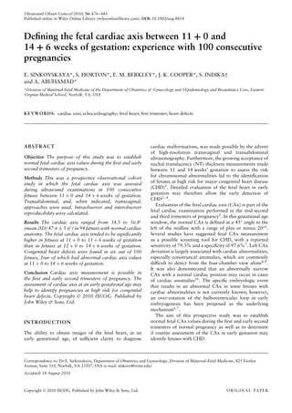
  • 2. Fetal cardiac axis 677 METHODS This study was approved by the Human Investigation Board of the Eastern Virginia Medical School (EVMS), and was conducted at EVMS’s Division of Maternal-Fetal Medicine ultrasound laboratories. After receiving writ- ten informed consent, a total of 100 consecutive women, ≥ 18 years of age and with singleton pregnancies between 11 + 0 and 14 + 6 weeks’ gestation, were enrolled in the study. Exclusion criteria included maternal obesity (body mass index (BMI) ≥ 30) and refusal to participate in the study. Transabdominal ultrasound was initially performed in all study patients to examine the fetus. The transvaginal approach was used if visualization of the fetus (because of its position) was insufficient or if suboptimal transabdom- inal images were obtained. All ultrasound examinations were performed using Voluson 730 Expert and Voluson E8 ultrasound equipment (GE Healthcare Ultrasound, Zipf, Austria) with a 4–8-MHz transabdominal trans- ducer and a 5–9-MHz transvaginal transducer. The ultrasound examination included a crown–rump length (CRL) measurement of the fetus followed by a NT measurement when requested by the patient. The CAx was assessed by obtaining an axial view of the fetal chest at the level of the four-chamber view with a single full rib visible on each side and then by measuring the CAx as the angle of two lines. The first line started at the spine posteriorly and ended in mid-chest anteriorly, bisecting the chest into two equal halves. The second line traced the long axis of the heart and ran through the interventricular septum (IVS) (Figure 1). Color or power Doppler was occasionally used to confirm the location of the IVS, which then guided the accurate placement of the intersecting angle line when the IVS was not clearly imaged on two-dimensional (2D) ultrasound (Figure 2). In each case one of the authors (E.S.) measured the CAx three times. The average of these three measurements was used to represent the CAx for each participant. In addition, the CAx was also measured by another author (S.H.). Both investigators were blinded to each other’s results. In addition to measurement of the CAx, an evaluation of the fetal heart was performed, which included imaging of the four-chamber view and the outflow tracts. All patients underwent ultrasound examination during the second and/or third trimesters, which included a targeted evaluation of fetal anatomy, fetal echocardiography and CAx measurement. Postnatal follow up was obtained for all patients by reviewing the medical records and by telephone interview. Statistical analysis Statistical analysis was performed using the SAS 9.1.3 software (SAS, Cary, NC, USA). Normal distribu- tion of continuous variables was assessed using the Kolmogorov–Smirnov test. Continuous variables are reported as mean ± SD or as median (range), depending on the data distribution. Categorical data are expressed Figure 1 Cardiac axis measurement in a fetus at 13 + 4 weeks’ gestation. The angle shown in this case is 43◦ (normal). LV, left ventricle; RV, right ventricle; S, spine. as frequencies and percentages. A P < 0.05 was consid- ered significant. The Tukey test was applied to examine the variance of the CAx at different gestational ages. The effect of CRL on the CAx was evaluated using regres- sion analyses. Repeated-measures ANOVA was used to assess intraobserver variations. Interobserver repro- ducibility was evaluated by calculating the limits of agreement using the Bland–Altman analysis11 and the coefficient of variation (CV). The following formula was used to assess the CV: CV (%) = (SD/mean of measure- ment (Observer 1; Observer 2)) × 100. RESULTS Demographic characteristics, gestational age and NT measurements at first presentation of the study patients are shown in Table 1. Seventy-two patients underwent first-trimester screening with NT measurement for chromosome abnormalities. Of the 72 fetuses in which NT thickness was measured, 2/72 had an NT of ≥ 3.5 mm and both had CHD. A transabdominal ultrasound alone was performed in 81/100 (81%) of cases, and a combined transabdominal and transvaginal approach was used in 19/100 (19%) of cases. The four-chamber view was visualized in all fetuses in early gestation. In 94 fetuses heart anatomy was normal. The CAx value in this group of normal fetuses ranged from 34.5 to 56.8◦ (mean (SD) 47.6 ± 5.6◦ ). Based on our results, a CAx of < 35◦ and > 60◦ represents 2 SD outside our mean measurement and should be considered abnormal. The relationship between CAx and gestational age is shown in Figure 3. The CAx showed a tendency to be significantly higher (levorotation) at a gestational age of 11 + 0 to 11 + 6 weeks compared with a gestational age of between 12 + 0 and 14 + 6 weeks (Table 2). Copyright  2010 ISUOG. Published by John Wiley & Sons, Ltd. Ultrasound Obstet Gynecol 2010; 36: 676–681.
  • 3. 678 Sinkovskaya et al. Figure 2 Assessment of the cardiac axis using high-definition power Doppler in a normal fetus at 12 + 6 weeks’ gestation. (a) Right and left ventricles are colored bright red and separated by a dark line, which represents the interventricular septum (arrows). (b) Cardiac axis measurement is shown. LV, left ventricle; RV, right ventricle; S, spine. Table 1 Demographics, gestational age and nuchal translucency (NT) measurements at first presentation (n = 100) Parameter Value Maternal age (years) 31.1 ± 6.4 Race Caucasian 59 African–American 31 Asian 8 Hispanic 2 Gravidity 3 (1–13) Parity 1 (0–4) Body mass index (kg/m2) 24.2 ± 3.5 Gestational age (weeks) 11 + 0 to 11 + 6 18 12 + 0 to 12 + 6 40 13 + 0 to 13 + 6 26 14 + 0 to 14 + 6 16 NT* (mm) 1.5 (0.9–4.6) Values given as mean ± SD, median (range) or %. *n = 72. Repeated-measures ANOVA showed no significant differences in the three separate measurements of CAx made by the same observer (P = 0.3). Figure 4 presents a Bland–Altman plot of interobserver reproducibility. The mean difference in CAx measurements performed by Observer 1 (E.S.) and Observer 2 (S.H.) was close to zero, and no significant difference was obtained. Based on the CV, the interobserver variation for CAx measurements was 2.8%. CHDs were diagnosed prenatally in six out of 100 fetuses and were confirmed postnatally or by autopsy. In four cases the CHDs were found during the initial scan at the first trimester and included heterotaxy syndrome with complex CHD, hypoplastic left heart syndrome, 40 35 40 45 50 55 50 60 CRL (mm) CAx(°) 70 80 90 Figure 3 Scatter plot presenting cardiac axis (CAx) measurement plotted against crown–rump length (CRL). Individual values for the CAx of normal fetuses and the reference range (mean, 5th and 95th centiles) are shown. tetralogy of Fallot and atrioventricular septal defect. All fetuses had abnormal CAx measurements. In three of these cases left deviation of the CAx (74, 97 and 68◦ ) was noted, and one fetus had mesocardia with the CAx = 0◦ (Table 3). Figures 5 and 6 show CAx measurements in two fetuses with left axis deviations in early gestation (12 + 2 and 13 weeks, respectively). In addition, two fetuses were first diagnosed with CHDs during fetal echocardiography in the second and third trimesters of pregnancy. In these two fetuses, CAx measurements in the first trimester were within the normal range. Copyright  2010 ISUOG. Published by John Wiley & Sons, Ltd. Ultrasound Obstet Gynecol 2010; 36: 676–681.
  • 4. Fetal cardiac axis 679 Table 2 Cardiac axis (CAx) measurements in fetuses with normal heart anatomy CAx (◦) GA (weeks) n Mean ± SD 95% CI 11 + 0 to 11 + 6* 17 52.0 ± 2.9 46.2–57.8 12 + 0 to 12 + 6 38 47.3 ± 2.4 42.4–52.2 13 + 0 to 13 + 6 24 48.8 ± 3.0 39.9–51.8 14 + 0 to 14 + 6 15 45.6 ± 5.0 35.6–55.7 *Multiple comparison using the Tukey test showed a significant (P < 0.05) difference between the 11 + 0 to 11 + 6 group compared with the three other groups (i.e. between 12 + 0 and 14 + 6 weeks’ gestation). GA, gestational age. DISCUSSION CHD is the most common congenital abnormality in the human fetus, and it accounts for more than half of the deaths from congenital abnormalities in childhood12 . Sev- eral risk factors for CHD, including maternal and fetal factors, have been reported13 . Most neonates born with CHD, however, have no preidentified risk factors14 . In fact, of all pregnancies referred for fetal echocardio- graphy, the highest rate of CHD (50%) is found in pregnancies with a suspected CHD on a routine ultra- sound examination15 . The four-chamber view of the heart is included in the basic obstetric ultrasound examination and has been pro- posed as a screen for CHD in the second trimester of pregnancy5 . Specialized ultrasound skills are not required because the heart is easily imaged in a transverse view of the fetal chest. Detection of an abnormal four-chamber view, axis or position of the fetal heart should be con- sidered as an indication for fetal echocardiography in the second trimester7,16. In recent years, fetal heart evaluation has become feasible in the first and early second trimesters of pregnancy because of improvements in the resolution of transvaginal and transabdominal probes. Measurement of NT is offered routinely in many countries and thickened 30 DifferenceinCAx(°) −3 −1 1 3 40 Mean CAx (°) 50 60 +2SD Mean −2SD Figure 4 Bland–Altman plot of interobserver variation (mean ± SD, 0.4 ± 1.1) in measurements of the fetal cardiac axis (CAx). NT is associated with cardiac anomalies. Recently- published data show that, in comparison to other views, the four-chamber view has the highest visualization rate at each gestational age and can be obtained in 85–100% of first-trimester ultrasound examinations17,18 . Based on our experience, a combined transabdominal and transvaginal approach allows visualization of the four-chamber view in all cases between 11 + 0 and 14 + 6 weeks of gestation. The normal CAx does not change significantly between 16 and 40 weeks of gestation and lies at a 45◦ angle to the left of the midline6. The present study shows the CAx to be significantly higher at 11 + 0 to 11 + 6 weeks of gesta- tion than later in pregnancy. The reason for a levorotated CAx in early gestation is currently unclear. Defining left axis deviation as > 75◦ , one study noted fetal anomalies in 76% of fetuses9 in the second trimester. In left CAx deviation, tetralogy of Fallot, coarctation of the aorta and Ebstein anomaly are the most common cardiac lesions, whereas double-outlet right ventricle, atrioventricular septal defect and common atrium are the most common cardiac lesions in right axis deviation8,10,19 . Our findings in early gestation were similar. Three Table 3 Cardiac axis (CAx) values in six fetuses diagnosed with congenital heart defect (CHD) First-trimester scan CAx at Case CAx (◦) NT (mm) second/third-trimester scan (◦) GA at diagnosis (weeks) Type of congenital heart defect 1 74 2.2 67 12 + 2 Tetralogy of Fallot 2 97 3.7 92 12 + 6 Hypoplastic left heart syndrome 3 68 4.6 79 13 + 0 AVSD, dominant RV 4 0 1.3 2 13 + 6 Heterotaxy syndrome, mesocardia, complex CHD (AVSD, common atrium, infracardiac TAPVC to the portal vein) 5 44 1.1 45 23 + 4 Muscular VSD 6 48 NM 68 33 + 2 Coarctation of the aorta, small VSD AVSD, atrioventricular septal defect; GA, gestational age; NM, not measured; RV, right ventricle; TAPVC, total anomalous pulmonary venous connection; VSD, ventricular septal defect. Copyright  2010 ISUOG. Published by John Wiley & Sons, Ltd. Ultrasound Obstet Gynecol 2010; 36: 676–681.
  • 5. 680 Sinkovskaya et al. Figure 5 Cardiac axis measurement in a fetus with tetralogy of Fallot at 12 + 2 weeks’ gestation. The angle shown in this case is 74◦ (left axis deviation). RV, right ventricle; S, spine. Figure 6 Cardiac axis measurement in a fetus with an unbalanced atrioventricular septal defect at 13 + 0 weeks’ gestation. Left axis deviation, with an angle of 68◦, is present. RV, right ventricle; S, spine. fetuses with left axis deviations had hypoplastic left heart syndrome, tetralogy of Fallot and unbalanced atrioventricular septal defect in our small series. Right deviation of CAx was found in the fetus with heterotaxy syndrome. Of note, two of four fetuses in our study which had CAx deviation in the first trimester and CHD had a normal NT measurement and thus CHD could have escaped detection by NT screening alone. In one case of coarctation of the aorta in our series, CAx was normal in the first trimester and left deviated in the third trimester. Isolated ventricular septal defect did not affect the CAx significantly. The interobserver reproducibility for measuring the CAx in our study was similar to that previously reported by Crane et al.7 in fetuses in the second and third trimesters (CV: 2.8% vs. 3%). Intraobserver agreement in measurement of the CAx was also noted in our study. Currently there are no approved indications for patient referral for early fetal echocardiography. Based upon our experience and that of others, an enlarged NT, the pres- ence of a major extracardiac malformation, the presence of reversed flow in the ductus venosus and the detection of tricuspid and/or mitral regurgitation or an abnor- mal CAx can be considered indications for early fetal echocardiography20–22. Limitations of the study Maternal body habitus and in utero fetal position play a critical role in the image obtained during the ultrasound examination in early pregnancy. The ability to perform an evaluation of the fetal CAx in difficult-to-image patients (BMI > 30) is challenging and remains to be determined. To our knowledge this is the first study to evaluate prospectively the CAx during the first and early second trimesters of pregnancy. The value of the CAx in early gestations for the prenatal diagnosis of CHD remains to be established in larger studies. However, our initial results are promising. In this study, we demonstrated the feasibility of CAx assessment in the first and early second trimesters of pregnancy and its potential clinical applica- bility. Further prospective studies in a clinical setting are needed to confirm the value of CAx measurement as a screening test for CHD in early gestation. REFERENCES 1. Johnson B, Simpson LL. Screening for congenital heart disease: a move toward earlier echocardiography. Am J Perinatol 2007; 24: 449–456. 2. Smrcek JM, Berg C, Geipel A, Fimmers R, Axt-Fiedner R, Diedrich K, Gembruch U. Detection rate of early fetal echocar- diography and in utero development of congenital heart defects. J Ultrasound Med 2006; 25: 187–196. 3. Huggon IC, Ghi T, Cook AC, Zosmer N, Allan LD, Nico- laides KN. Fetal cardiac abnormalities identified prior to 14 weeks gestation. Ultrasound Obstet Gynecol 2002; 20: 22–29. 4. Haak MC, van Vugt JM. Echocardiography in early pregnancy: review of literature. J Ultrasound Med 2003; 22: 271–280. 5. Cardiac screening examination of the fetus: guidelines for performing the ‘basic’ and ‘extended basic’ cardiac scan. Ultrasound Obstet Gynecol 2006; 27: 107–113. 6. Comstock CH. Normal fetal heart axis and position. Obstet Gynecol 1987; 70: 255. 7. Crane JM, Ash K, Fink N, Desjardins C. Abnormal fetal cardiac axis in the detection of intrathoracic anomalies and congenital heart disease. Ultrasound Obstet Gynecol 1997; 10: 90–93. 8. Shipp TD, Bromley B, Hornberger LK, Nadel A, Benacer- raf BR. Levorotation of the fetal cardiac axis: a clue for the presence of congenital heart disease. Obstet Gynecol 1995; 85: 97–102. Copyright  2010 ISUOG. Published by John Wiley & Sons, Ltd. Ultrasound Obstet Gynecol 2010; 36: 676–681.
  • 6. Fetal cardiac axis 681 9. Smith RS, Comstock CH, Kirk JS, Lee W. Ultrasonographic left cardiac axis deviation: a marker for fetal anomalies. Obstet Gynecol 1995; 85: 187–191. 10. Comstock CH, Smith R, Lee W, Kirk JS. Right fetal cardiac axis: clinical significance and associated findings. Obstet Gynecol 1998; 91: 495–499. 11. Bland JM, Altman DG. Applying the right statistics: analyses of measurement studies. Ultrasound Obstet Gynecol 2003; 22: 85–93. 12. Hoffman JIE, Christianson R. Congenital heart disease in a cohort of 19,502 births with long-term follow-up. Am J Cardiol 1978; 42: 641–647. 13. Small M, Copel JA. Indications for fetal echocardiography. Pediatr Cardiol 2004; 25: 210–222. 14. Allan LD, Sharland GK, Milburn A, Lockhart SM, Groves AM, Anderson RH, Cook AC, Fagg NL. Prospective diagnosis of 1,006 consecutive cases of congenital heart disease in the fetus. J Am Coll Cardiol 1994; 23: 1452–1458. 15. Copel JA, Pilu G, Green J, Hobbins JC, Kleinman CS. Fetal echocardiographic screening for congenital heart disease: the importance of the four-chamber view. Am J Obstet Gynecol 1987; 157: 648–655. 16. Allan LD, Lockhart S. Intrathoracic cardiac position in the fetus. Ultrasound Obstet Gynecol 1993; 3: 93–96. 17. Haak MC, Twisk JWR, van Vugt JMG. How successful is fetal echocardiographic examination in the first trimester of pregnancy? Ultrasound Obstet Gynecol 2002; 20: 9–13. 18. Smrcek JM, Berg C, Geipel A, Fimmers R, Diedrich K, Gem- bruch U. Early fetal echocardiography: heart biometry and visualization of cardiac structures between 10 and 15 weeks’ gestation. J Ultrasound Med 2006; 25: 173–182. 19. Abuhamad A, Chaoui R. Fetal cardiac axis. In A Practical Guide to Fetal Echocardiography: Normal and Abnormal Hearts. Lippincott Williams & Wilkins: Philadelphia, PA, 2010; 34–36. 20. Matias A, Huggon I, Areias JC, Montenegro N, Nicolaides KH. Cardiac defects in chromosomally normal fetuses with abnormal ductus venosus blood flow at 10–14 weeks. Ultrasound Obstet Gynecol 1999; 14: 307–310. 21. Martinez JM, Comas M, Borrell A, Bennasar M, Gomez O, Puerto B, Gratacos E. Abnormal first-trimester ductus venosus blood flow: a marker of cardiac defects in fetuses with normal karyotype and nuchal translucency. Ultrasound Obstet Gynecol 2010; 35: 267–272. 22. Smrcek JM, Krapp M, Axt-Fliedner R, Kohl T, Geipel A, Diedrich K, Gembruh U, Berg C. Atypical ductus venosus blood flow pattern in fetuses with severe tricuspid valve regurgitation. Ultrasound Obstet Gynecol 2005; 26: 180–182. Copyright  2010 ISUOG. Published by John Wiley & Sons, Ltd. Ultrasound Obstet Gynecol 2010; 36: 676–681.