1. Molecular Plant • Volume 1 • Number 1 • Pages 15–26 • January 2008
Leaf Positioning of Arabidopsis in Response to
Blue Light
Shin-ichiro Inouea, Toshinori Kinoshitaa,2, Atsushi Takemiyaa, Michio Doib and Ken-ichiro Shimazakia,1
a Department of Biology, Faculty of Science, Kyushu University, Ropponmatsu, Fukuoka, 810-8560 Japan
b Research and Development Center for Higher Education, Kyushu University, Ropponmatsu, Fukuoka, 810-8560 Japan
ABSTRACT Appropriate leaf positioning is essential for optimizing photosynthesis and plant growth. However, it has not
been elucidated how green leaves reach and maintain their position for capturing light. We show here the regulation of leaf
positioning under blue light stimuli. When 1-week-old Arabidopsis seedlings grown under white light were transferred to
red light (25 mmol m22 s21) for 5 d, new petioles that appeared were almost horizontal and their leaves were curled and
slanted downward. However, when a weak blue light from above (0.1 mmol m22 s21) was superimposed on red light, the
Downloaded from http://mplant.oxfordjournals.org/ by guest on June 8, 2012
new petioles grew obliquely upward and the leaves were flat and horizontal. The leaf positioning required both photo-
tropin1 (phot1) and nonphototropic hypocotyl 3 (NPH3), and resulted in enhanced plant growth. In an nph3 mutant, neither
optimal leaf positioning nor leaf flattening by blue light was found, and blue light-induced growth enhancement was dras-
tically reduced. When blue light was increased from 0.1 to 5 mmol m22 s21, normal leaf positioning and leaf flattening were
induced in both phot1 and nph3 mutants, suggesting that phot2 signaling became functional and that the signaling was
independent of phot1 and NPH3 in these responses. When plants were irradiated with blue light (0.1 mmol m22 s21) from the
side and red light from above, the new leaves became oriented toward the source of blue light. When we transferred these
plants to both blue light and red light from above, the leaf surface changed its orientation to the new blue light source
within a few hours, whereas the petioles initially were unchanged but then gradually rotated, suggesting the plasticity of
leaf positioning in response to blue light. We showed the tissue expression of NPH3 and its plasma membrane localization
via the coiled-coil domain and the C-terminal region. We conclude that NPH3-mediated phototropin signaling optimizes the
efficiency of light perception by inducing both optimal leaf positioning and leaf flattening, and enhances plant growth.
INTRODUCTION cotyl growth inhibition in response to blue light (Ahmad and
Plants respond appropriately to ever-changing environments Cashmore, 1993), and later it turned out to act as an animal
by morphogenesis, movement, changes in cellular compo- blue light receptor to regulate the circadian clock and other
nents, and metabolic activity, thereby optimizing growth in responses (Cashmore et al., 1999). Cryptochromes (cry1 and
natural environments. Plants respond by sensing changes in cry2) in plants act together with the red/far-red light receptor
light, gravity, temperature, salt, and water status through in- phytochromes to regulate photomorphogenic responses
dividual receptors. Light is the most important factor influenc- based on multiple gene expression (Lin, 2002; Nemhauser
ing plant life, and wide ranges in wavelength from UV-A to far- and Chory, 2002; Wang and Deng, 2002).
red light are perceived by several photoreceptors to recognize Phototropin1 (phot1) was identified as a plant-specific blue
the light environment. Blue light induces various developmen- light receptor using an Arabidopsis mutant that showed im-
tal and movement responses, including phototropic bending, paired phototropic bending in response to blue light (Liscum
cotyledon opening, photoperiodic flowering, leaf flattening, and Briggs, 1995; Huala et al., 1997). Phototropin is a serine/
de-etiolation, stomatal opening, chloroplast movements, threonine protein kinase in the C-terminus, with two LOV
anthocyanin accumulation, gene expression, and the inhibi-
tion of hypocotyl elongation (Cashmore et al., 1999; Briggs
1
and Christie, 2002; Lin, 2002; Wang and Deng, 2002). In Arabi- To whom correspondence should be addressed. E-mail kenrcb@mbox.nc.
kyushu-u.ac.jp, fax 81-92-726-4758.
dopsis plants, three classes of major blue light receptors—cryp- 2
Present address: Division of Biological Science, Graduate School of Science,
tochromes, phototropins, and FKF1/ZTL/LKP2 (Imaizumi et al.,
Nagoya University, Chikusa, Nagoya, 464-8602, Japan.
2003)—are responsible for the responses mentioned above.
ª The Author 2007. Published by Oxford University Press on behalf of CSPP
Cryptochrome was identified as the first plant blue light re- and IPPE, SIBS, CAS.
ceptor using an Arabidopsis mutant that did not show hypo- doi: 10.1093/mp/ssm001, Advance Access publication 7 June 2007
2. 16 | Inoue et al. d Blue Light-Mediated Leaf Positioning
(light, oxygen, voltage) domains as the binding sites of the plants each had a pair of open cotyledons and undeveloped
chromophore flavin mononucleotide (FMN) in the N-terminus. first true leaves (data not shown). We then transferred these
Later, phototropin2 (phot2) was found as a photoreceptor that green plants to red light from above at 25 lmol mÀ2 sÀ1 with or
mediates the photoavoidance response of chloroplasts to pre- without low-intensity blue light (0.1 lmol mÀ2 sÀ1) and kept
vent strong light from damaging the photosynthetic machin- them growing for 5 d to allow the appearance of new first true
ery (Jarillo et al., 2001; Kagawa et al., 2001; Kasahara et al., leaves. Slightly arched new petioles grew nearly horizontally,
2002). In general, phot1 functions under a low intensity of blue and the first true leaflets slanted down without blue light (Fig-
light, and phot2 under a relatively high intensity. Phot1 and ure 1A, left). However, straight new petioles grew obliquely
phot2 act redundantly and cover wide ranges of light intensity upward, and the new leaflets faced toward the light source
in phototropism, chloroplast accumulation, stomatal opening, when the blue light was supplemented with red light (Figure
and leaf flattening (Kagawa et al., 2001; Kinoshita et al., 2001; 1A, right). These results suggest that blue light from above ori-
Sakai et al., 2001; Sakamoto and Briggs, 2002). Furthermore, ented the leaf surface perpendicular to the light direction by
phot1 alone acts as a blue light receptor in the rapid inhibition inducing both the straight and upward growth of petioles. We
of hypocotyl elongation, followed by the cryptochrome action refer to these responses as leaf positioning.
in the much slower response (Folta and Spalding, 2001), and We measured the angle of a petiole of a first true leaf from
is required for blue light-mediated destabilization of Lhcb the horizontal (h), illustrated in Figure 1B, to express an index of
and rbcL transcripts at high intensities (Folta and Kaufman, leaf positioning. The angles were nearly 45° in the presence of
2003). All these responses probably serve to optimize photosyn- blue light and ,10° in the absence of blue light. The blue light-
Downloaded from http://mplant.oxfordjournals.org/ by guest on June 8, 2012
thesis, and a dramatic plant growth enhancement mediated by dependent leaf positioning increased the area of light inter-
phototropin is demonstrated under a low intensity of photo- ception 2-fold in each first leaf when the blue light was pro-
synthetically active radiation (PAR) (Takemiya et al., 2005). vided together with red light from the top (Figure 1C and D).
Extensive studies on phototropism were done using etio- We next illuminated the plants with blue light (0.1 lmol mÀ2
À1
lated hypocotyls and coleoptiles as model systems, and in s ) from the side but red light from the top as before. New
many cases blue light was provided from the lateral side be- petioles emerged and the new leaflets became oriented toward
cause it is easy to measure and analyze the responses (Fank- the blue light source, but the face of the leaf was not completely
hauser and Casal, 2004; Vandenbussche et al., 2005). These perpendicular to that source (Figure 1E, solid arrowheads). The
investigations have provided detailed information on photo- surface of a pair of open cotyledons became partially oriented to
tropic bending at the physiological and biochemical levels. the blue light (Figure 1E, open arrowheads).
Although phototropism, together with other phototropin- From these results, we conclude that the plant determines
mediated responses, has an important role in maximizing light the orientation of a newly developed leaf through the percep-
capture by green leaves, most of the experimental work has tion of blue light.
been done without considering green leaf behavior and devel-
opment. Therefore, it becomes important to elucidate the Phototropin1 (phot1) Mediates the Optimal Leaf
functional roles of blue light in more developed stages of Positioning Under Low Blue Light
plants with green leaves. However, the behavior of green Phototropins optimize photosynthesis and promote plant
leaves in response to blue light has not been investigated, growth by inducing blue light-mediated multiple physiologi-
nor has an attempt been made to formulate the optimal po- cal responses at the same time (Briggs and Christie, 2002; Take-
sition to maximize photosynthesis in response to blue light miya et al., 2005). We thus expected that phototropins might
when leaves are newly developed. function in the leaf-positioning response shown above. To test
In this study, we established the experimental conditions this hypothesis, we grew phototropin mutant plants under the
that allow the appearance of new leaves, and investigated same growth conditions. As expected, the optimal leaf posi-
blue light’s effects on the development of green leaves when tioning for capturing light was not found in either a phot1
the light was provided from above. We showed that, in phot2 double mutant (phot1-5 phot2-1) or a phot1 mutant
response to a weak blue light, newly emerged leaves exhibit (phot1-5), but was found in phot2 (phot2-1) and cry1 cry2 dou-
the appropriate positioning and leaf flattening to increase ble mutants (hy4-3 cry2-1) (Figure 2B and C). Without blue
light capturing efficiency. We also showed that these light, none of these plants showed the normal leaf positioning
responses are mediated by nonphototropic hypocotyl 3 and their leaves slanted down (Figure 2A). These results indi-
(NPH3) via the phot1 pathway and probably enhance growth. cate that the blue light-induced leaf positioning is mediated
by phot1, and neither phot2 nor cryptochromes are involved
RESULTS in the response under our growth conditions.
Blue Light-Dependent Leaf Positioning Increases NPH3 Mediates Phot1-Dependent Leaf Positioning
Light Capture Since blue light-dependent leaf positioning is mediated by
We grew Arabidopsis seedlings under white light at 50 lmol phot1, we wished to identify the components downstream
mÀ2 sÀ1 for 7 d and induced de-etiolation. The de-etiolated of phot1 by isolating the mutants that lack the upward petiole
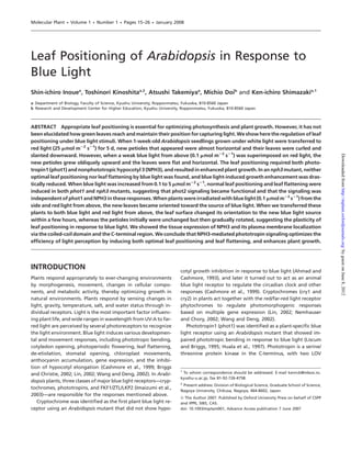
3. Inoue et al. d Blue Light-Mediated Leaf Positioning | 17
Figure 1. Leaf Positioning in Response to a Very
Low Intensity of Blue Light.
Wild-type (Col-0) plants of Arabidopsis were
grown under white light (50 lmol mÀ2 sÀ1) for
7 d and then transferred to red light (25 lmol
mÀ2 sÀ1) with or without blue light (0.1 lmol
mÀ2 sÀ1). The plants were further grown for 5 d.
The supplemental blue light was applied from
above (A–D) or from the side (E). White solid
arrowheads show the first true leaves. White
open arrowheads show cotyledons. White arrows
Downloaded from http://mplant.oxfordjournals.org/ by guest on June 8, 2012
show the direction of blue light.
(A) Side view of plants after growth for 5 d with
or without blue light. The white bar represents
1 cm.
(B) Angles (h) of petioles from the horizontal line.
Values presented are means of 25 seedlings with
standard errors.
(C) Pictures taken from above. The black bar rep-
resents 1 cm.
(D) Area of light perception in the first leaf. Areas
of projections by the first leaves were measured
by taking pictures from above. Bars represent
means 6 SE (n = 32).
(E) Side view of plants after growth for 5 d. Side
view is perpendicular to the applied blue light.
Right view is from the same direction as the blue
light source.
growth. We obtained two mutant lines: an ethylmethane sul- sitioning with upward petioles (Figure 3D). The results demon-
fonate (EMS)-mutagenized plant and a T-DNA insertional strate that our mutants are allelic to the nph3 mutant and that
plant, both of which showed impairment in the upward pet- NPH3 functions as a signal component in phot1-mediated leaf
iole growth (Figure 3A). By crossing the two mutants, we positioning. We thus named the EMS-mutagenized and the
found that the two mutations are allelic to each other. T-DNA insertional mutants as nph3-201 and nph3-202, respec-
To identify the mutated gene, we performed thermal asym- tively (Figure 3).
metric interlaced (TAIL)-PCR using the genomic DNA prepared
from the T-DNA insertional mutant. We found that T-DNA was Expression of NPH3
in the fifth exon of the NPH3 gene and confirmed that this line We investigated the expression of NPH3 mRNA by RT-PCR using
was a null nph3 mutant by reverse transcription (RT)-PCR (Fig- wild-type Arabidopsis plants. The NPH3 mRNA was highly
ure 3B and C). Because the EMS-mutagenized mutant is allelic expressed in mesophyll cells, leaves, stems, and roots, but only
to the T-DNA insertional line, we cloned and sequenced the a small amount was expressed in guard cells (Figure 4A). The
full-length NPH3 cDNA from the EMS-mutagenized mutant results agree with observations that NPH3 functions mainly in
and found that the mutant had a single nucleotide substitu- the leaf and petiole (Figure 3A), and that NPH3 does not act in
tion of cytosine to thymine in the last exon (Figure 3B). This stomata (Inada et al., 2004).
substitution produced a stop codon on Gln681 in the coiled-
coil domain of the NPH3 protein. Subcellular Localization of NPH3
We tested the functional complementation of the nph3 mu- To investigate the subcellular localization of NPH3 protein, we
tation by the wild-type genomic NPH3 gene. A 5400 bp geno- transiently expressed NPH3 fused with green fluorescent pro-
mic NPH3 fragment containing the 5’ and 3’ noncoding tein (GFP) in epidermal cells and guard cells of Vicia faba by par-
regions was introduced into the two distinct mutants. The ticle bombardment. The fluorescence from full-length NPH3
transformed lines in the T3 generation restored normal leaf po- was found on the periphery of both epidermal and guard cells,
4. 18 | Inoue et al. d Blue Light-Mediated Leaf Positioning
Downloaded from http://mplant.oxfordjournals.org/ by guest on June 8, 2012
Figure 2. Leaf Positioning Mediated by phot1.
Wild-type (gl1 and WS), phot1-5, phot2-1, phot1-5 pho2-1, and hy4-3
cry2-1 plants were grown and transferred as described in Figure 1.
(A) Plants grown under red light at 25 lmol mÀ2 sÀ1.
(B) Plants grown under red light with blue light at 0.1 lmol mÀ2 sÀ1.
(C) Angles of petioles in these plants. The measurements were done
as in Figure 1. Values are the means of 25–38 seedlings with stan-
dard errors. White bars represent 1 cm.
Figure 3. Involvement of NPH3 in Leaf Positioning.
suggesting plasma membrane localization of NPH3 as described (A) Isolation of mutants impaired in upward petiole growth under
previously (Motchoulski and Liscum, 1999; Lariguet et al., 2006; the low blue light condition. The picture shows mutant plants
grown under red light with low blue light. The white bar represents
Figure 4B, full length). We then investigated the localization in
1 cm.
more detail using guard cells because the transient expression of (B) Determination of the mutated gene in the isolated mutants. The
NPH3 is much easier in them than in epidermal cells. The fluores- genomic structure of NPH3 on chromosome 5 is shown. Black boxes
cence of mutant NPH3-201 protein from nph3-201 was observed and bold lines represent exons and introns, respectively. An nph3-
as many particles in cytosolic compartments (Figure 4B, NPH3- 201 mutant has a C-to-T nucleotide substitution in the last exon.
This nucleotide change causes the substitution of Gln681 by the
201). Since the mutant NPH3-201 protein may lack a C-terminal
stop codon. T-DNA insertion in nph3-202 was identified in the fifth
region downstream from the coiled-coil domain (Figure 3B), it is exon.
possible that this region is required for the membrane localiza- (C) Expression of NPH3 and TUB2 (b-tubulin) mRNAs analyzed by
tion of NPH3. To test this, we expressed the NPH3 C-terminal RT-PCR in 2-week-old seedlings of wild-type (Col and WS) plants
fragment containing the coiled-coil domain (coiled-coil-C) and of two nph3 mutants (nph3-201 and nph3-202).
(D) Functional complementation of nph3-201 and nph3-202 mutants
fused with GFP. As expected, the fluorescent signal of this region
with wild-type genomic NPH3 genes. Plants of nph3-201, nph3-201
was found on the plasma membrane (Figure 4B, coiled-coil-C). transformed with wild-type genomic NPH3 (201-G), nph3-202, and
We then divided this coiled-coil-C into a coiled-coil domain nph3-202 transformed with wild-type genomic NPH3 (202-G) were
(coiled-coil) and a C-terminal region (C-terminus) and expressed grown as in Figure 1. The white bar represents 1 cm.
these as above. The GFP fluorescence of the coiled-coil domain
was detected mainly in the plasma membrane and slightly in the
cytoplasm (Figure 4B, Coiled-coil). The fluorescence of the C-ter- and the membrane localization may be needed for the function
minus was found in both the cytosol and the plasma membrane of NPH3 (Figure 3A).
(Figure 4B, C-terminus), and the distribution was different from
that of GFP alone, which showed a clear cytosolic localization Recovery of Leaf Positioning in nph3 Mutants Under
(Figure 4B, sGFP). These observations suggest that both the con- High Intensity Blue Light
served coiled-coil domain and the C-terminal region probably We found that the petioles in nph3-201 and nph3-202 grew
function to localize NPH3 protein on the plasma membrane, upward and exhibited almost wild-type leaf positioning when
5. Inoue et al. d Blue Light-Mediated Leaf Positioning | 19
supplemented blue light was increased to 5 lmol mÀ2 sÀ1 from
0.1 lmol mÀ2 sÀ1 (Figure 5A). Quantitative data indicate that
phot1-5, nph3-201, and nph3-202 largely restored the wild-
type leaf positioning at relatively high fluence rates of blue
light, whereas phot1-5 phot2-1 did not (Figure 5B). These
results suggest that phot2 becomes functional and mediates
the leaf positioning in response to the higher intensity of blue
light. They also suggest that NPH3 functions principally
through the phot1-dependent pathway in the response.
NPH3 Mediates Leaf Flattening Only Under Low
Blue Light
Under our low blue light growth conditions (25 lmol mÀ2 sÀ1
red light with 0.1 lmol mÀ2 sÀ1 blue light), leaves of nph3-201
and nph3-202 curled, as did leaves of phot1-5 and phot1-5
phot2-1 mutants. This phenotype became more prominent
when these plants were further grown for another 5 d (Figure
6A). In contrast, gl1, Col, WS, and phot2-1 exhibited flattened
Downloaded from http://mplant.oxfordjournals.org/ by guest on June 8, 2012
leaves under the same conditions. All of these plants showed
curled leaves under red light alone (data not shown). These
results suggest that NPH3 functions in leaf flattening through
the phot1-mediated pathway.
When the intensity of supplemental blue light was in-
creased to 5 lmol mÀ2 sÀ1, leaves of nph3-201, nph3-202,
and phot1-5 became flattened, but those of the phot1-5
phot2-1 double mutant remained curled (Figure 6B). These
results indicate that leaf flattening is mediated by phot2 under
Figure 4. Expression of NPH3 mRNAs and Subcellular Localization
of NPH3 Protein.
(A) Expression of NPH3 mRNAs in guard cell protoplasts (GCPs), me-
sophyll cell protoplasts (MCPs), leaves, stems, and roots from 4-week-
old plants analyzed by RT-PCR. The purities of GCPs and MCPs were
98 and 99%, respectively, on a cell number basis. ACT8 was used as
an internal standard for cDNA amounts. Two separate experiments
gave similar results.
(B) Transient expression of NPH3–GFP proteins in Vicia epidermal
cells and guard cells. The primary structure of NPH3 protein and
structures of fusion proteins are illustrated. Four conserved domains
in the NPH3/RPT2 family are shown in light gray open blocks as de-
scribed in Liscum (2002). The BTB (broad complex, tramtrack, bric a `
brac)/POZ (pox virus and zinc finger) domain and the coiled-coil do- Figure 5. Rescue of Leaf Positioning Under a Relatively High Inten-
main are shown in the dark gray block and black block, respectively. sity of Blue Light in phot1-5 and nph3 Mutants.
The full length and fragments of NPH3 proteins were fused in-frame Wild-type (gl1, Col-0, and WS) plants and phot1-5, phot2-1, phot1-5
to the N-terminal end of sGFP and were expressed transiently by par- phot2-1, nph3-201, and nph3-202 plants were grown under white
ticle bombardment under the control of the CaMV 35S promoter. light at 50 lmol mÀ2 sÀ1 from fluorescent lamps for 7 d and then
Full length, full-length NPH3 protein fused to GFP; NPH3-201, transferred under red light (25 lmol mÀ2 sÀ1) with blue light and
NPH3 fragment of the N-terminus fused to GFP on Met680; allowed to grow for an additional 5 d for the determination of the
Coiled-coil-C, NPH3 fragment of Phe645 to the C-terminus fused petiole angles.
to GFP; Coiled-coil, NPH3 fragment from Phe645 to Ser696 fused (A) Pictures indicate the leaf positioning in the mutant plants under
to GFP; C-terminus, NPH3 fragment from Thr693 to the C-terminus 5 lmol mÀ2 sÀ1 of blue light.
fused to GFP; sGFP, GFP protein. Epidermal cells and guard cells (B) Angles of petioles were measured under 0.1 or 5 lmol mÀ2 sÀ1 of
expressing these proteins were inspected by GFP fluorescence using blue light as in Figure 1. Values are means of 21–28 seedlings with stan-
a confocal laser microscope. All pictures are cross-sectional. dard errors.
6. 20 | Inoue et al. d Blue Light-Mediated Leaf Positioning
Figure 6. Leaf Flattening in Wild Type and Various Mutants in Re-
sponse to Low and High Intensities of Blue Light.
Plants of the wild types (gl1, Col-0, and WS), phot1-5, phot2-1,
phot1-5 phot2-1, nph3-201, and nph3-202 were initially grown un-
der white light at 50 lmol mÀ2 sÀ1 from fluorescent lamps for 7 d.
The plants were then transferred under red light (25 lmol mÀ2 sÀ1)
with blue light of two different intensities and allowed to grow for
an additional 10 d to determine the leaf flattening.
Downloaded from http://mplant.oxfordjournals.org/ by guest on June 8, 2012
(A) Leaf flattening of the wild types and mutants with blue light at
0.1 lmol mÀ2 sÀ1.
(B) Leaf flattening of wild-types and mutants with blue light at
5 lmol mÀ2 sÀ1. White bars represent 1 cm.
a relatively high intensity of blue light, and that this phot2-
dependent leaf flattening is not mediated by NPH3.
Contribution of NPH3 to Growth Enhancement Under
Low Blue Light
NPH3 mediates both horizontal leaf positioning and leaf flat-
tening in response to very weak blue light (Figures 3A and 6A),
but does not mediate chloroplast movement or stomatal open-
ing (Inada et al., 2004). All these blue light responses are
known to increase photosynthesis and plant growth in
a low-light environment in particular (Takemiya et al.,
2005). Taking advantage of the properties of nph3 mutants,
we evaluated the contributions of leaf positioning and flatten-
ing to growth enhancement. We measured the fresh weights
of the wild type (gl1) and of nph3-201, nph3-6, and phot1-5
mutants that had been grown under our conditions for 5
weeks. As shown in Figure 7A and B, the wild-type plants
showed 2.5-fold growth enhancement by the addition of Figure 7. Growth Enhancement, Chloroplast Accumulation, and
0.1 lmol mÀ2 sÀ1 blue light to the red light, but no actual Stomatal Opening in Response to Low Intensity of Blue Light.
growth enhancement was found in the phot1-5 mutant. Inter- Wild-type (gl1), phot1-5, nph3-201, and nph3-6 plants were grown
estingly, the nph3-201 and nph3-6 mutants showed slight but for 5 weeks under red light (25 lmol mÀ2 sÀ1) with or without blue
light (0.1 lmol mÀ2 sÀ1). The growth was determined as fresh
significant growth enhancement in response to very weak blue
weight of green tissues.
light (Figure 7B). This slight growth enhancement may have (A) Growth enhancement by blue light in wild-type and mutant
been brought about by both chloroplast movement and sto- plants. Plants grown under red light (left) and red light with blue
matal opening, because in the nph3 mutants chloroplasts light (right).
gathered at the surface of mesophyll cells and stomata opened (B) Fresh weights of green tissues of plants. Bars represent means 6
SE (n = 25). Asterisks show significant statistical differences by t-test
in response to blue light (Figure 7C and D; Inada et al., 2004).
(P ,0.05) in fresh weights.
The growth difference between wild-type plants and nph3 (C) Distribution of chloroplasts in mesophyll cells of wild-type and
mutants is probably provided by the leaf positioning and leaf mutant leaves under our growth conditions.
flattening that were mediated by NPH3. These results further (D) Stomatal aperture in leaves of the wild type and mutants under
suggest that growth enhancement in response to a weak blue our growth conditions. Apertures are expressed as the ratio of
width to length of the guard cell pair, as described in Takemiya
light is brought about mainly through the function of NPH3, as
et al. (2005). Bars represent means 6 SE (n = 25).
both responses tend to maximize light interception.
7. Inoue et al. d Blue Light-Mediated Leaf Positioning | 21
Reversibility of Leaf Positioning in Response to Blue Light these plants to both red (25 lmol mÀ2 sÀ1) and blue (0.1 lmol
It is unclear whether the leaf positioning responses shown mÀ2 sÀ1) light from above and kept them growing for another
above are reversible or not. To test this, we utilized plants that 5 d. After the second transfer, the leaf surface began to orient
had been grown under irradiation with blue light from the rapidly toward the new blue light source with a time delay of
side and red light from above, as indicated in Figure 1E. The 20 min (Figure 8B, leaf angle in left graph; hL), and began a rel-
surfaces of the first true leaves of the plants were oriented to- atively slower phase after about 2 h (Figure 8A and B, 2 h).
ward the source of blue light (Figure 8A, 0 h). Such leaf orien- Then, the leaf surface gradually approached the maximum an-
tation in response to blue light was not found in the mutants gle within 8 h (Figure 8B, leaf angle in left graph), and main-
of phot1-5 or nph3-201 (data not shown). Then, we transferred tained this position thereafter with a very slight change
Figure 8. Changes in Leaf Position in Re-
sponse to Blue Light.
Wild-type (gl1) plants were grown under
Downloaded from http://mplant.oxfordjournals.org/ by guest on June 8, 2012
white light (50 lmol mÀ2 sÀ1) for 7 d
and then transferred to red light (25
lmol mÀ2 sÀ1) from above with blue light
(0.1 lmol mÀ2 sÀ1) from the plant side, and
were grown for 5 d, as indicated in Figure
1E. The plants were then transferred again
and irradiated with blue light (0.1 lmol
mÀ2 sÀ1) from above under the red light,
and growth was allowed for an additional
5 d.
(A) Side view of the plants after the second
transfer. Pictures were taken at the indi-
cated times from the perpendicular to
the direction of the first applied blue light,
which had been derived from the left (up-
per panels), and taken from the same di-
rection of the blue light (lower panels).
White solid arrowheads show the first
true leaves. White open arrowheads show
cotyledons. The black arrow indicates the
direction of the first blue light treatment.
The white arrow shows the direction of
the second blue light treatment.
(B) Angle of the first leaf from the vertical
(hL) and that of the first leaf petiole from
the vertical (hP). Typical changes in these
angles in response to blue light are shown.
The left illustration indicates the change
of angles during 8 h with high time reso-
lution. The right illustration shows the
change of angles during 5 d. Gray ovals
represent the first leaves. White ovals
show the cotyledons.
(C) Rotation of the first leaves which oc-
curred after the initial leaf orientation.
Pictures were taken at the indicated times
from above. White solid arrowheads show
the first true leaves. Black arrows indi-
cate the direction of blue light applied
previously.
(D) Petiole rotation. Typical changes in
the angles of petioles (hR) in response to
blue light are shown. Gray ovals repre-
sent the first leaves. White ovals show
the cotyledons.
8. 22 | Inoue et al. d Blue Light-Mediated Leaf Positioning
(Figure 8A and B, right graph). The petiole angle in the pro- this study, we grew plants for 5 d under definite conditions and
jected image of the first leaf became almost zero in the time determined the positions of newly emergent leaves (Figure 1).
course, similar to the light behavior of the leaflet (Figure 8A However, these experimental conditions did not produce
and B, petiole angle; hP). The rate of the rapid leaflet orienta- a rapid change in position in response to blue light. To monitor
tion was 15° hÀ1, which is almost the same value as that for the changes, we investigated the leaf positioning by moving
solar-tracking responses as reported for Lavatera cretica leaves the blue light source: plants that had been irradiated from
(Koller et al., 1985; Koller and Levitan, 1989; Koller, 2000). Our the side were now irradiated from above (Figure 8). We found
results suggests that the rapid leaflet orientation might be a so- that the leaf changed its direction to the new blue light source
lar-tracking response in Arabidopsis, and is mediated by phot1. within several hours, followed by a slow change in petiole di-
During the leaf repositioning responses, the petiole was rection after 24 h. These results suggest that the plants pref-
arch-shaped from 4 to 24 h, a conformation that facilitated ori- erentially change leaf direction, and that such rapid regulation
enting the leaf surface perpendicular to the blue light from of leaf direction is suitable for maximizing light interception.
above. The petiole subsequently became straight after 48 h The rapid leaf orientation Arabidopsis seems to be identical
(Figure 8A). Although the leaf itself became oriented to the to the response reported as solar tracking in Lavatera leaves
blue light source within 8 h, the petiole remained unchanged (Figure 8; Koller, 2000).
and the adaxial side was still toward the original source of blue We recently reported that phototropins mediate the leaf
light during this time (Figure 8A, 8 h; and C, 12 h). Afterwards, movement of kidney bean and that the response greatly in-
the petiole gradually rotated from 24 to 96 h, and completed creased the light absorption of leaves (Inoue et al., 2005).
Downloaded from http://mplant.oxfordjournals.org/ by guest on June 8, 2012
its rotation within 120 h (Figure 8D). The petioles with leaves The movement response is reversible and is completed in
finally became aligned directly opposite each other (Figure 8C, a short time (1.5 h), which is achieved by the water transport
120 h; and D). These results suggested that the leaf positioning in specialized motor cells of the pulvinus (Inoue et al., 2005).
is plastic in response to blue light and is comprised of both a rel- Although the physiological roles of both plant responses seem
atively rapid leaf orientation response (within 0.3–8 h) and to be similar (i.e. the enhancement of photosynthesis), and al-
a slow petiole rotation response (within 24–120 h). In contrast though the responses are mediated by the same photorecep-
to the first true leaves, cotyledons maintained their original tors, the mechanisms between leaf positioning and leaf
angles irrespective of the change in blue light direction (Figure movement may differ, since the complete Arabidopsis leaf po-
8A, 0–8 h). sitioning probably requires at least a few days to complete
(Figure 8).
Very recently it was shown that Arabidopsis petioles move
DISCUSSION upward and that the leaf surface becomes more vertical
when the plants are placed in the dark. This movement is sug-
Blue Light-Mediated Leaf Positioning Promotes
gested to be a shade-avoidance role in reaction to shading by
Light-Capturing Efficiency
neighboring leaves (Mullen et al., 2006); it is regulated by
Plants control leaf position in response to environmental stim- phytochrome action (Mullen et al., 2006) and/or negative grav-
uli, such as light, gravity, and the circadian rhythm, to optimize itropism (Mano et al., 2006), and is distinct from the responses
their photosynthetic performance. However, it has not been shown here.
elucidated how a plant maintains a leaf position that is opti- Interestingly, the three distinct responses (two movements
mal for capturing light energy efficiently for photosynthesis. In and positioning) mentioned above have a similar physiological
this study, we found that blue light induced the leaf surface role of increasing the light capture efficiency (Figure 1C–E;
into a perpendicular orientation to the light source and that Mullen et al., 2006), but the reactions are induced by at least
the response increased the light interception (Figure 1). We two different stimuli (blue light and darkness). It is likely that
also demonstrated that the response is mediated by phototro- the appropriate leaf positioning is very important for plant
pins (Figures 2 and 5). The leaf positioning was achieved by the survival and is finely controlled by the integration of various
regulation of the position of new emergent petioles and environmental stimuli including blue light, red/far-red light,
leaves (Figure 1A and E). When the source of blue light was and gravity in natural environments through movements
changed from above to the side without changing the source and morphogenic processes.
of red light, plants oriented the new leaf surface to the source
of blue light (Figure 1E). These results suggest that plants uti- Involvement of NPH3 in Leaf Positioning and
lize blue light to determine leaf direction. Leaf Flattening
It has been demonstrated that NPH3 and its ortholog CPT1 are
Importance of Leaf Positioning as a Means of responsible for hypocotyl and coleoptile phototropism in Ara-
Capturing Light bidopsis and Oryza, respectively (Motchoulski and Liscum,
The Arabidopsis leaf positioning might comprise both rapid 1999; Haga et al., 2005). Another example of NPH3 involve-
movement and a slow growth process, requiring a long time ment is phot1-mediated destabilization of Lhcb and rbcL tran-
(several days) to establish the response (Figures 1A and 8). In scripts (Folta and Kaufman, 2003). In the present study, we
9. Inoue et al. d Blue Light-Mediated Leaf Positioning | 23
found for the first time that NPH3 mediated both leaf position- ment and stomatal opening contribute only slightly to the en-
ing and leaf flattening in the phot1-dependent pathway (Fig- hancement of photosynthesis, particularly under the low light
ures 5 and 6). In accord with these functional roles of NPH3, we environments.
showed that NPH3 is localized on the plasma membrane,
on which phot1 also localizes (Sakamoto and Briggs, 2002), Signaling Mechanism of Leaf Positioning and
via the coiled-coil domain and the C-terminus (Figure 4B). Leaf Flattening
The co-localization of NPH3 and phot1 on the same mem- Without blue light, petioles were arched (Figure 1A, left). This
brane may facilitate phot1–NPH3 complex formation and sig- suggests that the upper side of the petiole might elongate
ing (Motchoulski and Liscum, 1999; Lariguet et al., 2006; more than the lower side. When blue light was superimposed
Figure 3A). on red light, the epinastic growth of petioles was inhibited and
NPH3 is suggested to function as a common signal com- caused the petioles to grow straight (Figure 1A, right). A sim-
ponent in both phot1- and phot2-dependent pathways in ilar differential growth between irradiated and shaded sides
phototropism, since nph3 mutants showed no hypocotyl was previously reported in the coleoptile phototropism in
phototropism under high irradiation with blue light (Sakai monocotyledons (Iino and Briggs, 1984; Haga et al., 2005).
et al., 2000; Inada et al., 2004). Unexpectedly, we found that Such differential growth is induced by a lateral translocation
the leaf positioning and leaf flattening responses were lost of auxin to the shaded side, and CPT1 is reported to function in
in nph3 mutants under a very low intensity of blue light (Fig- this process (Friml et al., 2002; Haga et al., 2005). Moreover, the
ures 3A and 6A), but both responses were restored by high- mutants defective in auxin sensitivity, such as msg1/nph4 and
Downloaded from http://mplant.oxfordjournals.org/ by guest on June 8, 2012
intensity blue light in both the nph3 and phot1 mutants axr4, have strongly curled leaves (Hobbie and Estelle, 1995;
(Figures 5A and B, and 6B). These results suggest that the Watahiki and Yamamoto, 1997), as has been found in the phe-
responses observed under a high blue light intensity might notype of the phot1 phot2 mutant (Sakai et al., 2001; Saka-
be mediated by phot2, and that an additional signal compo- moto and Briggs, 2002). The leaf curling of the msg1/nph4
nent other than NPH3 must be involved downstream from mutant is attributed to the differential growth between the
phot2. upper and lower sides (Stowe-Evans et al., 1998). It is likely that
the leaf positioning and leaf flattening shown in this study are
Contribution of Responses to Phot1-Mediated also achieved by the differential growth in both the petioles
Growth Enhancement and leaves, which might be achieved via the alteration of auxin
We demonstrated that the leaf positioning and leaf flattening distribution. Further studies are needed to clarify the partici-
responses actually contribute to blue light-dependent growth pation of auxin in these responses using transgenic plants in
enhancement by increasing the amount of light captured which auxin distribution can be visualized (Friml et al., 2002).
(Figures 1C–E and 7). Our findings add a means by which to
optimize photosynthesis through phototropin functions, in
addition to an understanding of the physiological and mor- METHODS
phological changes in photosynthetic tissues under various
Plant Materials and Growth Conditions
light environments (Niklas and Owens, 1989; Ballare and ´
Scopel, 1997). Arabidopsis thaliana wild-type and mutants plants were
In a previous work we demonstrated that phot1 dramati- grown under white fluorescent lamps at 50 lmol mÀ2 sÀ1
cally enhances plant growth in response to a very low intensity for 7 d under a 14/10 h light–dark cycle. The plants were then
of blue light, and that the enhancement is achieved by inte- transferred to red light (25 lmol mÀ2 sÀ1) with or without blue
grating phot1-mediated responses, including those of chloro- light (0.1 or 5 lmol mÀ2 sÀ1) under continuous light. All plants
plast accumulation (Jarillo et al., 2001; Kagawa et al., 2001; were grown at 24°C with a relative humidity of 55–75% in
Sakai et al., 2001), stomatal opening (Kinoshita et al., 2001; growth rooms. To determine growth, plants were grown
Doi et al., 2004), and leaf flattening (Sakamoto and Briggs, under red light (25 lmol mÀ2 sÀ1) with or without blue light
2002; Takemiya et al., 2005). Although we suggested that leaf (0.1 lmol mÀ2 sÀ1). The T-DNA insertional mutant pool
flattening was the largest factor responsible for growth en- CS22830, of M. Sussman and R. Amasino, was obtained from
hancement, we could not evaluate the contributions to the Arabidopsis Biological Research Center (The Ohio State
growth by these distinct responses. In the present study, we University, Columbus, OH, USA). We used nph3-6 as a null mu-
found that NPH3 mediates leaf positioning and flattening tant instead of the WS background nph3-202 mutant to com-
but does not mediate chloroplast movement or stomatal open- pare growth on the Col background (Motchoulski and Liscum,
ing. Taking advantage of this property of the nph3 mutant, we 1999; Figure 7).
showed that this mutant slightly enhanced plant growth un-
der our growth conditions, with active chloroplast movement Isolation of Mutants Lacking Blue Light-Induced
and stomatal opening in the mutant (Figure 7). These results Leaf Positioning
indicate that leaf flattening and positioning play an important We screened 34 000 EMS-mutagenized Arabidopsis seedlings
role in maximizing photosynthesis, and that chloroplast move- of the M2 population and 30 000 T-DNA insertion seedlings
10. 24 | Inoue et al. d Blue Light-Mediated Leaf Positioning
by isolating the mutant lacking upward petiole growth under were designed at the right border of the T-DNA region on
our experimental conditions. We obtained 32 mutants (23 lines the pD991 vector. For arbitrary degenerate primers, 5#-NTC-
of the EMS-mutagenized population and nine lines of the T- GASTWTSGWGTT-3#, 5#-NGTCGASWGANAWGAA-3#, 5#-WGT-
DNA insertional population) that showed horizontal petiole GNAGWANCANAGA-3#, 5#-TGWGNAGWANCASAGA-3#, 5#-
growth. Of these, 11 lines were fertile and heritable pheno- AGWGNAGWANCAWAGG-3#, 5#-CAWCGICNGAIASGAA-3#, 5#-
types in M3 generations. We found that one EMS mutant TCSTICGNACITWGGA-3#, and 5#-GTNCGASWCANAWGTT-3’
and one T-DNA mutant expressed wild-type levels of phot1 were used. The amplified genomic DNA fragments were
protein by immunoblotting using these mutants. The phot1 obtained by nested PCR twice, and were cloned into a pCR4-
proteins in these two mutants exhibited autophosphorylation TOPO vector (Invitrogen) and sequenced.
in response to blue light, and no mutation in the genomic
PHOT1 of either mutant was found (data not shown). When Construction of Plant Transformation Vector
the two mutants were crossed with each other, upward petiole To complement our nph3 mutants with the wild-type NPH3
growth was impaired in all of the obtained F1 seedlings (data gene, we constructed a gene transfer vector bearing the geno-
not shown), suggesting that the two mutations are allelic to mic NPH3 gene under the control of the native NPH3 promoter.
each other. After three backcrosses to the wild type (Col-0 The genomic NPH3 gene, including 5’ and 3’ noncoding
and WS, respectively), these two mutants were used in all sequences, was partially amplified by PCR from genomic
experiments. DNA of the wild type (Col-0) using oligonucleotide primers
5#-CCGGGAGCTCTCTCGCTAGCATAACCATAAACCCC-3’ and 5#-
Downloaded from http://mplant.oxfordjournals.org/ by guest on June 8, 2012
Preparation of Protoplasts from Guard Cells and TTGTTCGAATTGCATCCCTACGCG-3’ (for the first half of
Mesophyll Cells NPH3), and 5#-CGTCTTCTTAGAGCAGCAAACATGC-3’ and 5#-
Protoplasts of guard and mesophyll cells from Arabidopsis CGCGGATCCGAAATCTGCAGACAGATAAGGCGTG-3’ (for the
were prepared enzymatically as reported by Ueno et al. second half of NPH3). These amplified DNA fragments were
(2005) with slight modifications. The amount of protein was treated with SacI, or SacI and BamHI, respectively, and sub-
determined as described previously (Bradford, 1976). cloned into pBluescript II KS (+) (Stratagene, La Jolla, CA,
USA), respectively. The latter half of the NPH3 fragment was
Expression of NPH3 Transcripts Determined by RT-PCR cloned into the gene transfer vector pCAMBIA1300 (Cambia,
Total RNAs were extracted from guard cell protoplasts, meso- Canberra, Australia) with SacI and BamHI sites. Then, the first
phyll cell protoplasts, leaves, stems, and roots of 4-week-old half of the NPH3 fragment was cloned into pCAMBIA1300 con-
plants with ISOGEN (Nippon Gene, Tokyo, Japan). First-strand taining the latter half of the NPH3 fragment with the SacI site.
cDNAs were synthesized from 5 lg of each total RNA by Super- The resulting vector was verified by DNA sequencing.
Script III reverse transcriptase using oligo(dT)12–18 primer (Invi-
trogen, Carlsbad, CA, USA). A 500 bp fragment of NPH3 cDNA Transformation of Arabidopsis
was amplified with the primers 5#-GGTTGGAGTTGGAGGTG- The gene transfer vector was introduced into Agrobacterium
GAG-3’ and 5#-GATCGTCGGGTCAGGATCTC-3#. As an internal tumefaciens (GV3101), and the Agrobacterium was trans-
standard, a 350 bp fragment of ACT8 cDNA was used with formed into the nph3-201 and nph3-202 mutants by an A.
the primers 5#-ACTTTACGCCAGTGGTCGTACAAC-3’ and 5#- tumefaciens-mediated method (Clough and Bent, 1998).
AAGGACTTCTGGGCACCTGAATCT-3#. The PCR was obtained Transformed plants were selected on a half-strength MS plate
after 27 cycles for Figure 4A. containing 2% (w/v) sucrose and 30 lg mlÀ1 hygromycin. The
For amplification of the full-length NPH3 cDNA from the complementation test was performed using independent
wild types (Col and WS) and from nph3-201 and nph3-202 transgenic lines from the T3 generation.
mutants, total RNAs were prepared and first-strand cDNAs
were synthesized as described above. For PCRs, two pairs of Transient Expression Assays by Particle Bombardment
oligonucleotide primers were used: 5#-TTCCCTTGGTCCTTTCT- The cDNAs encoding the full-length, NPH3-201 fragment, and
TGCTTCC-3’ and 5#-CTATCACTTCATGAAATTGAGTTCCTCC-3’ coiled-coil-C fragment of NPH3 protein were amplified by
(for NPH3), and 5#-CTCAAGAGGTTCTCAGCAGTA-3’ and 5#- RT-PCR using the total RNA from wild-type seedlings with
TCACCTTCTTCATCCGCAGTT-3’ (for TUB2). oligonucleotide primers 5#-CCATGGGGGAATCTGAGAGCGAC-3’
and 5#-CCGGCCATGGCTGAAATTGAGTTCCTCCATCGTCTTG-3’
Thermal Asymmetric Interlaced (TAIL)-PCR (for full length), 5#-CCATGGGGGAATCTGAGAGCGAC-3’ and
To identify the T-DNA insertion site of the nph3-202 mutant, 5#- CCGGCCATGGCCATCACTTCCATCTCGTTCTGAAGC-3’ (for
we performed TAIL-PCR using genomic DNA from the mutant NPH3-201), and 5#-CCGGCCATGGCCTTTCAGGAAGGATGGGCT-
seedlings. The PCR and thermal cycler programs were GCAG-3’ and 5#- CCGGCCATGGCTGAAATTGAGTTCCTCCATC-
performed according to the method of Liu et al. (1995) with GTCTTG-3’ (for coiled-coil-C). The obtained cDNAs were
a minor modification. For the gene-specific primers, 5#-CCTA- cloned into the CaMV35S-sGFP(S65T)-NOS3’ vector with NcoI
TAAATACGACGGATCG-3#, 5#-ATAACGCTGCGGACATCTAC-3#, (Niwa et al., 1999). Plasmids expressing the coiled-coil and
and 5#-TGATCCATGTAGATTTCCCG-3’ were used. The primers C-terminus fragments were constructed from the plasmid of
11. Inoue et al. d Blue Light-Mediated Leaf Positioning | 25
coiled-coil-C by inverse PCR with oligonucleotide primers 5#- Cashmore, A.R., Jarillo, J.A., Wu, Y.J., and Liu, D. (1999). Crypto-
GCCATGGTGAGCAAGGGC-3’ and 5#-AGAAGATGGCGTGTTCTT- chromes: blue light receptors for plants and animals. Science
CACTTTCC-3’ (for coiled-coil), and 5#-ACGCCATCTTCTTCGGC- 284, 760–765.
TTGGACC-3’ and 5#-CCATCCTTCCTGAAAGGCCATGG-3’ (for Clough, S.J., and Bent, A.F. (1998). Floral dip: a simplified method
C-terminus). After the inverse PCR, reaction mixtures were for Agrobacterium-mediated transformation of Arabidopsis
treated with DpnI for the degradation of template DNA and thaliana. Plant J. 16, 735–743.
then with T4 polynucleotide kinase for phosphorylation of Doi, M., Shigenaga, A., Emi, T., Kinoshita, T., and Shimazaki, K.
the 5’ ends. The phosphorylated linear DNAs were self-ligated. (2004). A transgene encoding a blue-light receptor, phot1,
restores blue-light responses in the Arabidopsis phot1 phot2
Plasmid DNAs were prepared for the particle bombardment
double mutant. J. Exp. Bot. 55, 517–523.
and transfected as described previously (Emi et al., 2005).
Emi, T., Kinoshita, T., Sakamoto, K., Mineyuki, Y., and Shimazaki, K.
The transfected Vicia leaves were kept in darkness for 6–10
(2005). Isolation of a protein interacting with Vfphot1a in guard
h at room temperature. Epidermal peels were obtained from
cells of Vicia faba. Plant Physiol. 138, 1615–1626.
the leaves, and epidermal cells and stomata were examined by
Fankhauser, C., and Casal, J.J. (2004). Phenotypic characterization
a confocal laser-scanning microscope (Digital Eclipse C1;
of a photomorphogenic mutant. Plant J. 39, 747–760.
Nikon, Tokyo, Japan).
Folta, K.M., and Kaufman, L.S. (2003). Phototropin1 is required for
high-fluence blue-light-mediated mRNA destabilization. Plant
Determination of Phototropin-Mediated Mol. Biol. 51, 609–618.
Physiological Responses
Downloaded from http://mplant.oxfordjournals.org/ by guest on June 8, 2012
Folta, K.M., and Spalding, E.P. (2001). Unexpected roles for crypto-
Growth enhancement, chloroplast distribution, and stomatal chrome 2 and phototropin revealed by high-resolution hypo-
apertures were measured according to a previous report cotyl growth analysis. Plant J. 26, 471–478.
(Takemiya et al., 2005). ´ ´
Friml, J., Wisniewska, J., Benkova, E., Mundgen, K., and Palme, K.
(2002). Lateral relocation of auxin efflux regulator PIN3 mediates
Light Source tropism in Arabidopsis. Nature 415, 806–809.
White light was produced by fluorescent lamps (FL 40S N-SDL; Haga, K., Takano, M., Neumann, R., and Iino, M. (2005). The
National, Tokyo, Japan), and both red and blue light were rice COLEOPTILE PHOTOTROPISM1 gene encoding an ortholog
produced by light-emitting photodiodes (LED-R, maximum in- of Arabidopsis NPH3 is required for phototropism of cole-
tensity at 660 nm; and Stick-B-32, maximum intensity at 470 optiles and lateral translocation of auxin. Plant Cell 17, 103–
115.
nm; Eyela, Tokyo, Japan). Photon flux densities were deter-
mined with a quantum meter (LI-250; Li-Cor, Lincoln, NE, Hobbie, L., and Estelle, M. (1995). The axr4 auxin-resistant
mutants of Arabidopsis thaliana define a gene important for
USA) equipped with a light sensor (LI-190 SA; Li-Cor).
root gravitropism and lateral root initiation. Plant J. 7, 211–
220.
Huala, E., Oeller, P.W., Liscum, E., Han, I.-S., Larsen, E., and
ACKNOWLEDGMENTS Briggs, W.R. (1997). Arabidopsis NPH1: a protein kinase with
a putative redox-sensing domain. Science 278, 2120–2123.
We thank M. Wada (National Institute for Basic Biology, Okazaki,
Iino, M., and Briggs, W.R. (1984). Growth distribution during first
Japan) for providing seeds of the nph3-6 mutant. This work was
positive phototropic curvature of maize coleoptiles. Plant Cell
supported by the Ministry of Education, Science, Sports, and Cul-
Environ. 7, 97–104.
ture of Japan (grant Nos 16207003, 17084005 to K.S. and
14704003 to T.K.). Imaizumi, T., Tran, H.G., Swartz, T.E., Briggs, W.R., and Kay, S.A.
(2002). FKF1 is essential for photoperiodic-specific light signal-
ling in Arabidopsis. Nature 426, 302–306.
Inada, S., Ohgishi, M., Mayama, T., Okada, K., and Sakai, T. (2004).
RPT2 is a signal transducer involved in phototropic response and
REFERENCES
stomatal opening by association with phototropin1 in Arabidop-
Ahmad, M., and Cashmore, A.R. (1993). HY4 gene of A. thaliana sis thaliana. Plant Cell 16, 887–896.
encodes a protein with characteristics of a blue-light photo- Inoue, S., Kinoshita, T., and Shimazaki, K. (2005). Possible involve-
receptor. Nature 366, 162–166. ment of phototropins in leaf movement of kidney bean in re-
´
Ballare, C.L., and Scopel, A.L. (1997). Phytochrome signaling sponse to blue light. Plant Physiol. 138, 1994–2004.
in plant canopies: testing its population-level implications Jarillo, J.A., Gabrys, H., Capel, J., Alonso, J.M., Ecker, J.R., and
with photoreceptor mutants of Arabidopsis. Funct. Ecol. 11, Cashmore, A.R. (2001). Phototropin-related NPL1 controls
441–450. chloroplast relocation induced by blue light. Nature 410,
Bradford, M.M. (1976). A rapid and sensitive method for the quan- 952–954.
titation of microgram quantities of protein utilizing the princi- Kagawa, T., Sakai, T., Suetsugu, N., Oikawa, K., Ishiguro, S., Kato, T.,
ple of protein–dye binding. Anal. Biochem. 72, 248–254. Tabata, S., Okada, K., and Wada, M. (2001). Arabidopsis NPL1:
Briggs, W.R., and Christie, J.M. (2002). Phototropins 1 and 2: versa- a phototropin homolog controlling the chloroplast high-light
tile plant blue-light receptors. Trends Plant Sci. 7, 204–210. avoidance response. Science 291, 2138–2141.
12. 26 | Inoue et al. d Blue Light-Mediated Leaf Positioning
Kasahara, M., Kagawa, T., Oikawa, K., Suetsugu, N., Miyao, M., and doi/10.1199/tab.0054, http://www.aspb.org/publications/
Wada, M. (2002). Chloroplast avoidance movement reduces pho- arabidopsis/
todamage in plants. Nature 420, 829–832. Niklas, K.J., and Owens, T.G. (1989). Physiological and morpholog-
Kinoshita, T., Doi, M., Suetsugu, N., Kagawa, T., Wada, M., and ical modifications of Plantago major (Plantginaceae) in response
Shimazaki, K. (2001). phot1 and phot2 mediate blue light regu- to light conditions. Am. J. Bot. 76, 370–382.
lation of stomatal opening. Nature 414, 656–660. Niwa, Y., Hirano, T., Yoshimoto, K., Shimizu, M., and Kobayashi, H.
Koller, D. (2000). Plants in search of sunlight. Adv. Bot. Res. 33, 35–131. (1999). Non-invasive quantitative detection and applications of
Koller, D., and Levitan, I. (1989). Diurnal phototropism in leaves of non-toxic, S65T-type green fluorescent protein in living plants.
Lavatera cretica L. under conditions of simulated solar-tracking. Plant J. 18, 455–463.
J. Exp. Bot. 40, 1059–1064. Sakai, T., Kagawa, T., Kasahara, M., Swartz, T.E., Christie, J.M.,
Briggs, W.R., Wada, M., and Okada, K. (2001). Arabidopsis
Koller, D., Levitan, I., and Briggs, W.R. (1985). The vectorial photo-
nph1 and npl1: blue light receptors that mediate both photot-
excitation in solar-tracking leaves of Lavatera cretica (Malva-
ropism and chloroplast relocation. Proc. Natl Acad. Sci. USA 98,
ceae). Photochem. Photobiol. 42, 717–723.
6969–6974.
Lariguet, P., et al. (2006). PHYTOCHROME KINASE SUBSTRATE 1 is
Sakai, T., Wada, T., Ishiguro, S., and Okada, K. (2000). RPT2: a signal
a phototropin 1 binding protein required for phototropism.
transducer of the phototropic response in Arabidopsis. Plant Cell
Proc. Natl Acad. Sci. USA 103, 10134–10139.
12, 225–236.
Lin, C. (2002). Blue light receptors and signal transduction. Plant
Sakamoto, K., and Briggs, W.R. (2002). Cellular and subcellular
Cell 14 (suppl.), S207–S225.
Downloaded from http://mplant.oxfordjournals.org/ by guest on June 8, 2012
localization of phototropin 1. Plant Cell 14, 1723–1735.
Liscum, E. (2002). Phototropism: mechanisms and outcomes. In
Stowe-Evans, E.L., Harper, R.M., Motchoulski, A.V., and Liscum, E.
The Arabidopsis Book, Somerville C.R. and Meyerowitz E.M.,
(1998). NPH4, a conditional modulator of auxin-dependent dif-
eds (Rockville, MD: American Society of Plant Biologists)
ferential growth responses in Arabidopsis. Plant Physiol. 118,
doi/10.1199/tab.0042, http://www.aspb.org/publications/
1265–1275.
arabidopsis/
Takemiya, A., Inoue, S., Doi, M., Kinoshita, T., and Shimazaki, K.
Liscum, E., and Briggs, W.R. (1995). Mutations in the NPH1 locus of
(2005). Phototropins promote plant growth in response to blue
Arabidopsis disrupt the perception of phototropic stimuli. Plant
light in low light environments. Plant Cell 17, 1120–1127.
Cell 7, 473–485.
Ueno, K., Kinoshita, T., Inoue, S., Emi, T., and Shimazaki, K. (2005).
Liu, Y.-G., Mitsukawa, N., and Whitter, R.F. (1995). Efficient
Biochemical characterization of plasma membrane H+-ATPase
isolation and mapping of Arabidopsis thaliana T-DNA insert junc-
activation in guard cell protoplasts of Arabidopsis thaliana in re-
tions by thermal asymmetric interlaced PCR. Plant J. 8, 457–463.
sponse to blue light. Plant Cell Physiol. 46, 955–963.
Mano, E., Horiguchi, G., and Tsukaya, H. (2006). Gravitropism in Vandenbussche, F., Verbelen, J.P., and Van Der Straeten, D. (2005).
leaves of Arabidopsis thaliana (L.) Heynh. Plant Cell Physiol. Of light and length: regulation of hypocotyl growth in Arabi-
47, 217–223. dopsis. BioEssays 27, 275–284.
Motchoulski, A., and Liscum, E. (1999). Arabidopsis NPH3: a NPH1 Wang, H., and Deng, X.W. (2002). Phytochrome signaling mecha-
photoreceptor-interacting protein essential for phototropism. nism. In The Arabidopsis Book, Somerville C.R. and Meyerowitz
Science 286, 961–964. E.M., eds (Rockville, MD: American Society of Plant Biolo-
Mullen, J.L., Weinig, C., and Hangarter, R.P. (2006). Shade avoidance gists) doi/10.1199/tab.0074, http://www.aspb.org/publications/
and the regulation of leaf inclination in Arabidopsis. Plant Cell arabidopsis/
Environ. 29, 1099–1106. Watahiki, M.K., and Yamamoto, K.T. (1997). The massugu1 muta-
Nemhauser, J., and Chory, J. (2002). Photomorphogenesis. In tion of Arabidopsis identified with failure of auxin-induced
The Arabidopsis Book, Somerville C.R. and Meyerowitz E.M., growth curvature of hypocotyl confers auxin insensitivity to hy-
eds (Rockville, MD: American Society of Plant Biologists) pocotyl and leaf. Plant Physiol. 115, 419–426.