Molecular identification of food poisoning pathogens in bulk tank milk
•
1 gefällt mir•226 views

This document summarizes a study on identifying food poisoning pathogens in bulk tank milk from 3 dairy farms in Egypt. Escherichia coli, Staphylococcus aureus and Listeria monocytogenes were detected in the milk samples. PCR was used to detect virulence genes in the isolated bacteria. For E. coli, one isolate encoded the heat-stable enterotoxin gene. For S. aureus, 2 isolates carried the sea gene. PCR confirmed the presence of L. monocytogenes in all 4 isolates obtained. The presence of these foodborne pathogens in bulk tank milk poses a potential health risk and controls are needed to limit bacterial growth in milk.

Empfohlen
Empfohlen
Weitere ähnliche Inhalte
Was ist angesagt?
Was ist angesagt? (20)
Ähnlich wie Molecular identification of food poisoning pathogens in bulk tank milk
Ähnlich wie Molecular identification of food poisoning pathogens in bulk tank milk (20)
Molecular identification of food poisoning pathogens in bulk tank milk
- 1. 29 MOLECULAR IDENTIFICATION OF SOME CONTAGIOUS MICROORGANISMS CAUSING FOOD POISONING FROM BULK TANK MILK IN GHARBIA GOVERNORATE Amal, M. Eid Food Hygiene Department, Animal Health Research Institute, Tanta Branch, Egypt A B S T R A C T This study was conducted to determine the prevalence of food poisoning pathogens in Bulk tank milk from 3 dairy herds in Gharbia Governorate, Egypt. Escherichia coli, Staphylococcal aureus and Listeria monocytogenes were detected in 20, 60, and 10 % in samples collected from farm I , in farm II with percentage of 40, 40 and 20 % and in farm III 20, 20 and 10 % of examined bulk tank milk samples, respectively. Polymerase chain reaction is a powerful technique for detection of pathogens in foods. It is a rapid procedure with both sensitivity and specificity for quick detection and identification of specific pathogenic bacteria from different sources. The eight E. coli isolates were screened for the presence of virulence associated genes (stx1, stx2), heat-stable enterotoxin gene (STa ) and only one (50%) isolate from farm I encoded the STa gene. The ability of Staphylococcus aureus to produce enterotoxins which is linked to Staphylococci enterotoxins SEs genes was investigated by using multiplex PCR, out of 12 Staph. aureus isolated from the examined BTM samples, 2 isolates were carrying sea gene, 1(16.6%) from farm I and 1(25%) from farm II. Listeria monocytogenes detection methods based on PCR amplification of the hly gene sequences specific for confirmation of L. monocytogenes and not any other type of Listeria have been used for identification of all four obtained isolates and the results obtained from isolation were in line with that of molecular diagnosis as PCR detected only the presence of L. monocytogenes. Since presence of these food poisoning microorganisms constitute a potential risk to public health, these findings underscore the need to control them and to limit bacterial multiplication in bulk tank milk. Key words: Bulk tank milk- Echerichia coli- Staph. auerus- L. monocytogenes –Toxic genes (BVMJ-27(2): 29-47 , 2014) 1.INTRODUCTION ilk is an excellent medium for the growth of numerous microbes which produce consequential spoilage of the milk and various milk products or infections in consumers Oliver, et al. (2005). According to the procedure of milk production, it is impossible to avoid contamination of milk with microorganisms therefore the microbial content of milk is a major feature in determining its quality Torkar and Teger (2008). The existence of food borne pathogens in raw milk may increases the threat of ingestion and transmission of food borne pathogens and ingestion of harmful toxins Srinu, et al (2012). Huge numbers of microbes can get access to milk and various milk products including these often listed pathogens in raw milk as Staphylococcus aureus, Escherichia coli, Salmonella spp., Shigella spp., Yersinia enterocolitica, Aeromonas hydrophila, Brucella abortus, Campylobacter jejuni, Bacillus cereus, and Listeria monocytogenes (Garbutt et al., 1997). Enteropathogenic Escherichia coli (ETEC) have been implicated in sporadic and epidemic outbreaks of diarrhea in both infants and adults in many parts of the word. ETEC produce one or both of two plasmid- M BENHA VETERINARY MEDICAL JOURNAL, VOL. 27, NO. 2:29-47, DECEMBER 2014
- 2. Amal, M. Eid (2014) 30 mediated enterotoxins: a heat-stable enterotoxin (ST) and a heat-labile enterotoxin (LT) Gyles et al., (1974); Smith and Halls (1968) . LT and ST toxin genes are the main pathogenic elements of ETEC strains. These strains are intestinal E. coli and cause diarrhea in infected individuals, also can cause urinary hemolytic syndrome which often happens after an intestinal infection Johnson et al., (2002). The most important causes of food borne diseases are shiga toxin producing E.coli (STEC) among the other seropathotypes of E.coli. Beutin and Stephan (2006). STEC produce various complications including diarrhea, haemlytic uremic syndrome (HUS) and haemorrhagic colitis (HC) Brett et al., (2003). Report indicate that consumption of raw milk and various milk products related with occurrence of 1 to 5 percent of food infections and among that 53per cent of cases produced by enteropathogenic E.coli (EPEC) Schrade and Yager (2001). Humans infected with STEC show symptoms, such as abdominal pain and watery diarrhea, and a number of patients develop a life- threatening disease, such as hemorrhagic colitis (HC) and hemolytic-uremic syndrome (HUS) Verweyen et al., (2000). The natural reservoirs of STEC are domestic and wild ruminant animals, which shed the bacteria with their feces into the environment Caprioli et al., (2005). STEC- infected animals normally do not show signs of disease and can be included in food production. As a consequence, products of animal origin, such as meat and milk, are at risk of contamination with STEC originating from animals Hussein and Sakuma (2005). Consumption of food containing STEC was identified as a major route of human infections with these pathogens in different countries Caprioli et al (2005); Hussein and Sakuma (2005); Mead et al., (1999). STEC strains can be divided into more than 200 E. coli serotypes. The For STEC, two major types of Shiga toxins, called Stx1 and Stx2, which share 56% homology to each other, have been described previously Paton and Paton (1998). Genetic variants were detected within members of the Stx1 and Stx2 families, and a growing number of toxin types were defined according to differences in toxicity, toxin receptor, and amino acid composition of StxA and StxB subunits Paton and Paton (1998); Scheutz et al., (2001). Some Stx types, such as Stx2 and the elastase (mucus)-activatable Stx2d type, are associated with the high virulence of STEC and with HC and HUS Bielaszewska et al., (2006); Boerlin et al., (1999). Staphylococcus aureus is one of the most common agents causing food poisoning. It is involved in intramammary infections in bovine causing economic losses and milk safety problems (Taverna et al., 2007). It produce a number of protein, toxins and extracellular virulence factors that one of the most important of them is enterotoxin that cause food poisoning (Orwin et al., 2003). The emetic staphylococcal enterotoxins (SE) are classified as members of the pyrogenic toxin superantigen family because of their biological activities and structural relatedness Dings et al., (2000); Bolaban and Rasooly (2000). Eleven major antigenic types of SEs have been recognised (SEA to SEJ) Monday and Bohach (1999); Tamarapu et al., (2001) and their corresponding genes have been reported Munson et al., (1998). More recently further SE toxins have been identified (SEK, SEL, SEM, SEN, SEO and SEU) Orwin et al., (2001) & Stephan et al., (2001) and the corresponding genes have also been described Letertre et al., (2003) & Omoe et al., (2002). It is known that about 95% of staphylococcal food poisoning outbreaks are caused by SE types SEA to SEE Bergdoll (1983). The remaining 5% of outbreaks may therefore be associated with other newly identified SEs. Staphylococcal enterotoxins are resistant to inactivation by gastrointestinal proteases such as pepsin. Heat resistance is one of their most important physical and chemical properties; their biological activity remains unchanged even after thermal processing of food
- 3. Molecular identification of some contagious microorganisms causing food poisoning 31 (Martin et al., 2004 & Chapaval et al., 2006). For the above mentioned reason, these toxins can cause epidemic gastroenteritis. Actually, SEB is the most important enterotoxin that causes gastroenteritis. The toxins enter from the alimentary tract into the blood circulation. They stimulate the vomiting center of the involuntary nervous system, causing nausea, vomiting, abdominal cramps and diarrhea (Rosec and Gigaud, 2002 & Letertre et al., 2003). Although Staph. aureus is not difficult to cultivate and easily identified, there is still need for rapid and sensitive DNA –based assay specific for detecting S. aureus (Saei et al., 2010).The polymerase chain reaction (PCR), which is a technique for the in vitro amplification of specific segments of DNA, offers a rapid, sensitive and specific identification method for the genes responsible for toxins produced by Staph. aureus (Mehrotra et al., 2000 & Anvari et al., 2008). Detection of SE-genes by PCR allows the determination of potentially enterotoxigenic S. aureus irrespective of whether the strain produces the toxin or not the inability to detect the enterotoxin by immunological methods may occur due either to low level production of enterotoxin or to mutation in the coding region or in a regulatory region. For this reason, PCR may be considered more sensitive than methods that determine SE-production as immunological methods Zschock et al., (2000) & Holeckova et al., 2002). PCR assays used to identify the pathogen and its enterotoxin genes in food samples can be made in hours rather than days, with high sensitivity and method accuracy, allowing for the detection of very low concentrations of micro-organisms. The PCR assay can detect not only live but also damaged and dead micro-organisms in food subjected to thermal processing Najera-Sanchez et al., (2003).Therefore, there is a need for greater characterization data of such strains from bovine bulk-tank milk because of little data are available in literature for strains in Egypt. listeria monocytogenes may reach bulk tanks as a result of exogenous contamination via the milking equipment, because of fecal contamination during milking, or, less frequently, by an intramammary route following generalized asymptomatic infection or mast Hassan et al., 2001). It is proved that L. monocytogenes grows into biofilms attached to the surfaces in food-processing plants Arizcun, et al., (1998) and Roberts and Wiedmann (2003) and milking systems in dairy farms. The common treatment of surfaces is not effective to eliminate this dangerous foodborne pathogen, and it easily can pass into raw milk. L. monocytogenes can cause a rare but serious disease called listeriosis, especially among pregnant women, the elderly, or individuals with a weakened immune system. L. monocytogenes is more likely to cause death than other bacteria that cause food poisoning. 20 to 30% of foodborne listeriosis infections in high-risk individuals may be fatal Ramaswamy et al., (2007). Detection of L. monocytogenes by molecular methods is very specific and can be as fast as the immunological assays Janzten et al., (2006). A number of PCR assays had been described for its detection in foods Levin, (2003). PCR methods had superior sensitivity when compared to standard nucleic acid probes or immunoassays. However, complex sample preparation methods and the use of gel electrophoresis endpoint detection have hampered the transition of these methods from research to routine use in food microbiology laboratories. Nevertheless, factors influencing the performance of conventional PCR in foods continue to be investigated Aznar and Alarcón, (2003). Moreover, recent studies have shown that a broad distribution of identical or closely related enterotoxin-producing S. aureus clones is found in bovine mastitis and bulk- tank milk samples (Annemüller et al., 1999; Stephan et al., 2002). Therefore, the objective of the present investigation is (i) to study the occurrence of food poisoning
- 4. Amal, M. Eid (2014) 32 causing microorganisms (Escherichia coli, Staphylococcus aureus and Liesteria monocytogens), (ii) molecular identification of toxigenic genes using polymerase chain reaction (PCR) in isolated strains obtained from bulk tank milk in Gharbia Governorate. 2.MATERIALS AND METHODS 2.1. Samples: A total of thirty bulk milk samples collected from 3 dairy farms in Gharbia Governorate and subjected to bacteriological examination of food poisoning microorganisms including enumeration of Staphylococcal aureus , Coliform count and isolation and identification of Escherichia coli and Liesteria monocytogen, the obtained isolates were subjected molecular typing of toxigenic genes 2.2. Enumeration of Total Coliform (MPN/g) ICMSF, (1978) Estimation of coliforms was done by using most probable number technique with MacConkey's broth tubes. A series of 3 fermentation tubes containing MacConkey;s broth and inverted Durham's tubes were inoculated with 1 ml from the previously prepared 10th fold serial dilutions. After thorough mixing, inoculated and control tubes were incubated at 37 °С 24-48 hours. Tubes showing acid and gas were considered as positive for the test. From the laboratory records, the most probable number (MPN) of coliforms/g. was calculated by matching with (MPN) table. Isolation and identification: Samples were processed to isolate the E. coli as per the standard Bacteriological Analytical Manual (BAM), U.S. Food and Drug Administration (USFDA) method Kumar et al., (2008). The samples were enriched in MacConkey broth, and then loopful of culture was inoculated into MacConkey agar. Pink colour colonies obtain from MacConkey agar were taken and inoculate on Eosin methelene blue agar. Greenish metallic sheen colonies obtain on EMB agar were regard as an E. coli. Various biochemical tests such as catalase test, Indole production, Methyl red, Voges proskauer, Simon's citrate agar, Urease production, Nitrate reduction etc. were done for the confirmation of E. coli as proposed by Edwards and Wing (1972). 2.3. Bacteriological examination of Staphylococcal aureus - Enumeration of Total Staphylococcal aureus Count: from each dilution 0.1 ml was spread onto a dry surface of double sets of Baird parker agar plate (OxoidCM 275, SR54). Inoculated plates were incubated at 37°C for 48hours. Typical colonies of S.aureus(black shining convex colonies, 1- 1.5 mm in diameter with narrow white margin and surrounded by a clear zone extending into opaque medium) were enumerated and the average number per gram was calculated APHA, (1992). 2.3.1.Identification The purified S. aureus isolates were identified through different biochemical tests [catalase test, coagulase test (tube test) Quinn, et al., (2002). 2.3.2.Detection of different viulence genes in isolated E.coli and Staph aureus strains by PCR. DNA extraction. DNA extraction from samples was performed using the QIAamp DNA Mini kit (Qiagen, Germany, GmbH) with modifications from the manufacturer’s recommendations. Briefly, 200 µl of the sample suspension was incubated with 10 µl of proteinase K and 200 µl of lysis buffer at 56O C for 10 min. After incubation, 200 µl of 100% ethanol was added to the lysate. The sample was then washed and centrifuged following the manufacturer’s recommendations. Nucleic acid was eluted
- 5. Molecular identification of some contagious microorganisms causing food poisoning 33 with 100 µl of elution buffer provided in the kit. Oligonucleotide Primer. Primers used were supplied from Metabion (Germany) are listed in table (1). PCR amplification. Primers were utilized in a 25- µl reaction containing 12.5 µl of Emerald Amp Max PCR Master Mix (Takara, Japan), 1 µl of each primer of 20 pmol concentrations, 4.5 µl of water, and 6 µl of DNA template. The reaction was performed in an applied biosystem 2720 thermal cycler. 2.3.3. Analysis of the PCR products. The products of PCR were separated by electrophoresis on 1-1.5% agarose gel (Applichem, Germany, GmbH) in 1x TBE buffer at room temperature using gradients of 5V/cm. For gel analysis, 20 µl of the products was loaded in each gel slot. A 100 bp plus DNA Ladder (Qiagen, Germany, GmbH) was used to determine the fragment sizes. 2.3.4. For multiplex PCR, used for toxigenic genes of Staph aureus: Primers were utilized in a 50- µl reaction containing 25 µl of EmeraldAmp Max PCR Master Mix (Takara, Japan), 1 µl of each primer of 20 pmol concentration, 7 µl of water, and 10 µl of DNA template. The gel was photographed by a gel documentation system (Alpha Innotech, Biometra) and the data was analyzed through computer software Table (1): Primers sequences, target genes, amplicon sizes and cycling conditions. Target gene Primers sequences Amplified segment (bp) Primary den. Sec den Ann. Ext. Final ext. Refer ence stx1 ACACTGGATGATCTCAGTGG 614 94˚C 5 min. 94˚C 1 min 58˚C 1 min 72˚C 1 min 72˚C 10 min Dipin eto et al., 2006 CTGAATCCCCCTCCATTATG stx2 CCATGACAACGGACAGCAGTT 779 CCTGTCAACTGAGCAGCACTTTG STa GAAACAACATGACGGGAGGT 229 94˚C 30 sec. 57˚C 30 sec. 72˚C 30 sec. Lee et al., 2008 GCACAGGCAGGATTACAACA Sea GGTTATCAATGTGCGGGTGG 102 94˚C 45 sec. 50˚C 45 sec. 72˚C 45 sec. Mehr otra et al., 2000 CGGCACTTTTTTCTCTTCGG Seb GTATGGTGGTGTAACTGAGC 164 CCAAATAGTGACGAGTTAGG Sec AGATGAAGTAGTTGATGTGTATGG 451 CACACTTTTAGAATCAACCG See AGGTTTTTTCACAGGTCATCC 209 CTTTTTTTTCTTCGGTCAATC Sed CCAATAATAGGAGAAAATAAAAG 278 94˚C 30 sec. 48˚C 30 sec. 72˚C 30 sec. ATTGGTATTTTTTTTCGTTC
- 6. Amal, M. Eid (2014) 34 2.4. Bacteriological examination of Listeria monocytogen One ml of milk sample inculcated in 9ml of Listeria enrichment broth (Difco), and incubated at 30°c for 48 hr. After incubation one loopful was subcultured on Listeria Oxford medium base. The plates were incubated at 35°c for 24-48 h. 2.4.1. - Identification: Four typical colonies were transferred from Listeria Oxford medium base to Trypticase soy agar with yeast extract for purification. Purified isolates were identified by the Gram- stain, Catalase test, motility test, biochemical tests and Christie- Atkins, Munch- Petersen; test of haemolysis (CAMP Test). Further confirmation of L. monocytogenes the isolates were inoculated in to 10% aqueous stock solution of Manitol, L. Rhamnose and D. Xylose FDA (2003). 2.4.2.Polymerase chain reaction (PCR) DNA Extraction: Boiling method (Bansal, 1996). Bacterial pellets were washed once with 1 ml phosphate buffered saline (PBS), pH 7.4, resuspended in a same volume of cold water and incubated in a boiling water bath for 10 min. The clear supernatants obtained after a 5 min centrifugation at 12000g were used for PCR reaction. Oligonucleotide Primers: in this study 1set of primer was used, hyl gene specific for confirmation of L. monocytogenes and not any other type of Listeria. The sequence, cycling conditions and amplicone size were described in table (2). The PCR products were visualized on 1.3% agarose gel in 1x TBE using GeneRuler 100 bp plus DNA Ladder (Fermentas Cat.No. #SM0323). Table (2) the sequence, cycling conditions and amplicon size of the used genes: Gene Sequence 5- 3 Cycling condition Product size Reference Hyl LM1 CCT-AAG-ACG- CCA-AT C-GAA LM2 CCT-AAG-ACG- CCA-AT C-GAA Initial denaturation 95o C for 5 min 30 cycle of 95 o C for 15 sec 57 o C for 2 sec 72 o C for 30 sec Final extention at 72 o C for 5 min 702 Mengaud et al. (1988) 3.RESULTS In the present study, table (3) presents the enumeration results for coliform and Staph. aureus counts giving an idea about the levels of the concerned pathogens in the 3 dairy farms under investigation. The mean values of total Coliform counts for farms I, II and III were 10.5x10³ + 3.1x10³ , 23.2x10³ + 17.4x10³ and 8x10³ + 3.2x10³ respectively. The mean values of total Staph. Count for farms I, II and III were 45.2x10³ + 6x10³ , 43.8x10³ + 3.3x10³ and 36.3x10³ + 12.2x10³ respectively. Also, the presence of food poisoning organisms and isolation rates of E. coli , S. aurus and L. monocytogenes have been reported in table (4) in examined BTM samples collected from the three dairy farms (10 samples from each). Incidence of E. coli: The incidence of E. coli was observed in the samples comprising of BTM was (20%), (40%) and (20%), in the concerned dairy farms. Prevalence of Staph. aureus was (60%), (40%) and (20%) in the three farms respectively, while L. monocytogenes was
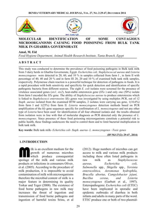
- 7. Molecular identification of some contagious microorganisms causing food poisoning 35 observed in percentage of (10%), (20%) and (10%) respectively. (Table, 4). Detection of virulence genes of E.coli: In this study, the obtained eight E. coli identified field isolates by biochemical tests were tested for the presence of STa gene. Only one strain obtained from farm I was positive for the presence of STa gene. Also, examined for presence of virulence genes (stx1, stx2) and none of which were found to be positive. Detection of enterotoxigenic genes of Staph aureus: Using multiplex PCR, out of 12 identified field isolates by biochemical tests were tested for the presence of enterotoxigenic gene (Sea, Seb, Sec, Sed and See). Two samples were positive to Sea gene (16.6 and 25%) one from each farm I and farm II, respectively. The two isolates gave one band at (102 bp) in agarose gel. All four samples were subjected to PCR from initial culture (Fig. 5) compared to L. monocytogenes reference strain, gave a characteristic band at 702 bp to hyl gene specific for L. monocytogenes. Table (3): Statistical analysis of coliform & staph.aureus counts in BTM samples in examined farms Total staph Mean SE T. coliform Mean SE 45.2x10³ +6x10³ 10.5x10³ +3.1x10³Farm I 43.8x10³ +3.3x10³ 23.2x10³ +17.4x10³Farm II 36.3x10³ +12.2x10³ 8x10³ +3.2x10³Farm III Table (4): Incidence of food poisoning microorganisms isolated from examined farms. Farm I Farm II Farm III No. % No. % No. % E. coli 2 20 4 40 2 20 Staph. aureus 6 60 4 40 2 20 Liesteria monocytogenes 1 10 2 20 1 10 Table (5) Incidence of virulence genes in E. coli isolates Farm E.coli isolates Positive E.coli isolates STa stx1 Stx2 No % I 2 1 50 0 0 II 4 0 0 0 0 III 2 0 0 0 0
- 8. Amal, M. Eid (2014) 36 Table (6) Incidence of enterotoxin genes in staph aureus isolates Farm No of S. aureus Positive S. aureus isolates for presence of enteric genes Sea Seb Sec Sed See No % I 6 1 16.6 0 0 0 0 II 4 1 25 0 0 0 0 III 2 0 0 0 0 0 0 Figure-1. Agarose gel showing PCR amplification of E. coli STa gene product (229 bp) Pos Positive control, Neg: Negative control, L: DNA Ladder, Lane 1: positive E. coli strains and Lane 2 to 8 : negative E. Coli strains Figure-2. Agarose gel showing PCR amplification of E. coli stx1, stx2 gene product (614, 779 bp) Pos: Positive control, Neg: Negative control, L: DNA Ladder, Lane 1 to 8: E. coli isolates Figure -3 Agarose gel electrophoresis showing the results of multiplex PCR for detection of enterotoxin genes among the S. aureus isolates. Lane L: 100 bp ladder DNA molecular weight marker (Qiagen ), Lane Pos: positive control for Sea, Seb, Sec and See genes, Lane Neg : negative control, Lane 1: positive Sea S. aureus isolated from farm I BTM sample, Lane 2: positive Sea S. aureus isolated from farm II BTM sample, Lanes 3 to 6 : no amplification.
- 9. Molecular identification of some contagious microorganisms causing food poisoning 37 Figure-4. Agarose gel showing monoplex PCR amplification for detection of enterotoxin gene sed among the S. aureus isolates. Lane L: 100 bp ladder DNA molecular weight marker (Qiagen ), Lane Pos: positive control for Sed gene ( 278 bp), Lane Neg : negative control, Lane 1 to 6: negative S. aureus isolates obtained from farm I BTM and farm II BTM samples for Sed gene : no amplification. Figure-5: Agarose gel electrophoresis of the amplification products of L. monocytogenes DNA obtained from tested BTM samples strains compared to Reference strain using hyl gene . Lane 1 - L. monocytogenes Reference strain (702bp), Lane 2: farm I positive band, Lane 3: Marker (Fermentas), Lane 4, 5: farm II positive bands, Lane 6: farm III positive band 4.Discussion The safety of milk is an important attribute of consumers of milk and dairy products. Milk pasteurization safeguards consumers from many potential food borne hazards in milk and milk products. Despite the pasteurization process, the quality and safety of raw milk are important in reducing the risk of food borne diseases associated with milk because raw milk is the starting point of the milk production-consumption chain. The presence of food poisoning organisms in raw milk generally comes from cows with mastitis, handlers or deficient hygiene. When found in milk, high levels of contamination can be reached quickly under favorable conditions. Its presence in foods can be a risk to human health, causing a public health problem, as these bacteria produces toxins that can cause toxic food infections (Quintana and Carneiro, 2006). In the present study total of 30 BTM samples 10 of each were collected from 3 dairy shops in Gharbia Governorate, Egypt. These samples were investigated bacteriologically to detect occurrence of E.coli, S. aureus and L. monocytogenes among the examined samples. Table (3) illustrates the mean values of total Coliform counts for farms I, II and III were 10.5x10³ + 3.1x10³ , 23.2x10³ + 17.4x10³ and 8x10³ + 3.2x10³ respectively, but this results came in contrast of results reported by Sobih, (2000) and Gillespie et al., (2012) who found higher findings of Coliform count . The current results were lower than those reported by Gihan, (1997) and Jayarao and Wang, (1999) but they were nearly similar to results reported by Hassan and Al-Sanjary, (1999). Colifrom counts of raw bulk tank milk should be routinely performed to identify bacteria that
- 10. Amal, M. Eid (2014) 38 originate from fecal contamination of milk. Colifrom bacteria can contaminate milk through poor udder preparation or unhygienic handling of the milking machines. The mean values of total Staph. Count for farms I, II and III were 45.2x10³ + 6x10³ , 43.8x10³ + 3.3x10³ and 36.3x10³ + 12.2x10³ , respectively, observed in Table (3). Nearly similar results were obtained by Gillespie et al., (2012). Staph. aureus is one of the causative agents of mastitis in dairy herds (Barkema et al., 2006). This disease involves inflammation of the mammary glands and a resultant sporadic shedding of Staph. aureus cells into the raw milk (Barkema et al., 2006). Therefore, the presence of large concentrations of Staph. aureus is indicative of mastitis in a dairy herd. From a food safety perspective, it is recognized that Staph. aureus is an enterotoxin-producing pathogen but that the concentration needs to exceed 105 cfu/ml for sufficient toxin to be produced to cause human illness (Hill, 1981; Jay, 2000). None of the raw milk samples in this study contained numbers of S. aureus that were close to this count Results in Table (4) showed the incidence of E. coli was observed in the samples comprising of BTM was (20%), (40%) and (20%), higher incidence of E. coli (52%) was observed in Virpari et al., (2013) and in Soomro et al., (2002) was 57%, while nearly similar results (26.4%) was reported by Bandyopadhyay et al., (2011) and (30.2%) by Farzan et al., (2012). Incidence of Staph. aureus was (60), (40) and (20) % in the three farms respectively. Similar results of Staphylococcus species isolation was observed in raw milk samples (56%) reported by EL-Jakee et al., (2013) and Stephan et al., (2001) showed only 32.4% Staph. aureus and Khudor et al. (2012) where S. aureus isolated from raw milk by percentage of 28.5% . Lower results of raw milk were observed with that of Rahimi and Alian (2013) as they isolate Staph. aureus from raw milk by percentage of 17.5%.on the other hand higher results were reported by Rall et al.(2008) isolated Staph. aureus from raw milk by percentage of 68% and 70.4% respectively. In the current study the isolated L. monocytogenes found in percentage of (10%), (20%) and (10%) respectively. Lower incidence of observed as 5.1% in raw milk samples (Kalorey et al., 2008). The source of L. monocytogenes in raw milk is mostly the gastrointestinal tract of animals and the environment, skin of the teats, in particular shedding of Listeria into milk due to chronic mastitis (O’Donnell, (1995) is less frequent. Waak et al. (2002) studied the incidence of Listeria species in raw whole milk from farm bulk tanks and from raw milk and L.monocytogenes was found in 1.0 % of 294 farm bulk tank milk. LT and ST toxin genes are the main pathogenic elements of ETEC strains. These strains are intestinal E. coli and cause diarrhea in infected individuals ,also can cause urinary hemolytic syndrome which often happens after an intestinal infection Johnson et al., (2002). In this study, results observed in table (5) revealed that, the obtained eight E. coli identified field isolates by biochemical tests were tested for the presence of STa gene. Only one strain obtained from farm I was positive for the presence of STa gene. Nearly similar results observed in Jung, (1999) identified 3 strains of E.coli containing only the STa gene and only one strain containing LT and STa . Also, the obtained eight E.coli strains examined for presence of virulence genes (stx1, stx2) and none of which were found to be positive Shiga toxin-producing E. coli are highly pathogenic in humans with low infectious doses Hussein and Sakuma, (2005). Among the STEC, O157:H7 is the classical serotype associated with enterohemorrhagic diseases. Non- O157 STEC strains are only recently becoming recognized as important human pathogens (Nataro and Kaper, 1998; Hussein and Sakuma, 2005). Consumption of raw milk has been implicated as the cause in several outbreaks of disease caused by E. coli O157:H7 and by non-O157 STEC
- 11. Molecular identification of some contagious microorganisms causing food poisoning 39 (Hussein and Sakuma, 2005). Shiga toxin- producing E. coli excrete potent Shiga toxins that are encoded by the stx1 and stx2 genes, respectively (Hussein and Sakuma, 2005). The STEC isolates in this study predominantly carried the stx2 gene. Epidemiological data suggest that stx2 is more important than stx1 in the development of hemolytic uremic syndrome, a life-threatening illness associated with STEC infection in children (Nataro and Kaper, 1998). Results in table (6) revealed that the suspected STEC isolates none of them harboring the sx1and sx2. On the contrary, a study conducted by Steele et al. (1997) reported that only 0.87% of the BTM samples in Ontario contained STEC. Four of the five isolates of E. coli encoded for shiga-toxin 2 gene while one strain encoded for shiga-toxin 1 gene. Also, Montenegro et al. (1990) reported that most of the STEC isolates of bovine origin encoded for shiga-toxin 1 gene. Virpari et al., (2013) reported that out of 80 E. coli isolates, 12 (15.00%) E. coli isolates found positive for stx1gene and 18 (22.50%) E. coli isolates found positive for stx2 gene. Similar finding of predominance of stx2 producing strains were reported by Sabry and Elmalt 2008) Milk is a good substrate for S. aureus growth and for enterotoxin production. In addition, enterotoxins retain positive their biological activity even after pasteurization Asao et al., (2003).The determination of staphylococcal enterotoxin type has a long history of successful use in epidemiological studies in both clinical and environmental microbiology studies. Oligonucleotide primers for specific detection of enterotoxin genes sea, seb, sec, sed, and see have previously been reported (Johnson et al.,1991& Tsen et al., 1994), these were used in individual PCR assays, thus requiring several PCRs for each sample to screen for the presence of all of the enterotoxin genes. Monday and Bohach (1999) have recently described a multiplex PCR assay for the detection of all of the staphylococcal enterotoxin genes, but again this assay uses separate primer pairs for each toxin gene to be detected. Generally, five classical staphylococci enterotoxin (SE) SEA to SEE were recognized. It was shown that about 95% of staphylococcal foodpoisoning outbreaks were caused by strains carrying the classical SE and the remaining 5% of coagulase positive species; S. hyicus and S. intermedius outbreaks were associated with other identified (Wang et al., 2012). Using multiplex PCR, out of 12 S. aureus isolated from the examined samples, 1 (16.6%) and 1(50%) isolates were positive for sea could produce enterotoxins as shown in table (6) isolated from farm I & II respectively. Others found sea but with different percentage than our study as Adwan et al.(2005); Rall et al.(2008); Rahimi et al. (2012) & ElJakee et al.(2013); 7.1%,41%, 12.7% and 40% respectively. While Veronica et al.(2011) & Khudor et al.(2012) didn't find sea at all. None of the 12 isolates harboring other SEs genes. For sec results, our results agree with Rahimi and Alian (2013) didn't find sec at all. While Sharma et al.(2000)& Khudor et al.(2012) found only sec by percentage of 11.1%, 19% and 18.5% respectively and none of these isolates harboring other SEs genes. The current study, there is no gene in genes coding for more than one enterotoxin in one sample while Veronica et al.(2011) found a combination between sea-sed-see by percentage of 1.1% and El-Jakee et al.(2013) found combination between seb- sed by percentage of 20%. On the other hand, enterotoxin A was the most commonly produced toxin. Moreover, enterotoxin A is most often implicated in cases of staphylococcal food poisoning (Shale et al., 2005). The dominance of S. aureus enterotoxin A isolates in our present study has been also reported by other researchers for Staph. aureus recovered from food samples (Tsen et al., 1998 & Bendahou et al., 2009). The SEs could be able to indicate the origin of the Staph.aureus strains because it was observed that a higher ratio of isolates from
- 12. Amal, M. Eid (2014) 40 bovine produced SEC and those from human produced mainly SEA (Ahari et al., 2009). The ability of Staph. aureus isolates to produce one or more SEs in food products is linked to staphylococcal food poisoning (Bennett, 2005). Enterotoxigenic strains of Staph. aureus have been reported to cause a number of diseases or food poisoning outbreaks in many countries because of ingestion of contaminated dairy products or milk with staphylococcal enterotoxins (Oliver et al., 2005; Ikeda et al., 2005 & ISfID, 2010). In the present work 83.3% of Staph. aureus isolates were negative to the five classical enterotoxin genes.This might be explained by the fact that these isolates either have not harboured any gene of enterotoxins or thy might have other types of SEs which are family of 18 serological types of heat stable enterotoxin (MacLauchlin et al., 2000; Ikeda et al., 2005; Rall et al., 2008 & Bhunia, 2008). Differences in SE type prevalence compared with the present study likely reflect the distinct origin of the isolates. It can be considered that food handlers are the most usual contamination source leading to food poisoning. Nevertheless, since these toxins resist heat treatment, the present findings indicate a potential public health hazard and underscore the need to establish both effective bovine mastitis control programs and proper milk cooling methods to limit Staph. aureus presence and multiplication in bulk tank milk. (Veronica etal., 2011). Rapid isolation and confirmation methods for L. monocytogenes in foods are still being sought (Beumer and Hazeleger, 2003). But there are certain strains of L. monocytogenes which behave phenotypically quite typical and inconspicuous but are non-pathogenic (Hof and Rocourt, 1992). Six Listeria species are known to exist an attempt to identify L. monocytogenes by PCR-based detection, potentially suitable oligonucleotide primer sequences complementary to the virulence gene hlyA (GenBank accession no. AF253320) and known to be specific for L. monocytogenes. Thus, it has been suggested that diagnosis of pathogenic Listeria spp. And listeric infection should ideally be based on virulence markers (Notermans et al., 1991). Moreover, the importance of PCR has been investigated for detection of L. monocytogenes from foods (Gouws and Liedemann, 2005). Thus, in the present study L. monocytogenes strains isolated from BTM samples were analyzed for the presence of virulence-associated gene hlyA of L. monocytogenes employing the monoplex PCR for rapid comparing and confirmation with conventional culture method. All four samples were subjected to PCR from initial culture (Fig. 5) compared to L. monocytogenes reference strain, gave a characteristic band at 702 bp to hylA gene specific for L. monocytogenes so the results obtained from isolation were in line with that of molecular diagnosis ( PCR), while (Gouws and Liedemann, 2005) reported that only 37 % of samples were confirmed to be positive for L. monocytogenes by PCR amplification of the hly gene (732 bp). PCR was able to eliminate the false positives and detect all L. monocytogenes in the food products, unlike the conventional methods used in the industry. In addition to the fact that the incidence of Listeria species was higher than L. monocytogenes on selective media, there was also the presence of Listeria-like organisms. These organisms had the typical appearance of Listeria on selective media, but were non-Listeria species. PCR proves to be a sensitive and rapid technique to be included in the procedure of detection of L. monocytogenes in food products. Dairy products contaminated with Listeria monocytogenes have been responsible for human listeriosis outbreaks Dalton et al., (1997). The serious consequences of listeriosis, such as a septicemic form of the illness in elderly and immune compromised people, and abortion in pregnant women or death of their newborn, constitute a serious threat to public health Even though the
- 13. Molecular identification of some contagious microorganisms causing food poisoning 41 symptoms may be relatively mild in the mother, the illness may be transferred to the fetus causing serious illness or fetal death. Some symptoms of Listeriosis may include meningitis, encephalitis, septicemia, spontaneous abortion, still birth, and influenza-like symptoms ( Sutherland et al., 2003; Azevedo et al., 2005). Antibiotic treatment of pregnant women or immunocompromised people who have eaten food contaminated by L.monocytogenesis can prevent the most serious consequences of listeriosis, but only if the infection is diagnosed in time. Another complication is that Listeria is able to grow well at law temperatures. Thus, refrigeration is not as effective in preventing growth of Listeria in food as it is for most other bacteria that cause food-born disease (Salyers and Whitt 2002) Conclusions: Current study supports the finding that BTM can be regarded as critical source of pathogenic E. coli, Staph. aureus and L. monocytogenes This explains the need of strict monitoring and surveillance for effective measures of hygiene and sanitary practice during production of milk in farms. 5.REFERENCES Adwan, G., Abu-Shanab,B., Adwan,K. 2005. Enterotoxigenic Staphylococcus aureus in Raw Milk in the North of Palestine. Turk.J. Biol.,29:229-232. Ahari, H., Shahbazzadeh, D., Misaghi, A. 2009. Selective amplify-cation of SEA, SEB and SEC genes by multiplex PCR for rapid detection of Staphylococcus aureus. Pak. J. of Nutrit. 8(8):1224-1228. Annemüller, C., Lämmler, Ch., Zschöck, M. 1999. Genotyping of S. aureus isolated from bovine mastitis. Vet. Microbiol. 69:217–224. Anvari, S.H., Sattari, M., Forozandehe- Moghadam, M. 2008. Detection of Staphylococcus aureus enterotoxins A to E from clinical sample by PCR. Res. J. Biol. Sci. 3(8): 826-29. APHA, 1992. Compendium of methods for the microbial examination of foods. 3rd Edn. American public health association. Washingtion. DC, USA. Arizcun, C., Vasseur, C., Labadie, J.C. 1998. “Effect of several decontamination procedures on Listeria monocytogenes growing in biofilms,” J. Food Prot. 61(6):731–734. Asao, T., Kumeda, Y., Kawai, T., Shibata, T., Oda, H., Haruki, K., Nakazawa, H., Kozaki, S. 2003. An extensive outbreak of staphylococcal food poisoning due to low fat milk in Japan: estimation of enterotoxin A in the incriminated milk and powdered skim milk. Epidemiol. Infect., 130(1):33-40. Azevedo, I., Regalo, M., Mena, C., Almeida, G., Carneiro, L., Teixeira, P., Hogg, T., Gibbs, PA. 2005. Incidence of Listeria spp. in domestic refrigerators in Portugal. Food Control; 16(2):121-124. Aznar, R., Alarcón, B. 2003. PCR detection of Listeria monocytogenes: a study of multiple factors affecting sensitivity. J. Appl. Microbiol., 95:958-966. Balaban N., Rasooly A. 2000. Review: staphylococcal enterotoxins. Int. J Food Microbiol; 61:1–10. Barkema, H.W., Schukken, Y.H., Zadoks, R.N. 2006. Invited review: the role of cow, pathogen, and treatment regimen in the therapeutic success of bovine Staphylococcus aureus mastitis. J. Dairy Sci. 89:1877-1895. Bandyopadhyay, S., Lodh, C., Rahaman, H., Bhattacharya, D., Bera, A. K., Ahmed, F. A., Mahanti, A., Samanta, I., Mondal, D. K., Sarkar, S., Dutta, T. K., Maity, S., Paul, V., Ghosh, M. K., Sarkar, M., Baruah, K. K. 2011. Characterization of shiga toxin producing (STEC) and enteropathogenic Escherichia coli (EPEC) in raw yak. (Poephagus grunniens) milk and milk products. http://dx.doi.org/10.1016/j.rvsc.2011.1 2.011.10/04/2013
- 14. Amal, M. Eid (2014) 42 Bansal, N.S. 1996. Development of a polymerase chain reaction assay for the detection of Listeria monocytogenes in foods. Lett. of Appl. Microbiol. 22:353–356. Bendahou,A., Abid, M., Bouteldoun, N., Catelejine, D., Lebbadi,M. 2009. Enterotoxigenic coagulase positive staphylococcus in milk and milk products, lben and jben, in northern Morocco. J Infect Developing Countries. 3(3):169-176. Bennett, R.W. 2005. Staphylococcal enterotoxin and its rapid identification in foods by enzyme-linked immunosorbent assay-based methodology. J. Food Prot. 68(6): 1264-1270. Bergdoll, M.S. Enterotoxins. In:Easton C.S.F., Adlam C., editors. 1983. Staphylococci and staphylococcal infections. London: Academic Press; pp. 559–98. Beumer, R.R., Hazeleger, W.C. 2003. Listeria monocytogenes: diagnostic problems, FEMS Immunol. Med. Microbiol. 35:191–197. Beutin, L., Stephan R. 2006. Escherichia coli O157 and non- O157 Shiga toxin- producing Escherichia coli in fecal samples of finished pigs at slaughter in Switzerland. J. Food Prot. 69:260-266. Bhunia, A.K. 2008. Food born bacterial pathogen. Springer. Purdue University West Lafayette, IN USA. Pp: 125-134. Bielaszewska, M., Friedrich, A.W., Aldick, T., Schurk-Bulgrin, R., Karch, H. 2006. Shiga toxin activatable by intestinal mucus in Escherichia coli isolated from humans: predictor for a severe clinical outcome. Clin. Infect. Dis. 43:1160–1167. Boerlin, P., McEwen, S.A., Boerlin- Petzold, F., Wilson, J.B., Johnson, R.P., Gyles, C.L. 1999. Associations between virulence factors of Shiga toxin-producing Escherichia coli and disease in humans. J. Clin. Microbiol.37:497–503 Brett, K.N., Hornitzky, M.A., Bettelheim, K.A., Walker, M.J., Djordjevic, S.P. 2003. Bovine non-O157 Shiga toxin 2- containing Escherichia coli isolates commonly possess stx2- edl933 and/or stx2vhb subtypes. J. Clin. Microbiol. 41:2716- 2722. Caprioli, A., Morabito, S., Brugereb, H., Oswald E. 2005. Enterohaemorrhagic Escherichia coli: emerging issues on virulence and modes of transmission. Vet. Res. 36:289–311. Chapaval, L., Moon, D.H., Gomes, J. E., Duarte, F. R., Tsai, S. M. 2006. Use of PCR to detect classical enterotoxin genes and toxic shock syndrome toxin- 1 gene in Staphylococcus aureus isolated from crude milk and determination of toxin productivities of S. aureus isolates harboring these genes. J Arq. Inst. Boil. 73:165-169. Dalton, C.B., Austin,C.C., Sobel, J., Hayes,P.S., Bibb,W.F., Graves, L.M., Swaminathan, B., Proctor, M.E., Griffin, P.M. 1997. An outbreak of gastroenteritis and fever due to Listeria monocytogenes in milk. N. Engl. J. Med. 336:100–105. Dinges, M.M., Orwin, P.M., Schlievert P.M. 2000. Enterotoxins of Staphylococcus aureus. Clin Microbiol Rev; 13:16–34. Dipineto, L., Santaniello, A., Fontanella, M., Lagos, K., Fioretti, A., Menna, L.F. 2006. Presence of Shiga toxin- producing Escherichia coli O157:H7 in living layer hens. Lett. in Appl. Microbiol. 43:293–295 Edwards, P.R., Ewing, W.H. 1972. Identification of samples. Enterobacteriaceae. Emerg. Infect. Dis. 12: 154–159. EL-Jakee, J., Marouf, S.A., Ata, S. Nagwa, Abdel-Rahman, H. Eman, Abd EL- Moez, I. Sherein, Samy, A.A., EL- Sayed, E., Walaa, E. 2013. Raoid method for detection of staphylococcus aureus enterotoxins in food. Global Veterinaria. 11(3):335- 341.
- 15. Molecular identification of some contagious microorganisms causing food poisoning 43 Farzan, R., Rahimi, E., Momtaz, H. 2012. Virulence properties of Shiga Toxin- Producing Escherichia coli isolated from Iranian raw milk and dairy products. Slov. Vet. Res. 49(4):159-66. FDA 2003. BAM, Detection and Enumeration of L. monocytogenes. 8th Ed. Revision A, 1998, Chapter 10, Author: Antony D. Hitchins. Garbutt, J. 1997. Essentials of Food Microbiology, Hodder Arnold, London, UK. Gihan, G.H. 1997. Studies of coliforms in farm milk and its environment. M.V.Sc. Thesis., Fac. Vet. Med., Zagazig Univ, Egypt. Gillespie, B.E., Levvis, M.J., Boonyayarta, S., Ma, M.L., Saxton, A., Oliver, S.P., Almeida, R.A. 2012. Evaluation of bulk tank milk microbiological quality of nine dairy farms in Tennessee. J. Dairy sci. 95:4275-4279. Gyles, C. L, So, M., Falkow, S. 1974. The enterotoxin plasmids of Escherichia coli. J. Infect. Dis. 130: 40-49. Hassan, A.A., Al-Sanjary, R.A. 1999. Incidence of enteropathogenic E. coli in raw milk. Iraq J. of Vet. Sci. 12(1): 103-108. Hof, H., Rocourt, J. 1992. Is any strain of Listeria monocytogenes detected in food a health risk? Int.l J. Food Microbiol., 16:173 182. Holeckova, B., Holoda, E., Fotta, E. 2002. Occurrence of entero-toxigenic Staphylococcus aureus in food. Ann. Agric. Environl. Med. 9:179-182. Hussein, H. S., Sakuma T. 2005. Prevalence of Shiga toxin-producing Escherichia coli in dairy cattle and their products. J. Dairy Sci. 88:450–465. Ikeda, T., Tamate, N., Yamaguchi,K., Makino, S. 2005. Mass outbreak of food poisoning disease caused by small amounts of staphylococcal enterotoxins A and H. J. Appl Environ Microbiol. 71(5):2793–2795. International Commission on Microbiological Specification for Foods (ICMSF), 1978. Microbial ecology of foods. Their significance and methods of enumeration 2nd Ed- Univ. of Toronto press, Toronto and Buffalo, Canada. ISfID, 2010. International Society for Infectious Diseases. http://www. eurosurveillance.org/ View Article. aspx? Article Id=19528. Jayarao, B.M., Wang L. 1999. A study on the prevalence of Gram-negative bacteria in bulk tank milk. J. Dairy Sci. 82(12):2620-4. Johnson, J.R, Jerome, C., Boster, D.R., Stapleton, A.E., Tarr, P.I. 2002. Analysis of Urinary Escherichia coli isolates for ability to produce shiga toxin. J. Clin. Microbiol., 40(6):2247- 2248. Johnson, W. M., Tyler, E. P., Ashton, F.E., Pollard, D.R., Rozee, K.R. 1991. Detection of genes for enterotoxins, exfoliative toxins, and toxic shock syndrome toxin 1 in Staphylococcus aureus by the polymerase chain reaction. J. Clin. Microbiol. 29:426– 430 Jung, H.K. 1999. Identification of serotype by use of serologic assay and detection of the enterotoxin gene of Escherichia coli by means of a polymerase chain reaction assay for isolates from pigs, chickens, and cows. Am J Vet Res., 60(4):468-72. Kalorey, D. R., Warke, S. R., Kurkure, N. V., Rawool, D. B., Barbuddhe, S. B. 2008. Listeria species in bovine raw milk: a large survey of Central India. Food Control.19: 109-112. Khudor, M.H., Abbas,B.A., Idbeis, H.I. 2012. Detection of enterotoxin genes of Staphylococcus aureus isolates from raw milk. Bas.J.Vet.Res. 11(1):254- 264. Kumar, R., Surendran, P. K., Thampuran, N. 2008. Evaluation of culture, ELISA and PCR assays for the detection of Salmonella in seafood. Letters in Applied Microbiol. 46(2):221-226. Lee, S.I., Kang, S.G., Kang, M.L., Yoo, H.S. 2008. Development of multiplex
- 16. Amal, M. Eid (2014) 44 polymerase chain reaction assays for detecting enterotoxigenic Escherichia coli and their application to field isolates from piglets with diarrhea. J Vet Diagn Invest 20:492–496. Letertre, C., Perelle, S., Dilasser F., Fach P. 2003. Identification of a newputative enterotoxin SEU encoded by the egc cluster of Staphylococcus aureus. J Appl Microbiol; 95:38–43. MacLauchlin, J., Narayanan, G.L., Mithani, V. 2000. The detection of enterotoxins and toxic shock syndrome toxin genes in S. aureus by polymerase chain reaction.J. Food prot. 63:479-488. Martin, M.C., Fueyo, J.M., Gonzales- Hevia, J.M., Mendoza, M.C. 2004. Genetic procedure for identification of enterotoxigenic strains of Staphylococcus aureus from three food poisoning outbreaks. J. Food Microbiol., 94:279-286. Mead, P. S., Slutsker, L., Dietz, V., McCaig, L.F., Bresee, J.S., Shapiro, C., Griffin, P.M., Tauxe, R.V. 1999. Food-related illness and death in the United States. Emerg. Infect. Dis. 5:607–625. Mehrotra, M., WANG, G., Johnson, W.M. 2000. Multiplex PCR for Detection of Genes for Staphylococcus aureus Enterotoxins, Exfoliative Toxins, Toxic Shock Syndrome Toxin 1, and Methicillin Resistance. JOURNAL OF CLINICAL MICROBIOLOGY. 38:3. Monday, S.R., Bohach, G.A. 1999. Use of multiplex PCR to detect classical and newly described pyrogenic toxin genes in staphylococcal isolates. J Clin Microbiol; 37:3411–3414. Montenegro, M.A., Trumpf, T., Aleksia, S., Reuter, G., Bulling, E. Helmuth. R. 1990. Detection and characterization of fecal verotoxin-producing Escherichia coli from healthy cattle. J. Clin. Microbiol. 28:1417–1421. Munson, S.H., Tremaine, M.T., Betley, M.J., Welch, R.A. 1998. Identification and characterisation of staphylococcal enterotoxin types G and I from Staphylococcus aureus. Infect Immun. 66:3337–3348. Najera-Sanchez, G., Maldonado- Rodriguez, R., Olvera P.R., Mota de la Garza L. 2003. Development of two multiplex polymerase chain reactions for the detection of enterotoxigenic strains of Staphylococcus aureus isolated from foods. J Food Prot 2003; 66:1055–1062. Nataro, J. P., J. B. Kaper J.B. 1998. Diarrheagenic Escherichia coli. Clin. Microbiol. Rev., 11:142–201. Notermans, S.H.W., Dufrenne, J., Leimeister-Wachter, M., Domann, E., Chakraborty, T. 1991. Phosphatidyl inositol-specific phospholipase C activity as a marker to distinguish between pathogenic and nonpathogenic Listeria species. Appl. Environ. Microbiol. 57: 2666-2670. O’Donnell, E.T. 1995. The Incidence of Salmonella and Listeria in raw milk from farm bulk tanks in Englandand. Int. J. Dairy Technol., 48: 125–129. Oliver, S.P., Jayarao, B.M., Almeida, R.A. 2005. Foodborne pathogens, mastitis, milk quality, and dairy food safety. The University ofTennessee, Knoxville. NMC Annual Meeting Proceedings. 3:27. Omoe, K., Ishikawa, M., Shimoda, Y., Hu, D.L., Ueda, S., Shinagawa, K. 2002. Detection of seg, seh and sei genes in Staphylococcus aureus isolates and determination of enterotoxin productivities of S. aureus isolates harboring seg, seh, or sei genes. J Clin. Microbiol. 40:857–862. Orwin, P.M., Fitzgerald, J.R., Leung, D.Y.M., Gutierrez, J.A., Bohach, G.A., Schlievert, P.M. 2003. Characterisation of Staphylococcus aureus enterotoxin L. Infect Immun. 71:2916–2919. Orwin, P.M., Leung, D.Y., Donahue, H.L., Novick, R.P., Schlievert, P.M. 2001. Biochemical and biological properties of staphylococcal enterotoxi K. Infect Immun; 69:360–366.
- 17. Molecular identification of some contagious microorganisms causing food poisoning 45 Paton, J.C., Paton, A.W. 1998. Pathogenesis and diagnosis of Shiga toxin producing Escherichia coli infections. Clin. Microbiol. Rev. 11: 450–479. Quinn, P.J., Markey, B.K., Carter, M.E., Donnelly, W.J.C., Leonard F.C., Maguire, D. 2002. Veterinary Microbiology and Microbial Diseases. 1st published Blackwell Science ltd. Rahimi, E., Mommtaz, H., Shakerian, A., Kavyani, H.R. 2012. The detection of classical enterotoxins of Staphylococcus aureus in raw cow milk using the ELISA method. Turk. J. Vet. Anim. Sci. 36(3):319-322. Rahimi, E., Alian,F. 2013. Presence of enterotoxigenic Staphylococcus aureus in cow, camel, sheep, goat, and buffalo bulk tank milk Veterinarski Arhiv 83 (1): 23-30. Rall, V.L.M., Vieira, F.P., Rall, R., Vieitis, R.L., Fernandes, A., Candeias, J.M.G., Cardoso, K.F.G., Araujo, J.P. 2008. PCR detection of staphylococcal enterotoxin genes in Staphylococcus aureus strains isolated from raw and pasteurized milk. J. Vet. microbiol. 132(4):408-413. Ramaswamy,V., Cresence,V.M., Rejitha, J.S. 2007. “Listeria—review of epidemiology and pathogenesis , Journal of Microbiology, Immu- nology and Infection. 40(1):4–13. Roberts, A.J., M. Wiedmann, M. 2003. “Pathogen, host and environmental factors contributing to the pathogenesis of listeriosis,” Cellular and Molecular Life Sciences. 60(5) 904–918. Rosec, J.P., Gigaud, O. 2002. Staphylococcal enterotoxin genes of classical and new types detected by PCR in France. Int J Food Microbiol. 77(1-2):61-70. Sabry A. H., Elmalt, L. M., 2008. Informally raw milk Prevalence of Shiga Toxin-producing Escherichia coli in and kareish cheese investigation on the occurrence of toxigenic Escherichia coli in qena city, egypt with emphasis on molecular characterization. Ass. Univ. Bull. Environ. Res. 11(2):35-42 Saei, H.D., Ahmadi, M., Mardani, K., Batavani, R.A. 2010. Genotyping of Staphylococcus aureus isolated from bovine mastitis based on PCR-RFLP analysis of the aroA gene.Comp Clin Pathol 19:163-168. Salyers, A.A., Whitt, D.D. 2002. Bacterial pathogenesis. A molecular approach. Second ed. Washington D.C: ASM press: pp.398-406. Scheutz, F., Beutin, L., Pierard, D., Smith, H. 2001. Nomenclature of verotoxins, p. 447–452. In G. Duffy, P. Garvey, and D. A. McDowell (ed.), Verocytotoxigenic E. coli. Food and Nutrition Press, Trumbull, CT. Schrade, J. P., Yager, J. 2001. Implication of milk and milk products in food disease in France and in different industrialized countries. Int. J. Food Microbiol. 67:1–17. Shale, K., Lues, J.F.R., Venter, P., Buys, E.M. 2005. The distribution of Staphylococcus spp. on bovine meat from abattoir deboning rooms. Food Microbiol 22:433–438. Sharma, N.K., Rees, C.D., Dodd, C.E.R. 2000. Development of a single-reaction multiplex PCR toxin-typing assay for Staphylococcus aureus strains. Appl. Environ Microbiol. 66:1347–1353. Smith, H.W., Halls, S. 1968. The transmissible nature of the genetic factor in Escherichia coli that controls enterotoxin production. J. Gen. Microbiol. 52:319-334. Sobeih Azza M.K. 2000. Studies on raw milk quality in Kaft El-Sheikh Ph.D. thesis, Faculty of Veterinary Medicine, Kafr El-Sheikh Branch, Tanta University. Soomro, A. H., Arain, M. A., Khaskheli, M., Bhutto, B. 2002. Isolation of Escherichia coli from raw milk and milk products in relation to public
- 18. Amal, M. Eid (2014) 46 health sold under market condition at Tandojam. Pak. J. Nutr. 13:151–152. Steele, M.L., McNab, W.B., Poppe, C., Griffiths, M.W., Chen, S., Degrades, S.A., Fruhner, L.C., Larkin, C.A., Lynch, J.A., Odumeru, J.A. 1997. Survey of Ontario bulk raw milk for foodbornepathogens. J. Food Prot. 60:1341–1346. Stephan, R., Annemüller, C., Hassan, A.A., Lammler, C. 2001. Characterization of enterotoxigenic S. aureus strains isolated from bovine mastitis in northeast Switzerland. Vet. Microbiol.78:373–382. Stephan, R., Buehler, K., Lutz, C. 2002. Prevalence of genesencoding enterotoxins, exfoliative toxins and toxic shock syndrome toxin 1 in Staphylococcus aureus strains isolated from bulk-tank milk samples in Switzerland. Milchwissenschaft 57: 502–504. Sutherland, P.S., Miles, D.W., Laboyrie, D.A. 2003. Listeria monocytogenes. In: Hocking AD (eds). Food borne Microorganisms of Public Health Significance. Sixth edition. Sydney: Australian Institute of Food Science and Technology Inc. 34. Tamarapu, S., McKillip, J.L., Drake, M. 2001. Development of a multiplexpolymerase chain reaction assay for detection and differentiation of Staphylococcus aureus in dairy products. J. Food Prot; 64:664–668. Taverna, F., Negri, A., Piccinini, R., Zecconi, A., Nonnis, S., Ronchi, S., Tedeschi, G. 2007. Characterization of cell wall associated proteins of a S. aureus isolated from bovine mastitis. Vet. Microbiol., 119(2– 4):240 – 247. Torkar, K.G., Teger, S.G. 2008. The Microbiological Quality of Raw milk after introducing the two days milk collecting system.African J. of Nutrit. 92(1): 61-74. Tsen, H.Y., Chen, T.R., Yu, G.K. 1994. Detection of B, C types of enterotoxigenic Staphylococcus aureus using polymerase chain reaction. J. Chinese Agric. Chem. Soc. 32:322. Tsen, H.Y., Yu, G.K., Wang, K.C.; Wang, S.J., Chang, M.Y., Lin L.Y. 1998. Comparison of the enterotoxigenic types, toxic shock syndrome toxin I (TSST-1) strains and antibiotic susceptibilities for enterotoxigenic Staphylococcus aureus strains isolated from food and clinical samples. Food Microbiol., 15:33-41. Veronica, E.N, Vilma, R.C, Luis, F.C. 2011. Presence of entero-toxigenic Staphylococcus aureus in bulk tank milk from Argentine dairy farms. Revista Argentina de Microbiología. 43:104-106. Verweyen, H.M., Karch, H., Brandis, M., Zimmerhackl. L.B. 2000. Enterohemorrhagic Escherichia coli infections: following transmission routes. Pediatr. Nephrol., 14:73–83. Virpari, P.K., Nayak, J.B., Brahmbhatt, M.N., Thaker, H.C. 2013. Study on isolation, molecular detection of virulence gene and antibiotic sensitivity pattern of Escherichia coli isolated from milk and milk products. Vet world, pp. 541-545. Waak, E., Tham, W., Danielsson, T.M.L. 2002. Prevalence and fingerprinting of Listeria monocytogenes strains isolated from raw whole milk in farm bulk tanks and dairy plant receiving tanks. Appl Environ Microbiol. 68:3366-3370. Wang, X. J., Meng, J., Zhang, T., Zhou, Y., Zhang, B., Yang, M. X., Xia, X. 2012. Characterization of S. aureus isolated from powdered infant formula milk and infant rice cereal in China. Int. J. Food Microbiol., 153(1-2):142-147. Zschock, M., Botzler, D., Blochler, S. 2000. Detection of genes for enterotoxins (ent) and toxic shock syndrome toxin-1 (tst) in mammary isolates of Staphylococcus aureus by polymerase-chain-reaction. Int. Dairy J. 10: 569-574.
- 19. Molecular identification of some contagious microorganisms causing food poisoning 47 ﺎت رو ﻟﻠﻣ ﺋﻲ اﻟﺟز اﻟﺗﺻﻧﯾﻒﺔ اﻟﻣﻌدﻪ اﻟﻐر ﻣﺣﺎﻓظﻪ ﻓﻲ اﻟﻣﺟﻣﻌﻪ ﺎن اﻻﻟ ﻓﻲ اﻟﻐذاﺋﻲ ﻟﻠﺗﺳﻣم ﻪ اﻟﻣﺳﺑ ﻣﺻطﻔﻲ اﻣﻞﻋﯾد ﺻﺣﺔ ﻗﺳمﺔ أﻏذ–ﺣوث ﻣﻌﻬدﺻﺣﺔانواﻟﺣﯾ–طﻧطﺎ–ﻣﺻر ﻲاﻟﻌر اﻟﻣﻠﺧص ﻫذﻩ اﺟرتاﺳﺔراﻟدوﺗم ﻪ اﻟﻐر ﻣﺣﺎﻓظﻪ عارزﻣ ﺛﻼث ﻓﻲ اﻟﺣﻠﯾب اﻧﺎتزﺧ ﻓﻲ اﻟﻐذاﺋﻲ اﻟﺗﺳﻣم ﺎت رو ﻣ اﻧﺗﺷﺎر ﻣد ﻟﺗﺣدﯾد ﺎ ﺷرﺷ اﻻ روب ﻣ ﻣن ﻞ اﺟدوﺗ ﻋن اﻟﻛﺷﻒ، ﻻوﻣوﻧوﺳﯾﺗوﺟﯾن ﺳﺗرﺎ اﻟﻠو اﻟذﻫﺑﻲ اﻟﻌﻧﻘود ور اﻟﻣﻓاﻧﺎتزﺧ ﻋﯾﻧﺎت ﻲ ﻪ اﻻﺗ ﺎﻟﻧﺳب اﻻوﻟﻲ ﻋﻪاﻟﻣزر ﻓﻲ اﻟﺣﻠﯾب20و60و40ﻋﻠﻰ،اﻟﻲواﻟﺗوﻓﻲﻋﺔاﻟﻣزرﺔ اﻟﺛﺎﻧﺎﻟﻧﺳبﺔ اﻷﺗ40و40و20ﻋﻠﻰ اﻟﻲواﻟﺗﻋﺔاﻟﻣزرواﻟﺛﺎﻟﺛﺔﺎﻟﻧﺳبﺔ اﻷﺗ20و20و10ﻋﻠﻰ.اﻟﻲواﻟﺗاﻟﻣ ﻩراﻟﺑﻠﻣ ﺗﻔﺎﻋﻞ ﻌﺗﺑرﻟﻠﻛﺷ دﻩ ﻣؤ ﻪ ﻗو ﻪ ﺗﻘﻧ ﺗﺳﻠﺳﻞﻒ ﻣن ﺎت رو اﻟﻣ ﻋﻠﻲ اﻟﺗﻌرف ﻓﻲ اﻟﺳرﻊ اﻟﻛﺷﻒو ﻪ اﻟﺣﺳﺎﺳو ﻪ ﺎﻟﺧﺻوﺻ ﯾﺗﻣﯾز اﻧﻪ ﻣﺎ اﻻطﻌﻣﻪ ﻓﻲ اضراﻻﻣ ﺎت ﻣﺳﺑ ﻋن ﻫذﻩ ﺎ ﺗرا ﻣد ﺎن ﻟﺑ اﻟﺛﻼث ﺎت رو اﻟﻣ ﻻتﻣﻌزو ﻓﻲ ﻪ اﻟﺳﻣ ﺟﯾﻧﺎت ﻋن اﻟﻛﺷﻒ اﺳﻪراﻟد ﻫذﻩ اﺳﺗﻬدﻓت ﻟذا ﻣﺧﺗﻠﻔﻪ ﻣﺻﺎدر ﺣﺎ ﻻتاﻟﻣﻌزوﺟﯾﻧﺎت ﻋن ﺎﻟﻛﺷﻒ ﺎن اﻻﻟ ﺗﻠك ﻣن اﻟﻣﺻﻧﻌﻪ ﺎن اﻻﻟ ﻣﻧﺗﺟﺎت اﺳﺗﻬﻼك ﻣن اﻟﻧﺎﺗﺟﻪ اﻟﻐذاﺋﻲ اﻟﺗﺳﻣم ﻻت ﻩرارﻟﻠﺣ اﻟﻣﻘﺎوﻣﻪ ﻪ اﻟﻣﻌو اﻟزﻔﺎﻧﺎتSTaاﻟ وﺟﯾﻧﺎتاﻓ ﻋن اﻟﻣﺳﺋوﻟﻪ اوﻩرﺿروب ﻣ ﻟﺳﻣوم ﻪ اﻟﻣﺷﺎ اﻟﺳﻣوم ازراﻟﺷﯾﺟﻼsx1, sx2ﻋﺗر وﺟدت وﻗد ﻪ اﻟﺛﻣﺎﻧ ﻻو ﺎ ﺷرﺷ اﻻ ﻻتﻣﻌزو ﻓﻲاﻟﺟﯾن ﻟوﺟود ﻪ اﯾﺟﺎﺑ اﺣدﻩو ﻩاﻟﺳ ازراﻓ ﻋن اﻟﻣﺳﺋولاﻟﻣﻘﺎوﻣﻪ ﻣوم ﻓﻲ ﻩرارﻟﻠﺣاﻻ ﻋﻪاﻟﻣزرﺟﯾﻧﺎت اﺟدوﺗﺗ وﻟم وﻟﻲsx1, sx2ﻓﻻتاﻟﻣﻌزو ﻣن ا ﻲﻻﻪ ﻟﺛﻣﺎﻧاﻟﺳﻣوم ﺗﻠك ازرﺎﻓ.ﺗم ﻣﺎ اﻟﺟﯾﻧﺎت ﻋن اﻟﻛﺷﻒاﻟﻣﺳﺋوﻟﺔاﻟ ازراﻓﺳﻣومﻓﻲ ﺔ اﻟﻣﻌوو اﻟﻣ روب ﻣرﻻ اﻟذﻫﺑﻲ اﻟﻌﻧﻘودﻪ اﻟﻣﻌو اﻟﺳﻣوم ﻧﺗﺎجSes.ﺎن ﺗﻔﺎﻋﻞ ﺎﺳﺗﺧدام اﻟﻛﺷﻒةراﻟﺑﻠﻣاﻟﻣﺗﻌوﻣن ددأﺻﻞ12ﻣﻌزوﻟﺔاﻟ روب ﻟﻣاﻟﺛﻼث عارزاﻟﻣ ﻋﯾﻧﺎت ﻣن اﻟذﻫﺑﻲ اﻟﻌﻧﻘود ور ﻠﻣ اﺣدﻩو ﻩرﻋﺗ ﺎﻧت)16,6%ﻣن (ﻋﺔاﻟﻣزرﻪ اﻟﺛﺎﻧ ﻋﻪاﻟﻣزر ﻣن ﻪ ﺛﺎﻧ ﻩروﻋﺗ اﻻوﻟﻲ)25%اﻟﺟﯾن ﺗﺣﻣﻞ (.ﺎر اﺧﺗ اﺳﺗﺧدم ﻣﺎ ﺟﯾن ﻋن ﺎﻟﻛﺷﻒ ﻣوﻧوﺳﯾﺗوﺟﯾن ﺳﺗرﺎ اﻟ روب ﻟﻣ اﻟﺟزﺋﻲ ص اﻟﺗﺷﺧ ﻓﻲ اﻟﻣﺗﺳﻠﺳﻞ ﻩراﻟﺑﻠﻣﻟﺗﺄﻛﯾداﻟﺗﻌرفﻋﻠﻰﺳﺗرﺎ اﻟﻠ اترﻋﺗ اﻻرﻊ ﺗﺎﻛﯾد ﺗم وﻗد ﻣوﻧوﺳﯾﺗوﺟﯾناﻟﺟﯾن ﻟﻬذا عارزاﻟﻣ ﻣن اﻟﻣﻌزوﻟﻪ.hylﻋن اﻟﻣﺳﺋوﻟﻪ ﺎت رو اﻟﻣ ﻫذﻩ اﺟدوﺗ ان م و ﻩرط اﻟﺳ اﻟﻲ اﻟﺣﺎﺟﻪ د ﺗؤ اﻟﻧﺗﺎﺋﺞ ﻫذﻩ ﻓﺎن اﻟﻌﺎﻣﻪ اﻟﺻﺣﻪ ﻋﻠﻲ ﻣﺣﺗﻣﻼ ارﺧط ﻞ ﺗﺷ عارزاﻟﻣ ﺎن اﻟ ﺎت ﺗﻧ ﻓﻲ اﻟﻐذاﺋﻲ اﻟﺗﺳﻣم ﻣن اﻟﺣدو ﻋﻠﯾﻬﺎﺗﻛﺎﺛراﻟﻣﺟﻣﻌ اﻟﺣﻠﯾب ﺎت ﺗﻧ ﻓﻲ ﺗرﺎ اﻟﺔ. ﻋدد :ﺔ طر اﻟﺑ ﺔ اﻟطﺑ ﻟﻠﻌﻠوم ﺑﻧﻬﺎ )ﻣﺟﻠﺔ27)2(, 47-29:ﺳﻣﺑ در2014( ﺔاﻟﺒﻴﻄﺮ اﻟﻄﺒﻴﺔﻟﻠﻌﻠﻮم ﻬﺎ ﺑ ﻣﺠﻠﺔﻋﺪد27)2:(29-47ﺮ ﺴﻤ د2014