osteomyelitis
•Als PPTX, PDF herunterladen•
10 gefällt mir•868 views

osteomyelitis's types and it's treatment

Empfohlen
Weitere ähnliche Inhalte
Was ist angesagt?
Was ist angesagt? (20)
Ähnlich wie osteomyelitis
Ähnlich wie osteomyelitis (20)
Kürzlich hochgeladen
Kürzlich hochgeladen (20)
osteomyelitis
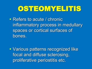
- 1. Refers to acute / chronic inflammatory process in medullary spaces or cortical surfaces of bones. Various patterns recognized like focal and diffuse sclerosing, proliferative periostitis etc. OSTEOMYELITIS
- 2. Predisposing factors of osteomyelitis Local Factors • Anatomical site of the disease: The mandibular bone has poor blood supply in comparison to that of the maxilla, besides this it has more compact bony pattern due to which, osteomyelitis occurs far more commonly in mandible than maxilla. • Pre-existing bone disease: Long standing bony disease like Paget’s disease of bone, fibrous dysplasia, cystic lesions, etc. SYSTEMIC FACTOR Immune deficiency states. Immunosuppression Diabetes mellitus. Malnutrition. Extremes of age. Certain predisposing factors play an important role in the onset and severity of the osteomyelitis, in addition to the virulence of microorganism.
- 6. Classification of osteomylitis Oteomyelitis can be classified into : Suppurative : A. Acute osteomyelitis B. Chronic osteomyelitis . Non suppurative A. Chronic Focal sclerosing ostitis B. Chronic Diffuse sclerosing ostitis C. Chronic osteomyelitis with proliferative periostitis ( Garr’s disease) D. Alveolar ostitis (dry socket). Osteonecrosis a Osteoradionecrosis . b. Bisphosphonate-associated osteonecrosis
- 7. ACUTE SUPPURATIVE OSTEOMYELITIS Acute osteomyelitis occurs when acute inflammation spreads through medullary spaces of bone. CLINICAL FEATURES: - Age incidence: Any age Sex incidence: Strong male predilection
- 10. Signs & symptoms: Fever, leukocytosis, lymphadenopathy and soft tissue swelling of affected area. X-rays can show large areas of radiolucencies in the jaw bone, with ill-defined, moth- eaten margins . Occasionally, fragments of necrotic bone can be seen separating from surrounding normal bone – Sequestrum. If sequestrum is surrounded by vital bone – Involucrum.
- 11. Treatment .. If obvious abscess formation is noted. the treatment of acute osteomyelitis consists of antibiotics and drainage. The antibiotics most frequently selected include penicillin. cephalexin. cefotaxime, and gentamicin.
- 12. CHRONIC OSTEOMYELITIS It can arise either de novo from the onset or as a continuation of acute osteomyelitis, if it is not resolved quickly. CLINICAL FEATURES: - Age incidence: Any age Sex incidence: Strong male predilection Site predilection: Mostly in mandible.
- 13. Signs & symptoms: Pain, swelling, purulent discharge, sinus formation, sequestrum formation, tooth loss. Frequent acute exacerbations may occur if infection continues for a long time. X-rays reveal ill defined, moth eaten radiolucency often showing a central radiopacity (sequestrum).
- 14. Treatment … Administration of antibiotics after bacterial culture and sensitivity testing. Surgical intervention in order to remove the sequestrum
- 15. DIFFUSE SCLEROSING OSTEOMYELITIS is a proliferative reaction in response to a low grade inflammation or infection in the jaw bone. The infections in such cases are usually wide spread or diffuse in nature and are derived either from the periodontal tissue or from the periapical tissue. CLINICAL FEATURES • Diffuse sclerosing osteomyelitis is usually seen among elderly people. • Mandible is mostly affected in diffuse sclerosing osteomyelitis especially in edentulous areas. The disease can affect the maxilla as well
- 16. RADIOGRAPHIC FEATURES: - Radiograph shows areas of diffuse or nodular sclerosis of the bone. The appearance may be similar to the “cottonwool” radiopacities seen in Paget’s disease of bone. The border between the sclerotic bone and the normal bone is not well- demarcated.
- 17. Treatment… No treatment is required as the disease is often asymptomatic and is too extensive for surgical removal. In case of acute exacerbations, surgical debribement and removal of the sequestrum is done along with antibiotic therapy.
- 18. FOCAL SCLEROSING OSTEOMYELITIS (Condensing osteitis) is non-suppurative inflammatory condition of bone characterized by sclerotic bone formation around the root apex of a nonvital tooth.
- 19. CLINICAL FEATURES: - may be found at any age but is typically discovered in young adults. Patients are usually asymptomatic, and most lesions are discovered on routine radiographic examination. A majority are found at the apices of mandibular first molars, with a minority associated with mandibular second molars and premolars.
- 20. Radiographically… Well-circumscribed radiopaque mass with uniform radiodensity; seen around the root apex of a nonvital tooth.
- 21. Treatment… The affected tooth should be treated endodontically or it should be removed. • No treatment required for the bony lesion. • Biopsy may be necessary to rule out the malignancy.
- 22. OSTEOMYELITIS WITH PROLIFERATIVE PERIOSTITIS Also called Periostitis ossificans or Garrѐ’s Osteomyelitis. It is a type of osteomyelitis it is characterized by focal gross thickening of the involved bone due to subperiosteal new bone deposition (duplication of the cortex).
- 23. CLINICAL FEATURES: - Affected patients are primarily children & young adults. Incidence is mean age of 13 years. No sex predominance is noted. Most cases arise in the premolar & molar area of mandible. Hyperplasia is located most commonly along lower border of mandible. Most cases are uni-focal, multiple quadrants may be affected.
- 25. Signs & symptoms: Swelling may be noted on lower border of mandible. Pain may / may not be present. Radiographs demonstrate radiopaque laminations roughly parallel to each other and the underlying cortical surface (onion skin appearance).
- 26. Radiographically… The standard occlusal radiograph reveals a smooth, convex, bony overgrowth on the outer cortex of the jaw. This is often called ‘duplication’ of the cortex. • Few newly formed bony trabeculae are often oriented perpendicular to the “onion skin” layers.
- 27. Treatment… • Elimination of the causative agent. • Extraction of the offending tooth and antibiotic therapy. • The cortical swelling undergoes spontaneous physiologic remodelling and does not required any additional surgical intervention
- 28. ALVEOLAR OSTEITIS (Dry socket / Fibrinolytic alveolitis) After extraction of a tooth, a blood clot is formed at the site. with eventual organization of the clot by granulation tissue, gradual replacement by coarse fibrillar bone, and, finally, replacement by mature bone. Destruction of the initial clot prevents appropriate healing and causes clinical syndrome known as alveolar osteitis . Extensive investigations have shown that the clot is lost secondary to transformation of plasminogen to plasmin, with subsequent lysis of fibrin and formation of kinins (fibrinolytic alveolitis)
- 29. PREDISPOSING FACTORS: - 1. Local trauma 2. Estrogens 3. Bacterial toxins 4. Inadequate irrigation of surgery site 5. Tobacco abuse.
- 30. CLINICAL FEATURES: - The frequency of alveolar osteitis is higher in the mandible and the posterior areas Typically, severe pain, foul odor, and (less frequently) swelling and lymphadenopathy develop 3 to 4 days after extraction of the tooth. The signs and symptoms may last from 10 to 40 days
- 32. Treatment … On evaluation of the patient complaining of post extraction pain, a radiograph should be taken of the affected area to rule out the possibility of a retained root tip or a foreign body. The socket is irrigated with warm saline, followed by thorough clinical inspection of the socket for any unexpected pathosis. Curettage of the socket is not recommended, because this typically increases the associated pain. Finally, the socket is packed with an obtundent and antiseptic dressing, such as iodoform gauze containing eugenol
- 33. Typically, the dressing is changed every 24 hours for the first 3 days, then every 2 to 3 days until granulation tissue covers the exposed bone. Because it acts as a foreign material. The dressing should be discontinued as soon as the patient is out of pain. After that time the patient should be given a plastic syringe with instructions for home irrigation. The irrigation should be continued until the socket no longer collects any debris (usually 3 to 4 weeks).
- 34. THANKS