

Empfohlen
Weitere ähnliche Inhalte
Was ist angesagt?
Was ist angesagt? (20)
Andere mochten auch
Andere mochten auch (18)
Ähnlich wie Pancreatitis.2012
Ähnlich wie Pancreatitis.2012 (20)
Mehr von Dr. Afzal Haq Asif
Mehr von Dr. Afzal Haq Asif (13)
Pancreatitis.2012
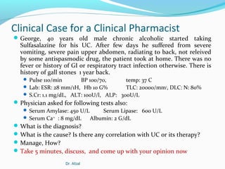
- 1. Clinical Case for a Clinical Pharmacist George, 40 years old male chronic alcoholic started taking Sulfasalazine for his UC. After few days he suffered from severe vomiting, severe pain upper abdomen, radiating to back, not releived by some antispasmodic drug, the patient took at home. There was no fever or history of GI or respiratory tract infection otherwise. There is history of gall stones 1 year back. Pulse 110/min BP 100/70, temp: 37 C Lab: ESR: 28 mm/1H, Hb 10 G% TLC: 20000/mm3, DLC: N: 80% S.Cr: 1.1 mg/dL, ALT: 100U/l, ALP: 300U/L Physician asked for following tests also: Serum Amylase: 450 U/L Serum Lipase: 600 U/L Serum Ca++ : 8 mg/dL Albumin: 2 G/dL What is the diagnosis? What is the cause? Is there any correlation with UC or its therapy? Manage, How? Take 5 minutes, discuss, and come up with your opinion now Dr. Afzal
- 3. ILO’s After completion of this module, student will be able to: Define acute and chronic pancreatitis Describe the epidemiology, etiology, and prognosis of acute pancreatitis., Identify medications with possible, probable, or definite associations with acute pancreatitis. Plan Treatment Goals for Acute Pancreatitis Construct Therapeutic Plan, (with importance of fluid therapy., opioids as analgesics, prophylactic antibiotics) Recommend appropriate nutritional support for patients with acute pancreatitis. Design follow up plan for patient of acute pancreatitis Dr. Afzal
- 4. Dr. Afzal
- 5. Acute Pancreatitis (AP) An inflammatory disorder of the pancreas characterized by severe pain in the upper abdomen Increased serum concentrations of pancreatic lipase and amylase. Complete recovery in most cases Severe AP is associated with local complications Acute fluid collection Pancreatic necrosis, Abscess Pseudo cyst. Exocrine and endocrine pancreatic functions may remain impaired for variable periods after an acute attack, AP rarely progresses to chronic pancreatitis (CP). Dr. Afzal
- 6. Classification Atlanta classification, acute pancreatitis can be divided into two broad categories : Interstitial edematous acute pancreatitis, : acute inflammation of the pancreatic parenchyma and peripancreatic tissues, but without tissue necrosis. Necrotizing acute pancreatitis: inflammation associated with pancreatic parenchymal necrosis and/or peripancreatic necrosis. According to the severity, acute pancreatitis is divided into the following: Mild acute pancreatitis: Absence of organ failure and local or systemic complications Moderately severe acute pancreatitis: No organ failure or transient organ failure (<48 hours) and/or local complications Severe acute pancreatitis: Persistent organ failure (>48 hours) that may involve one or multiple organs Dr. Afzal
- 7. Epidemiology 3% of all cases of abdominal pain admitted to hospital. 40 cases per year per 100,000 adults. [International] Ranges between 5 and 80 per 100,000 population The highest incidence recorded in the United States and Finland In 80% of cases: mild and resolves without serious prob. Sex No predilection exists. Age- 35-64 years Dr. Afzal
- 8. Dr. Afzal
- 9. Causes by demographic The most common causes of pancreatitis, are as follows : Western countries - chronic alcoholism and gallstones accounting for more than 85% of all cases Eastern countries - gallstones Children - trauma Adolescents and young adults - mumps Dr. Afzal
- 10. Etiology: Causes Most common causes Gallstone Ethanol use Unknown (idiopathic pancreatitis). Causes of pancreatitis spells.doc Many medications Medications Associated with Acute Pancreatitis.doc Definite Association Probable Association 5-Aminosalicylic acid, Azathioprine Mercaptopurine Tetracycline Sulfonamides Metronidazole Didanosine Asparaginase Estrogens Furosemide Thiazides Methyldopa Sulindac Valproic acid/salts Octreotide Dr. Afzal Ampicillin ACEI, Losartan Chlorthalidone Ifosfamide Cisplatin Cytarabine Zalcitabine (HIV) Clozapine Corticosteroids Interferon alfa-2b Piroxicam Salicylates Procainamide
- 11. Pathophysiology-1 AP is initiated by: Premature activation of pancreatic zymogens (inactive enzymes) within the acinar cells, Pancreatic ischemia, Pancreatic duct obstruction. Release of active pancreatic enzymes directly causes local or distant tissue damage. Trypsin digests cell membranes and leads to the activation of other pancreatic enzymes. Lipase damages fat cells, producing noxious substances that cause further pancreatic and per pancreatic injury. Release of cytokines injures the acinar cell and enhances the inflammatory response Dr. Afzal
- 12. Pathophysiology-2 Injured acinar cells liberate chemoattractants Attract neutrophils, macrophages, and other cells to the area of inflammation Increase vascular permeability promotes tissue edema. Pancreatic infection may result from colonic bacteria. Complications: Local complications: acute fluid collection, pancreatic necrosis, abscess, pseudo cyst formation, and pancreatic ascites. Systemic complications include cardiovascular, renal, pulmonary, metabolic, hemorrhagic, and central nervous system abnormalities. Dr. Afzal
- 13. Clinical Presentation The initial presentation: PAIN: Moderate abdominal discomfort to excruciating pain, Occurs in 95% of patients and is usually epigastric, often radiating to the upper quadrants or back. The onset is usually sudden Tends to be steady and usually persists for several days Nausea and vomiting occur in 85% of patients and usually follow the onset of pain Shock, Respiratory distress. Clinical signs Marked epigastric tenderness, Abdominal distention, Hypotension, and low-grade fever. In severe disease, Bowel sounds are diminished or absent. Dyspnea and tachypnea are signs of acute respiratory complications. Dr. Afzal
- 14. Physical signs Following finding may benoticed according to severity of the disease Fever (76%) and tachycardia (65%); hypotension Abdominal tenderness, muscular guarding (68%), and distention (65%); diminished or absent bowel sounds Jaundice (28%) Dyspnea (10%); tachypnea; basilar rales, especially in the left lung In severe cases, hemodynamic instability (10%) and hematemesis or melena (5%); pale, diaphoretic, and listless appearance Occasionally, extremity muscular spasm secondary to hypocalcemia Dr. Afzal
- 15. Typical signs if necrosis Cullen sign (bluish discoloration around the umbilicus resulting from hemoperitoneum) Grey-Turner sign (reddish-brown discoloration along the flanks resulting from retroperitoneal blood dissecting along tissue planes); more commonly, patients may have a ruddy erythema in the flanks secondary to extravasated pancreatic exudate Erythematous skin nodules, usually no larger than 1 cm and typically located on extensor skin surfaces; polyarthritis Dr. Afzal
- 16. Grey Turner’s sign Dr. Afzal
- 18. Diagnosis (guidelines 2013) Two of the following: characteristic (severe) abdominal pain, serum amylase and/or lipase exceeding 3 times the upper limit of normal, and/or characteristic abdominal imaging findings (strong recommendation, moderate quality of evidence). Dr. Afzal
- 19. DIAGNOSIS A definitive diagnosis by Surgical examination of the pancreas Pancreatic histology If not possible then recognition of an etiologic factor clinical signs and symptoms abnormal laboratory tests imaging techniques LAB: Increase Serum amylase (20-110 U/L) and Serum lipase (0-160 U/L) concentration more than 3 times the normal upper limits TLC: 10-30,000 Hypocalcaemia (9-11mg/mL) Why there is hypocalcaemia in Acute pancreatitis.doc hyperglycemia, hypoalbuminemia, mild hyperbilirubinemia, and elevations in serum alkaline phosphatase and hepatic transaminases Dr. Afzal
- 20. Labs Serum amylase and lipase Liver-associated enzymes Blood urea nitrogen (BUN), creatine, and electrolytes Blood glucose Serum cholesterol and triglyceride Complete blood count (CBC) and hematocrit; NLR C-reactive protein (CRP) Arterial blood gas values Serum lactic dehydrogenase (LDH) and bicarbonate Immunoglobulin G4 (IgG4 Dr. Afzal
- 21. Diagnosis-2 Contrast-enhanced computed tomography (CT) distinguishes interstitial from necrotizing pancreatitis. Endoscopic retrograde cholangiopancreatography (ERCP) used to visualize and remove bile duct stones in patients with gallstone pancreatitis. Dr. Afzal
- 22. Treatment guidelines Contrast-enhanced computed tomography (CT) scanning and/or magnetic resonance imaging (MRI) of the pancreas should performed only in the absence of clinical improvement Assessment of the patient’s hemodynamic status immediately upon presentation, Patients with systemic inflammatory response syndrome (SIRS) and/or organ failure should, be admitted to an intensive care unit (ICU) or an intermediary care setting Dr. Afzal
- 23. Treatment guidelines All patients should receive aggressive hydration, unless heart or renal disease most effective within the first 12-24 hours, with possibly little benefit derived from its administration after this point Within 24 hours of admission, patients with concurrent acute cholangitis should undergo endoscopic retrograde cholangiopancreatography (ERCP); in high-risk patients, The risk of severe post-ERCP pancreatitis should be reduced through the use of postprocedure rectal nonsteroidal anti-inflammatory drug (NSAID) suppositories and/or pancreatic duct stents Dr. Afzal
- 24. Treatment guidelines The guidelines recommend against routinely using prophylactic antibiotics in cases of severe acute pancreatitis and/or sterile necrosis; however, intervention in patients with infected necrosis may be delayed through the use of antibiotics that penetrate the necrosis In mild cases of acute pancreatitis with no nausea and vomiting, oral feeding can be initiated immediately; Enteral nutrition should be used in severe cases to prevent infectious complications, and parenteral nutrition should be avoided Dr. Afzal
- 25. Treatment guidelines Regardless of lesion size, location, and/or extension, intervention is not necessary for asymptomatic pancreatic and/or extrapancreatic necrosis and/or pseudocysts Surgical, radiologic, and/or endoscopic drainage in stable patients with infected necrosis should be postponed (for 4 weeks if possible) to permit a wall to develop around the necrosis Dr. Afzal
- 26. Who should be treated in ICU APACHE II score >8 in the first 24 hours of admission (calculator 1) Persistent (>48 hours) SIRS Elevated hematocrit (>44 percent), blood urea nitrogen (BUN) (>20 mg/dL), or creatinine (>1.8 mg/dL) Age >60 years Underlying cardiac or pulmonary disease, obesity Dr. Afzal
- 28. SIRS: systemic inflammatory response syndrome Two or more of the following conditions: Temperature >38.3°C or <36.0°C Heart rate of >90 beats/minute Respiratory rate of >20 breaths/minute or PaCO2 of <32 mmHg WBC count of >12,000 cells/mL, <4000 cells/mL, or >10 percent immature (band) forms Dr. Afzal
- 29. Initial Management For severe or mild with vomiting: NPO: to minimize exocrine stimulation of the pancreas. Naso-gastric (NG) aspiration in patients with profound pain, severe disease, paralytic ileus, and severe vomiting Enteral or parenteral nutrition if oral nutrition will be withheld for more than 1 week Fluid Therapy: all patients 5 to 10 mL/kg per hour of isotonic crystalloid solution (eg, normal saline or lactated Ringer’s solution) If dehydration: hypotension and tachycardia 20 mL/kg of intravenous fluid given over 30 minutes followed by 3 mL/kg/hour for 8 to 12 hours If AP is due to hypercalcemia, only NS Stop fluid if goals achieved: heart rate <120 beats/minute, mean arterial pressure between 65 to 85 mmHg), urine output (>0.5 to 1 cc/kg/hour) reduction in hematocrit (goal 35 to 44 percent) and BUN over 24 hours Dr. Afzal
- 30. Pain control Patient controlled analgesia: opioids Fentanyl better, safety profile, especially in renal impairment. Bolus regimen ranges from 20 to 50 micrograms with a 10minute lock-out period (time from the end of one dose infusion to the time the machine starts responding to another demand). Alternate: Parenteral meperidine (50 to 100 mg) every 3 to 4 hours. less spasm of the sphincter of Oddi. not as effective as other opioids , contraindicated in renal failure. Parenteral morphine 10-15 mg iv , cause spasm of the sphincter of Oddi, increase serum amylase and rarely pancreatitis. Hydromorphone, longer half-life than meperidine, parenterally by a patient-controlled analgesia (PCA) pump. Dr. Afzal
- 31. Dr. Afzal
- 32. Antibiotics are not recommended Prophylactic antibiotics 20 percent of patients develop an extra-pancreatic infection bloodstream infections, Pneumonia urinary tract infections Administer antibiotic according to site and C/S report If severe necrotizing AP: Broad-spectrum antibiotics: Start within the first 48 hours and continued for 2 to 3 weeks. IMIPENEM-CILASTATIN (500 mg every 8 hours) may be most effective Ciprofloxacin, Levofloxacin) with metronidazole should be considered for penicillin-allergic patients Antifungal: Prophylactic antifungal therapy not recommended occur in approximately 9 percent of necrotizing pancreatitis. However, it is not clear if they are associated with higher mortality Dr. Afzal
- 33. Miscellaneous Protease inhibitors: anti trypsin DROTRECOGIN ALFA may benefit patients with pancreatitis and systemic inflammatory response syndrome Recombinant form of human activated protein C that has anti- thrombotic, anti-inflammatory, and pro-fibrinolytic properties. Used mainly in intensive care medicine as a treatment for severe sepsis Octreotide, 0.1 mg subcutaneously every 8 hours, decrease sepsis, length of hospital stay, and mortality Morbidity did not differ significantly between the groups. This study did not demonstrate an inhibitory effect of octreotide on exocrine pancreatic secretion. Based on these results, the routine use of octreotide after PD cannot be recommended: HPB (Oxford). 2013 May;15(5):392-9 Insulin if hyperglycemia. surgical intervention in severe necrotizing pancreatitis. Dr. Afzal
- 34. IMPORTANT During the FIRST TWO weeks after a SEVERE ATTACK Intensive critical care (ICU): To support the cardiopulmonary (heart and lung), liver and kidneys that may fail due to RELEASE OF LARGE AMOUNTS TOXINS FROM THE DEAD PANCREAS in the abdomen. Almost all patients require intravenous nutrition. Surgical treatment for severe acute pancreatitis Only in a tertiary medical center by experienced surgeon pancreatitis Dr. Afzal
- 35. Complications in Acute pancreatitis Local complications Pancreatic necrosis -Infected necrosis is almost always fatal without intervention Acute Fluid Collections are common in patients with severe pancreatitis (occurring in 30%-50%). Pancreatic abscess is a collection of pus adjacent to pancreas presenting several months after attack. Acute pseudocyst rupture or haemorrhage in pseudocyst. Pancreatic ascites occurs when a pseudo-cyst collapses into peritoneal cavity or major pancreatic duct breaks down and releases pancreatic juices into peritoneal cavity.
- 36. Complications in Acute pancreatitis Systemic complications Respiratory:Pulmonary oedema/Pleural effusions Consolidation/ARDS Cardiovascular:Hypovolaemia/Shock/arrhythmias Disseminated intravascular coagulopathy (DIC) Renal dysfunction due to hypovolaemia, intra-vascular coagulation. Usually avoided by adequate fluid replacement plus/minus low-dose dopamine but acute tubular or cortical necrosis can follow. GIT: Haemorrhage/Ileus
- 37. Complications in Acute pancreatitis Metabolic: Hypocalcaemia Hypomagnesaemia Hyperglycaemia
- 38. Evaluation of Therapeutic Outcomes Periodic Assessment of: Pain control, Fluid and electrolyte status, and Nutrition should be assessed periodically depending on the degree of abdominal pain and fluid loss. For sever cases in ICU, monitor for: Vital signs, fluid and electrolyte status, white blood cell count, blood glucose, lactate dehydrogenase, aspartate aminotransferase, Serum albumin, hematocrit, blood urea nitrogen, serum creatinine, and international normalized ratio (INR). Arterial blood gas. Serum lipase, amylase, and bilirubin require less frequent monitoring. Signs of infection, relief of abdominal pain, and adequate nutritional status. Dr. Afzal
- 40. Chronic pancreatitis (CP) A syndrome of destructive and inflammatory conditions Characterized by irreversible fibrosis and destruction of exocrine and endocrine tissue Resulting from long-standing pancreatic injury. Variably progressive. Most patients have periods of intractable abdominal pain. Progressive pancreatic insufficiency leads to MALDIGESTION and DIABETES MELLITUS Dr. Afzal
- 41. Pathophysiology Causes: in United States Prolonged ethanol consumption accounts for 70% idiopathic 20% other causes 10%: gall stones, C.F etc Chronic alcohol ingestion,….. intraductal protein plugs that BLOCK SMALL DUCTULES. ………progressive structural damage in the ducts and acinar tissue. Calcium complexes with the protein plugs, ….destruction of pancreatic tissue. Inflammation leads to cellular necrosis, ……..fibrosis Malabsorption of protein and fat due to …decreased lipase, trypsin Reduced bicarbonate secretion : duodenal pH less than 4. Complications Pancreatic pseudocyst, abscess, and ascites or common bile duct obstruction leading to cholangitis or secondary biliary cirrhosis. Dr. Afzal
- 42. Clinicalpain, malabsorption, weight loss, and diabetes. Jaundice occurs Presentation Abdominal in about 10% of patients. PAIN: dull epigastric or abdominal pain radiates to the back. consistent or episodic deep-seated, positional, frequently nocturnal unresponsive to medication Nausea and vomiting often accompany the pain. Severe attacks last from several days to weeks aggravated by eating and relieved by abstinence from alcohol. Steatorrhea (excessive loss of fat in the feces) with diarrhea and bloating Azotorrhea (excessive loss of protein in the feces) are seen in most patients. Weight loss may occur. Diabetes usually a late due to pancreatic calcification. Neuropathy is sometimes seen. Dr. Afzal
- 43. Diagnosis History Heavy ethanol use Attacks of recurrent upper abdominal pain. CLASSIC TRIAD calcification, steatorrhea, and diabetes Malabsorption of Fat can be detected by Sudan staining of the feces or a 72-hour quantitative measurement of fecal fat Imaging techniques, Ultrasound, abdominal CT Surgical biopsy of pancreas is the gold standard for diagnosis of CP. ERCP is the most sensitive and specific diagnostic test, Serum amylase and lipase concentrations usually remain normal unless the pancreatic duct is blocked or a pseudo-cyst is present. The white blood cell count, fluid balance, and electrolyte concentrations usually remain normal unless vomiting and diarrhea. Dr. Afzal
- 44. Treatment Abstinence from alcohol is the most important Small and frequent meals (6 meals/day) and a diet restricted in fat (50 to 75 g/day) PAIN: Acetaminophen or nonsteroidal anti-inflammatory drugs before meals to prevent postprandial exacerbation of pain If not effective tramadol or adding a low-dose opioid (e.g., acetaminophen and codeine) If pain persists, pancreatic enzymes Opioids: oral, if no effect, parentral Modulators of chronic pain (e.g., selective serotonin reuptake inhibitors, tricyclic antidepressants MALABSORPTION: The combination of pancreatic enzymes (lipase, amylase, and protease) 30,000 IU of lipase and 10,000 IU of trypsin ENZYME CONTENT OF SELECTED PANCREATIC ENZYME PREPARATIONS.doc a reduction in dietary fat (to less than 25 g/meal) H2 receptor antagonists and PPI’s Dr. Afzal
- 45. Evaluation of Therapeutic Outcomes Analgesic control: Periodic assessment of: The severity and frequency of abdominal pain The effectiveness of pancreatic enzyme: improvement in body weight and stool consistency or frequency. The 72-hour stool test for fecal fat Serum uric acid and folic acid yearly Blood glucose must be monitored carefully in diabetic patien Dr. Afzal