SlideShare ist ein Scribd-Unternehmen logo
1 von 46
Downloaden Sie, um offline zu lesen
Lecture 3. Movement

Three principal kinds of movement:
  –
      ameboid
  –
      ciliary and flagellar
  –
      muscular
Ameboid Movement
–
    amebas and other unicellular forms
–
    white blood cells
–
    embryonic mesenchyme cells
–
    other mobile cells
Fig. 11.5a
Fig. 11.5c
Consensus model to explain extension and withdrawal
        of pseudopodia and ameboid crawling:


      1. hyaline cap appears
Consensus model to explain extension and withdrawal
        of pseudopodia and ameboid crawling:


      2. endoplasm flows toward hyaline cap
Consensus model to explain extension and withdrawal
        of pseudopodia and ameboid crawling:


     3. actin subunits attach to regulatory proteins
Consensus model to explain extension and withdrawal
        of pseudopodia and ameboid crawling:


      4. endoplasm fountains out to the periphery
Consensus model to explain extension and withdrawal
        of pseudopodia and ameboid crawling:


      5. actin subunits released and polymerized
Consensus model to explain extension and withdrawal
        of pseudopodia and ameboid crawling:


        6. microfilaments cross-linked
Consensus model to explain extension and withdrawal
        of pseudopodia and ameboid crawling:


        7. Ca2+ activate actin-severing protein
Consensus model to explain extension and withdrawal
        of pseudopodia and ameboid crawling:


   8. myosin associate with and pull on microfilaments
Ciliary and Flagellar Movement
Cilia
   –
        minute, hairlike, motile processes
   –
        occur in large numbers
   –
        ciliate protistans
   –
        found in all major groups of animals
   –
        move organisms through aquatic environment
   –
        propel fluids and materials across surfaces
Ciliary and Flagellar Movement
Flagella
  –
      whiplike
  –
      present singly or in small numbers
  –
      occur in unicellular eukaryotes
  –
      animal spermatozoa
  –
      sponges
• both cilia and flagella have the same ultrastructure


  – a core of microtubules sheathed by the plasma
    membrane
• both cilia and flagella have the same ultrastructure


  – “9 + 2” pattern
  – flexible “wheels” of proteins connect outer doublets to
    each other and to the core
• both cilia and flagella have the same ultrastructure

   – outer doublets are
     connected by
     motor proteins

   – anchored in the
     cell by a basal
     body
•
    The bending of cilia and flagella is driven by the
    arms of a motor protein, dynein.
•
    Addition to dynein of a phosphate group from
    ATP and its removal causes conformation changes
    in the protein.

•
    Dynein arms alternately grab, move, and release
    the outer microtubules.
•
    Protein cross-links limit sliding and the force
    is expressed as bending.
•
    A flagellum has an undulatory movement
    –
        force is generated parallel to the flagellum’s axis
•
    Cilia move more like oars with alternating
    power and recovery strokes
    –
        generate force perpendicular to the cilia’s axis
Invertebrate Muscle
Bivalve molluscan muscles
  – 2 kinds of fibers:
   • fast muscle fibers = striated, can contract rapidly
   • smooth muscle = capable of slow, long-lasting
     contractions
Invertebrate Muscle
Insect flight muscles (fibrillar muscle)
  – wings of small flies operate at 1000 beats/sec
  – limited extensibility; shorten only slightly
Vertebrate Muscle

Types
1. Striated
2. Smooth
3. Cardiac
Structure of Striated Muscle
Sliding Filament Model


• Actin filaments at both ends of sarcomere
   – one end of each filament attached to a Z-plate at one end
     of the sarcomere
   – other end suspended in sarcoplasm
Sliding Filament Model


• Myosin filaments suspended in between Z-plates
   – myosin filaments contain cross-bridges which pull the actin filaments
     inward
   – causes Z-plates to move toward each other
   – shortens sarcomere
   – sarcomeres stacked together in series and cause myofiber to shorten
Sliding Filament Model

• Working muscles require ATP
  – myosin breaks down ATP
  – sustained exercise
     • requires cellular respiration
     • regenerates ATP
35


                    Muscle Innervation
     •
         Neuromuscular junction
         –
             the synaptic contact between a nerve fiber and a
             muscle fiber
         –
             nerve impulses bring about the release of a
             neurotransmitter that crosses the synaptic cleft
         –
             signals the muscle fiber to contract
Human Muscular System

•
    Skeletal muscles
    –
        attached to the skeleton by cable-like fibrous
        connective tissue called tendons
    –
        arranged in antagonistic pairs
         •   can only contract, cannot push
         •   when one muscle contracts, it stretches its
             antagonistic partner
•
    a muscle at “rest” exhibits tone (minimal
    contraction)
•
    a muscle in tetany is at maximum sustained
    contraction
43
Muscle Performance
– slow oxidative fibers (red muscles)
 • for slow, sustained contractions without
   fatigue
 • contain extensive blood supply
 • high density of mitochondria
 • abundant stored myoglobin
 • important in maintaining posture in terrestrial
   vertebrates
Muscle Performance
fast fibers
  1. fast glycolytic fiber (white muscles)
    • lacks efficient blood supply
    • pale in color
    • function anaerobically
    • fatigue rapidly
  2. fast oxidative fiber
    • extensive blood supply
    • high density of mitochondria and myoglobin
    • function aerobically
    • for rapid, sustained activities
Energy for Contraction
– ATP, immediate source of energy
– glucose broken down during aerobic metabolism
– glycogen stores can supply glucose
– muscles have creatine phosphate, an energy
  reserve
– slow and fast oxidative fibers rely heavily on
  glucose and oxygen
– fast glycolytic fibers rely on anaerobic glycolysis
– muscles incur oxygen debt during anaerobic
  glycolysis

Weitere ähnliche Inhalte

Was ist angesagt?

Classification of bacteria
Classification of bacteriaClassification of bacteria
Classification of bacteriaMAULIK CHAUDHARI
 
Introduction to immunology
Introduction to immunologyIntroduction to immunology
Introduction to immunologypromotemedical
 
Cell cycle & cell division
Cell cycle & cell divisionCell cycle & cell division
Cell cycle & cell divisionDr Sara Sadiq
 
Digestive system of a frog
Digestive system of a frogDigestive system of a frog
Digestive system of a frogNikki Villarete
 
Immunology
ImmunologyImmunology
Immunologystudent
 
Archeabacteria presentation
Archeabacteria presentationArcheabacteria presentation
Archeabacteria presentationHina Zamir Noori
 
Immunity and its types
Immunity and its typesImmunity and its types
Immunity and its typesSrishtiPatel
 
Primary and Secondary Immune Responses
Primary and Secondary Immune Responses Primary and Secondary Immune Responses
Primary and Secondary Immune Responses AhmedRiyadh17
 
Classification of virus
Classification of virusClassification of virus
Classification of virusAman Ullah
 
Metagenesis-in-Obelia-sp.pptx
Metagenesis-in-Obelia-sp.pptxMetagenesis-in-Obelia-sp.pptx
Metagenesis-in-Obelia-sp.pptxSnigdhaRaniPanda5
 
Cell membrane (Plasma Membrane) & Cellular Junctions
Cell membrane (Plasma Membrane) & Cellular JunctionsCell membrane (Plasma Membrane) & Cellular Junctions
Cell membrane (Plasma Membrane) & Cellular JunctionsPradeep Singh Narwat
 
Plasma membrane -
Plasma membrane -Plasma membrane -
Plasma membrane -Ashok Katta
 
Adaptive immunity
Adaptive immunityAdaptive immunity
Adaptive immunityShanzaay
 
Ch22 Intro to Plants
Ch22 Intro to PlantsCh22 Intro to Plants
Ch22 Intro to Plantsmrsroy
 

Was ist angesagt? (20)

Classification of bacteria
Classification of bacteriaClassification of bacteria
Classification of bacteria
 
Introduction to immunology
Introduction to immunologyIntroduction to immunology
Introduction to immunology
 
Mitochondria structure
Mitochondria structureMitochondria structure
Mitochondria structure
 
Cell cycle & cell division
Cell cycle & cell divisionCell cycle & cell division
Cell cycle & cell division
 
Digestive system of a frog
Digestive system of a frogDigestive system of a frog
Digestive system of a frog
 
Immunology
ImmunologyImmunology
Immunology
 
Archeabacteria presentation
Archeabacteria presentationArcheabacteria presentation
Archeabacteria presentation
 
Fungi
FungiFungi
Fungi
 
12th - the flower - biology
12th - the flower - biology 12th - the flower - biology
12th - the flower - biology
 
Immunity and its types
Immunity and its typesImmunity and its types
Immunity and its types
 
Primary and Secondary Immune Responses
Primary and Secondary Immune Responses Primary and Secondary Immune Responses
Primary and Secondary Immune Responses
 
ZO 211 Week 4 lecture
ZO 211 Week 4 lectureZO 211 Week 4 lecture
ZO 211 Week 4 lecture
 
Classification of virus
Classification of virusClassification of virus
Classification of virus
 
Metagenesis-in-Obelia-sp.pptx
Metagenesis-in-Obelia-sp.pptxMetagenesis-in-Obelia-sp.pptx
Metagenesis-in-Obelia-sp.pptx
 
Classification of Algae
Classification of AlgaeClassification of Algae
Classification of Algae
 
Cell membrane (Plasma Membrane) & Cellular Junctions
Cell membrane (Plasma Membrane) & Cellular JunctionsCell membrane (Plasma Membrane) & Cellular Junctions
Cell membrane (Plasma Membrane) & Cellular Junctions
 
Cytoplasm
CytoplasmCytoplasm
Cytoplasm
 
Plasma membrane -
Plasma membrane -Plasma membrane -
Plasma membrane -
 
Adaptive immunity
Adaptive immunityAdaptive immunity
Adaptive immunity
 
Ch22 Intro to Plants
Ch22 Intro to PlantsCh22 Intro to Plants
Ch22 Intro to Plants
 

Andere mochten auch

Andere mochten auch (20)

Support System in Animals
Support System in AnimalsSupport System in Animals
Support System in Animals
 
Biology 20.2
Biology 20.2Biology 20.2
Biology 20.2
 
Automation
AutomationAutomation
Automation
 
31 Lecture Ppt
31 Lecture Ppt31 Lecture Ppt
31 Lecture Ppt
 
20.Locomotion and Movement
20.Locomotion and  Movement20.Locomotion and  Movement
20.Locomotion and Movement
 
17 Lecture Ppt
17 Lecture Ppt17 Lecture Ppt
17 Lecture Ppt
 
GAS EXCHANGE IN ANIMALS
GAS EXCHANGE IN ANIMALSGAS EXCHANGE IN ANIMALS
GAS EXCHANGE IN ANIMALS
 
Fish locomotion Notes
Fish locomotion NotesFish locomotion Notes
Fish locomotion Notes
 
Animal Digestive System
Animal Digestive SystemAnimal Digestive System
Animal Digestive System
 
29 Lecture Ppt
29 Lecture Ppt29 Lecture Ppt
29 Lecture Ppt
 
Locomotion of cell
Locomotion of cellLocomotion of cell
Locomotion of cell
 
Start here_ch03_lecture
 Start here_ch03_lecture Start here_ch03_lecture
Start here_ch03_lecture
 
Movements/Locomotion in Protists and animals by Saghir Ahmad
Movements/Locomotion in Protists and animals by Saghir AhmadMovements/Locomotion in Protists and animals by Saghir Ahmad
Movements/Locomotion in Protists and animals by Saghir Ahmad
 
How animals move
How animals moveHow animals move
How animals move
 
Flagella
FlagellaFlagella
Flagella
 
32 Lecture Ppt
32 Lecture Ppt32 Lecture Ppt
32 Lecture Ppt
 
04 lecture ppt cell structure and function
04 lecture ppt cell structure and function04 lecture ppt cell structure and function
04 lecture ppt cell structure and function
 
Biology - Chp 20 - Protists - PowerPoint
Biology - Chp 20 - Protists - PowerPointBiology - Chp 20 - Protists - PowerPoint
Biology - Chp 20 - Protists - PowerPoint
 
Animal skeletal system
Animal skeletal systemAnimal skeletal system
Animal skeletal system
 
Cilia and flagella
Cilia and flagellaCilia and flagella
Cilia and flagella
 

Ähnlich wie Lecture 3 movement 2nd sem 2008-2009

Ähnlich wie Lecture 3 movement 2nd sem 2008-2009 (20)

MUSCLE.ppt
MUSCLE.pptMUSCLE.ppt
MUSCLE.ppt
 
A and P Mod5
A and P Mod5A and P Mod5
A and P Mod5
 
MUSCLES (THREE TYPES OF MUSCLES IN THE BODY)
MUSCLES (THREE TYPES OF MUSCLES IN THE BODY) MUSCLES (THREE TYPES OF MUSCLES IN THE BODY)
MUSCLES (THREE TYPES OF MUSCLES IN THE BODY)
 
10 muscular tissue
10 muscular tissue10 muscular tissue
10 muscular tissue
 
Skeletal muscle
Skeletal muscleSkeletal muscle
Skeletal muscle
 
Muscles and Muscle Tissue
Muscles and Muscle TissueMuscles and Muscle Tissue
Muscles and Muscle Tissue
 
Animal Movement.pptx
Animal Movement.pptxAnimal Movement.pptx
Animal Movement.pptx
 
150 ch9 muscle
150 ch9 muscle150 ch9 muscle
150 ch9 muscle
 
skeletal muscle.pdf
skeletal muscle.pdfskeletal muscle.pdf
skeletal muscle.pdf
 
Muscle funccellularlevel animal systems
Muscle funccellularlevel animal systemsMuscle funccellularlevel animal systems
Muscle funccellularlevel animal systems
 
Structure of skeletal muscle
Structure of skeletal muscleStructure of skeletal muscle
Structure of skeletal muscle
 
150 ch9 muscle
150 ch9 muscle150 ch9 muscle
150 ch9 muscle
 
Unit 6 muscular system
Unit 6 muscular systemUnit 6 muscular system
Unit 6 muscular system
 
muscular ppt.pptx
muscular ppt.pptxmuscular ppt.pptx
muscular ppt.pptx
 
3.pdf
3.pdf3.pdf
3.pdf
 
skeletal, cardiac & smooth Muscles by Thiru Murugan.pptx
skeletal, cardiac & smooth Muscles by Thiru Murugan.pptxskeletal, cardiac & smooth Muscles by Thiru Murugan.pptx
skeletal, cardiac & smooth Muscles by Thiru Murugan.pptx
 
Lecture 6.dr hameed alsarraf
Lecture 6.dr hameed alsarrafLecture 6.dr hameed alsarraf
Lecture 6.dr hameed alsarraf
 
skeletal musclemicroemulsions microemulsions microemulsions microemulsions mi...
skeletal musclemicroemulsions microemulsions microemulsions microemulsions mi...skeletal musclemicroemulsions microemulsions microemulsions microemulsions mi...
skeletal musclemicroemulsions microemulsions microemulsions microemulsions mi...
 
Locomotion and movement
Locomotion and movement Locomotion and movement
Locomotion and movement
 
Muscular system
Muscular systemMuscular system
Muscular system
 

Mehr von Jonathan Chan

Lecture 8 regulatory mechanisms part 1
Lecture 8 regulatory mechanisms part 1Lecture 8 regulatory mechanisms part 1
Lecture 8 regulatory mechanisms part 1Jonathan Chan
 
Lecture 8 regulatory mechanisms part 1
Lecture 8 regulatory mechanisms part 1Lecture 8 regulatory mechanisms part 1
Lecture 8 regulatory mechanisms part 1Jonathan Chan
 
Lecture 6 transport circulation_part 1
Lecture 6 transport circulation_part 1Lecture 6 transport circulation_part 1
Lecture 6 transport circulation_part 1Jonathan Chan
 
Lecture 7 excretion and osmoregulation
Lecture 7 excretion and osmoregulationLecture 7 excretion and osmoregulation
Lecture 7 excretion and osmoregulationJonathan Chan
 
Lecture 6 transport circulation_part 2
Lecture 6 transport circulation_part 2Lecture 6 transport circulation_part 2
Lecture 6 transport circulation_part 2Jonathan Chan
 
Lecture 8 regulatory mechanisms part 2
Lecture 8 regulatory mechanisms part 2Lecture 8 regulatory mechanisms part 2
Lecture 8 regulatory mechanisms part 2Jonathan Chan
 
Lecture 4 digestion and nutrition 2nd sem 2008-2009
Lecture 4 digestion and nutrition 2nd sem 2008-2009Lecture 4 digestion and nutrition 2nd sem 2008-2009
Lecture 4 digestion and nutrition 2nd sem 2008-2009Jonathan Chan
 
Lecture 2 support and protection 2nd sem 2008-2009
Lecture 2 support and protection 2nd sem 2008-2009Lecture 2 support and protection 2nd sem 2008-2009
Lecture 2 support and protection 2nd sem 2008-2009Jonathan Chan
 
Lecture 1 animal cell types and tissues
Lecture 1 animal cell types and tissuesLecture 1 animal cell types and tissues
Lecture 1 animal cell types and tissuesJonathan Chan
 
Lecture 5 gas exchange
Lecture 5 gas exchangeLecture 5 gas exchange
Lecture 5 gas exchangeJonathan Chan
 
Bio 11 third_exam_post_labs
Bio 11 third_exam_post_labsBio 11 third_exam_post_labs
Bio 11 third_exam_post_labsJonathan Chan
 

Mehr von Jonathan Chan (12)

Surprise!
Surprise!Surprise!
Surprise!
 
Lecture 8 regulatory mechanisms part 1
Lecture 8 regulatory mechanisms part 1Lecture 8 regulatory mechanisms part 1
Lecture 8 regulatory mechanisms part 1
 
Lecture 8 regulatory mechanisms part 1
Lecture 8 regulatory mechanisms part 1Lecture 8 regulatory mechanisms part 1
Lecture 8 regulatory mechanisms part 1
 
Lecture 6 transport circulation_part 1
Lecture 6 transport circulation_part 1Lecture 6 transport circulation_part 1
Lecture 6 transport circulation_part 1
 
Lecture 7 excretion and osmoregulation
Lecture 7 excretion and osmoregulationLecture 7 excretion and osmoregulation
Lecture 7 excretion and osmoregulation
 
Lecture 6 transport circulation_part 2
Lecture 6 transport circulation_part 2Lecture 6 transport circulation_part 2
Lecture 6 transport circulation_part 2
 
Lecture 8 regulatory mechanisms part 2
Lecture 8 regulatory mechanisms part 2Lecture 8 regulatory mechanisms part 2
Lecture 8 regulatory mechanisms part 2
 
Lecture 4 digestion and nutrition 2nd sem 2008-2009
Lecture 4 digestion and nutrition 2nd sem 2008-2009Lecture 4 digestion and nutrition 2nd sem 2008-2009
Lecture 4 digestion and nutrition 2nd sem 2008-2009
 
Lecture 2 support and protection 2nd sem 2008-2009
Lecture 2 support and protection 2nd sem 2008-2009Lecture 2 support and protection 2nd sem 2008-2009
Lecture 2 support and protection 2nd sem 2008-2009
 
Lecture 1 animal cell types and tissues
Lecture 1 animal cell types and tissuesLecture 1 animal cell types and tissues
Lecture 1 animal cell types and tissues
 
Lecture 5 gas exchange
Lecture 5 gas exchangeLecture 5 gas exchange
Lecture 5 gas exchange
 
Bio 11 third_exam_post_labs
Bio 11 third_exam_post_labsBio 11 third_exam_post_labs
Bio 11 third_exam_post_labs
 

Lecture 3 movement 2nd sem 2008-2009

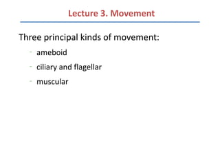
  • 1. Lecture 3. Movement Three principal kinds of movement: – ameboid – ciliary and flagellar – muscular
  • 2. Ameboid Movement – amebas and other unicellular forms – white blood cells – embryonic mesenchyme cells – other mobile cells
  • 3.
  • 6. Consensus model to explain extension and withdrawal of pseudopodia and ameboid crawling: 1. hyaline cap appears
  • 7. Consensus model to explain extension and withdrawal of pseudopodia and ameboid crawling: 2. endoplasm flows toward hyaline cap
  • 8. Consensus model to explain extension and withdrawal of pseudopodia and ameboid crawling: 3. actin subunits attach to regulatory proteins
  • 9. Consensus model to explain extension and withdrawal of pseudopodia and ameboid crawling: 4. endoplasm fountains out to the periphery
  • 10. Consensus model to explain extension and withdrawal of pseudopodia and ameboid crawling: 5. actin subunits released and polymerized
  • 11. Consensus model to explain extension and withdrawal of pseudopodia and ameboid crawling: 6. microfilaments cross-linked
  • 12. Consensus model to explain extension and withdrawal of pseudopodia and ameboid crawling: 7. Ca2+ activate actin-severing protein
  • 13. Consensus model to explain extension and withdrawal of pseudopodia and ameboid crawling: 8. myosin associate with and pull on microfilaments
  • 14. Ciliary and Flagellar Movement Cilia – minute, hairlike, motile processes – occur in large numbers – ciliate protistans – found in all major groups of animals – move organisms through aquatic environment – propel fluids and materials across surfaces
  • 15.
  • 16. Ciliary and Flagellar Movement Flagella – whiplike – present singly or in small numbers – occur in unicellular eukaryotes – animal spermatozoa – sponges
  • 17. • both cilia and flagella have the same ultrastructure – a core of microtubules sheathed by the plasma membrane
  • 18. • both cilia and flagella have the same ultrastructure – “9 + 2” pattern – flexible “wheels” of proteins connect outer doublets to each other and to the core
  • 19. • both cilia and flagella have the same ultrastructure – outer doublets are connected by motor proteins – anchored in the cell by a basal body
  • 20. The bending of cilia and flagella is driven by the arms of a motor protein, dynein.
  • 21. Addition to dynein of a phosphate group from ATP and its removal causes conformation changes in the protein. • Dynein arms alternately grab, move, and release the outer microtubules.
  • 22. Protein cross-links limit sliding and the force is expressed as bending.
  • 23. A flagellum has an undulatory movement – force is generated parallel to the flagellum’s axis
  • 24. Cilia move more like oars with alternating power and recovery strokes – generate force perpendicular to the cilia’s axis
  • 25. Invertebrate Muscle Bivalve molluscan muscles – 2 kinds of fibers: • fast muscle fibers = striated, can contract rapidly • smooth muscle = capable of slow, long-lasting contractions
  • 26.
  • 27. Invertebrate Muscle Insect flight muscles (fibrillar muscle) – wings of small flies operate at 1000 beats/sec – limited extensibility; shorten only slightly
  • 28.
  • 31.
  • 32. Sliding Filament Model • Actin filaments at both ends of sarcomere – one end of each filament attached to a Z-plate at one end of the sarcomere – other end suspended in sarcoplasm
  • 33. Sliding Filament Model • Myosin filaments suspended in between Z-plates – myosin filaments contain cross-bridges which pull the actin filaments inward – causes Z-plates to move toward each other – shortens sarcomere – sarcomeres stacked together in series and cause myofiber to shorten
  • 34. Sliding Filament Model • Working muscles require ATP – myosin breaks down ATP – sustained exercise • requires cellular respiration • regenerates ATP
  • 35. 35 Muscle Innervation • Neuromuscular junction – the synaptic contact between a nerve fiber and a muscle fiber – nerve impulses bring about the release of a neurotransmitter that crosses the synaptic cleft – signals the muscle fiber to contract
  • 36.
  • 37.
  • 38.
  • 39.
  • 40.
  • 41. Human Muscular System • Skeletal muscles – attached to the skeleton by cable-like fibrous connective tissue called tendons – arranged in antagonistic pairs • can only contract, cannot push • when one muscle contracts, it stretches its antagonistic partner • a muscle at “rest” exhibits tone (minimal contraction) • a muscle in tetany is at maximum sustained contraction
  • 42.
  • 43. 43
  • 44. Muscle Performance – slow oxidative fibers (red muscles) • for slow, sustained contractions without fatigue • contain extensive blood supply • high density of mitochondria • abundant stored myoglobin • important in maintaining posture in terrestrial vertebrates
  • 45. Muscle Performance fast fibers 1. fast glycolytic fiber (white muscles) • lacks efficient blood supply • pale in color • function anaerobically • fatigue rapidly 2. fast oxidative fiber • extensive blood supply • high density of mitochondria and myoglobin • function aerobically • for rapid, sustained activities
  • 46. Energy for Contraction – ATP, immediate source of energy – glucose broken down during aerobic metabolism – glycogen stores can supply glucose – muscles have creatine phosphate, an energy reserve – slow and fast oxidative fibers rely heavily on glucose and oxygen – fast glycolytic fibers rely on anaerobic glycolysis – muscles incur oxygen debt during anaerobic glycolysis