SlideShare ist ein Scribd-Unternehmen logo
1 von 55
Radiological imaging of upper limb ischemia.
Dr/ ABD ALLAH NAZEER. MD.
Introduction:
Upper limb ischemia is an uncommon entity which presents less
commonly as compared to lower limb ischaemia, and has a wide
range of etiology and controversies in preoperative investigations
and management. Less than 2% of patients with upper limb ischaemia
presents with rest pain, gangrene or ulcer. Although upper limb loss
is less as compared to lower limb, it can result in severe functional
impairment and disability if there should be any delay in diagnosis
and treatment.
The single most common etiology is thromboembolic events secondary
to atherosclerosis followed by trauma.
Nevertheless, differential diagnoses such as arteritis, thoracic outlet
obstruction and aneurysms need to be considered.
The signs and symptoms of presentation ranged from nonspecific
slight numbness and weakness to pain, pallor and pulselessness. The
most common combination of presenting complaints was cold,
pulselessness and painful upper limb.
Upper extremity occlusive disease is relatively uncommon and
accounts for only 5% of all extremity ischemia. Unlike occlusive
disease in the lower extremities, which is generally atherosclerotic
in origin, the etiology of upper extremity occlusive disease is more
varied and can include atherosclerotic disease, arteritis, connective
tissue disorders, occupational injuries, and Vasospastic diseases.
Etiology
Upper extremity occlusive disease can involve the large and small
arteries of the arm or a combination. Atherosclerosis is the most
common cause of large artery disease and generally involves the
subclavian artery, but disease may extend more distally, especially in
the setting of diabetes. Patient risk factors are similar to those of other
vascular beds. Tobacco use is almost universal. Hypercholesterolemia,
hypertension, diabetes and age are also important risk factors.
Inflammatory diseases (giant cell and Takayasu’s arteritis) can also
involve the large vessels. Small vessel disease due to diabetes can lead
to severe complications such as digital ulceration and gangrene.
Raynaud’s disease is a Vasospastic disorder that produces a tri-phasic
colour change in the hands or less commonly the feet, in response to cold or
emotional stimuli. The hallmark of this disorder is pallor, cyanosis and post
ischemic hyperemia of a digit or digits, although all three may not always be
present. The disorder is classified as primary when no identifiable cause is
evident or secondary when it is due to either a more proximal occlusion or
other disease process resulting in digital artery vasospasm . Patients whose
occupations involve the use of vibrational tools or who use their hands as a
hammer (especially the hypothenar eminence) can present with Raynaud’s or
frank digital ischemia from either hand-arm vibration syndrome or
hypothenar hammer syndrome. Occlusive lesions in the arm and hand
secondary to scleroderma, rheumatoid arthritis, systemic lupus and other
autoimmune disorders can lead to severe upper extremity ischemia
manifesting as pain, ulceration and gangrene. Thromboangiitis obliterans
(Buerger’s disease) is an inflammatory disease that can produce pain and
ulceration in the fingers and toes. The patient is usually a young man with
a long history of tobacco use.
Rare causes of upper extremity occlusive disease include iatrogenic injury,
trauma, malignancy and frostbite.
Common Conditions Associated with Upper Extremity Ischemia:
• Thromboembolism
• Atheromatous Embolization
• Atherosclerosis
• Connective Tissue Diseases( • Scleroderma, • Rheumatoid Arthritis,
• Sjörgren’s Syndrome, • Systemic Lupus Erythematosus)
• Large Artery Vasculitis( • Takayasu’s Disease, • Giant Cell Arteritis)
• Small Artery Vasculitis(• Thromboangitis Obliterans (Buerger’s
disease).
• Blood Dyscrasias
• Myeloproliferative Disorders
• Thoracic Outlet Syndrome
• Latrogenic Injury
• Occupational Exposure(• Hand-arm Vibration Syndrome,
• Hypothenar Hammer Syndrome).
• Vasospasm( • Raynaud’s Disease, • Cocaine Use, • Amphetamine Use).
Acute ischemia in the upper extremity
constitutes 10–15% of all acute extremity ischemia. The etiology
is emboli in 90% of the patients. The reason for this higher rate
compared with the leg is that atherosclerosis is less common in
arm arteries. Emboli have the same origins as in the lower
extremity and usually end up obstructing the brachial artery.
Sometimes plaques or an aneurysm in the subclavian or axillary
arteries is the primary source of emboli. Embolization to the
right arm is more common than to the left due to the vascular
anatomy. For the 10% of patients with atherosclerosis and acute
thrombosis as the main cause for their arm ischemia, the primary
lesions are located in the brachiocephalic trunk or in the
subclavian artery. Such pathologies are usually asymptomatic
due to well-developed collaterals around the shoulder joint until
thrombosis occurs, and they cause either micro- or
macroembolization.
Clinical Presentation
Acute arm ischemia is usually apparent on the basis of
the physical examination. The symptoms are often relatively
discreet, especially early after onset. The explanation for this is the
well developed collateral system circumventing the brachial artery
around the elbow, which is the most common site for embolic
obstruction. The "six Ps” – pain, pallor, paresthesia, paralysis,
pulselessness, poikilothermia– are applicable also for acute arm
ischemia, but coldness and color changes are more prominent than
for the legs. Accordingly, the most common findings in the physical
examination are a cold arm with diminished strength and disturbed
hand and finger motor functions. Tingling and numbness are also
frequent. The radial pulse is usually absent but is pounding in the
upper arm proximal to the obstruction. Gangrene and rest pain
appear only when the obstruction is distal to the elbow and affects
both of the paired arteries in a finger or in the lower arm. Ischemic
signs or symptoms suggesting acute digital artery occlusion in
only one or two fingers, imply microembolization.
Chronic symptomatic ischemia in the upper extremity
is the result of blood flow that is nutritionally inadequate to fulfill
metabolic requirements. Symptoms may also result from
inappropriate thermoregulatory function. Ischemia may be
secondary to inappropriate vessel response when vessels are unable
to vasodilate in response to stress or inappropriate vasoconstriction
(i.e., vasospastic disease); intima/media injury resulting in
aneurysm, embolism, and thrombosis (occlusive disease); or a
combination of both (vaso-occlusive disease). Occlusive events may
occur secondary to low-flow states in vasospastic disease that
precipitate thrombosis development (i.e., secondary occlusion) or
secondary to ischemia from occlusive disease that may produce
autonomic dysfunction and secondary vasospasm. This cross-over
phenomenon creates difficulty in evaluation and management. The
goal of this article is to assess contemporary methods of evaluation,
classification, and management of upper extremity ischemia.
Radiological Imaging:
X-Ray to show any evidence of cervical rib.
Duplex ultrasound is rapid, can be performed at the bedside, and has near
100% sensitivity for diagnosing complete arterial occlusion. Ankle-brachial
index (ABI) will be near zero for patients with acute limb ischemia.
CTA has the benefit of rapid availability and high-quality imaging, which
allows for precise planning of revascularization. CTA provides imaging of the
entire arterial tree from the aortic inflow to the digital level. CTA typically
requires 150 mL of iodinated contrast and therefore has to be used with
caution in patients with baseline renal insufficiency (glomerular filtration rate
<40). For patients with renal insufficiency, aggressive hydration before and
after examination with sodium bicarbonate is recommended for CTA or
invasive angiography.
MRA has a limited role in acute limb ischemia, as the examination can be
lengthy (45–60 minutes), is less often available outside regular work hours,
and generally has poorer arterial imaging than 64 (or greater) slice CTA.
Invasive angiography has the advantage of allowing for simultaneous
percutaneous revascularization with both mechanical thrombectomy and
thrombolytic therapy.
Acute Upper Limb Ischemia with right
subclavian and brachial thrombus.
Upper limb acute ischemia with recent jugular vein thrombosis.
With significant external compression with the probe, a visualized
thrombus (blue arrow) is seen within a non-compressible brachial artery
The distal (right side subclavian shows no flow.
Thrombosis of the brachial artery.
Vessels and nerve trauma. Biker’s hammer. (A) Longitudinal and (B) axial colour Doppler
US obtained over the palmar aspect of the hypothenar region. Images show a nearly
complete thrombosis (black arrowheads) of the superficial branch of the ulnar artery.
Some small persistent internal flow signal can be detected (white arrowheads). Note
increase in size of the artery and thickening of its wall. HH=hook of the hamate.
Left Cervical rib with upper limb chronic ischemia.
CT angiography using modern MDCT scanners has evolved into a highly accurate
noninvasive diagnostic tool for the evaluation of patients with pathologic
abnormalities of the upper extremity arterial system. upper extremity CTA can
effectively evaluate for stenosis, occlusion, aneurysm, or embolic events,
especially when they affect vessels proximal to the wrist.
Atherosclerotic disease affects the upper extremities disproportionately less
than the lower extremities. However, proximal involvement of the upper
extremities is not infrequent. Risk factors are no different than those for other
sites of atherosclerotic involvement and include hypertension, dyslipidemia,
diabetes mellitus, age, and a history of smoking . Claudication is the most
common presenting symptom, though most patients are asymptomatic because
of collateral formation. Blood pressure differences between the upper extremities
can frequently be found in these patients. Symptoms are often chronic, with more
acute symptoms suggesting acute thrombosis or an embolic event. In the setting
of subclavian involvement, upper extremity symptoms may be masked or
overshadowed by symptoms related to compromised flow to the ipsilateral
vertebral artery due to subclavian steal, particularly syncope and presyncope. In
patients with left or right internal mammary bypass grafts, subclavian artery
disease can cause myocardial ischemia. Atherosclerotic disease, when identified
on imaging, should prompt inquiry into concomitant sites of involvement
processes—namely, cardiovascular and cerebrovascular involvement.
CT Angiography of the Upper Extremity Arterial System:
Axial subvolume maximum intensity
projection better outlines stenosis
(arrowheads). Axillofemoral bypass
(asterisk) is depicted end-on.
Volume-rendered image shows
axillofemoral bypass graft (arrow) with
stenosis proximal and distal to anastomosis
in native left axillary artery (arrowheads).
Volume-rendered CT angiography shows long occlusion of
brachial artery, which was caused by acute thromboembolism.
Old woman with acute type A thoracic aortic dissection who presented with acute
chest pain and syncope. Physical examination revealed weak right upper extremity
pulse. CT angiography of right upper extremity, chest, abdomen, and pelvis was
performed to evaluate for aortic dissection and upper extremity arterial pathology.
Oblique multiplanar reconstruction image reveals ascending aortic dissection, with
intimal flap (arrowheads) covering origins of all supraaortic branch vessels.
49-year-old man in chronic phase of Takayasu
disease involving arch and supraaortic
vessels. A, Volume-rendered image shows
right subclavian artery aneurysm (arrow) and
left subclavian artery stent (arrowhead)
implanted for steno-occlusive disease.
49-year-old man in chronic phase of
Takayasu disease involving arch and
supraaortic vessels. B, Axial image shows
concentric thickening (arrowheads) of wall
of origins of all three supraaortic branches.
64-year-old woman with giant cell arteritis who presented with fever,
malaise, and bilateral upper extremity weakness. CT angiography using
axial curved multiplanar reconstruction algorithm shows typical irregular
wall thickening (arrowheads) of bilateral axillary and subclavian arteries.
29-year-old male heavy
smoker with thromboangiitis
obliterans who presented with
ischemic symptoms of both
upper and lower extremities.
A, Upper extremity CT
angiography (CTA) shows
distal ulnar artery occlusion
(short thick arrow) and distal
radial artery contour
irregularity (arrowheads) in
right upper extremity. Note
typical corkscrew collaterals
(long thin arrow).
Stenoocclusive disease was
also present in left ulnar and
interosseous arteries.
50-year-old woman with
history of embolic disease
to left arm and
fibromuscular dysplasia
involving bilateral carotid,
renal, and external iliac
arteries (not shown) who
also had involvement of
left brachial artery with
medial fibrosis, which
shows typical “string-of-
beads” sign (arrowheads).
17-year-old girl with thoracic
outlet syndrome who was
evaluated at outside institution
for embolic event to left hand.
She was found to have
aneurysm of left subclavian
artery, which was subsequently
treated with covered stent.
Several months after
procedure, patient presented
to our institution with
heaviness and coolness of left
arm and hand. A, Volume-
rendered image shows
subclavian artery stent, with
proximal occlusion
(arrowhead) and presence of
left cervical rib (single
asterisk). First rib is marked
with double asterisks. Clavicle
was excluded for clarity.
17-year-old girl with thoracic outlet syndrome who was evaluated at outside institution for
embolic event to left hand. She was found to have aneurysm of left subclavian artery, which was
subsequently treated with covered stent. Several months after procedure, patient presented to
our institution with heaviness and coolness of left arm and hand. B, Curved multiplanar
reconstruction of left subclavian artery shows completely thrombosed stent (asterisks). Patient
underwent subclavian artery vein graft placement and resection of left cervical and first ribs.
Volume-rendered image shows vein graft (asterisks) and resection of left cervical and first ribs.
Curved multiplanar reconstruction confirms vein graft patency (asterisks).
23-year-old man with arteriovenous malformation in right axilla. B, Maximum
intensity projection of upper extremity CT angiography shows malformation
(arrowhead), with lateral thoracic artery as main feeder, and early venous
drainage, Volume-rendered image shows arteriovenous malformation and
anomalous high origin of radial artery (arrowhead), which is normal variant.
17-year-old boy with known diagnosis of Ehlers-Danlos syndrome type 4 who presented
to emergency department with extremely painful acute onset swelling of right axilla
and Pulseless right upper extremity. A, CT of chest and upper extremity was performed.
Unenhanced CT shows large right axillary hematoma (asterisk). B, Maximum intensity
projection CT angiography shows extravasation from spontaneously ruptured right
axillary artery (arrow). Emergent proximal surgical ligation was performed.
Angiogram of the right-
upper extremity. (A)
Axillary artery occlusion
caused by a distal
segment. (B) The injured
artery and the extensive
collateralization.
23-year-old man with dialysis fistula who presented with reduced flows while undergoing
hemodialysis and ischemic symptoms in fingers of left hand. B, Subvolume maximum
intensity projection (MIP) shows patent arterial anastomosis but diseased radial artery (A)
with occlusion distally (arrowheads). There was also stenosis in vein (V) immediately distal
to anastomosis (arrow). A, Upper extremity CT angiography volume-rendered view shows
dialysis fistula with aneurysm formation (asterisk) on venous side.
23-year-old man with dialysis fistula who presented with reduced flows while
undergoing hemodialysis and ischemic symptoms in fingers of left hand. More
central subvolume MIP shows critical stenosis of left subclavian vein (arrow).
59-year-old woman who underwent stent placement in left subclavian artery at outside
institution presented with recurrent left upper extremity claudication. CT angiography
shows stent fracture (arrow) and severe stenosis (arrowheads) due to intimal hyperplasia.
Completion angiogram of the right upper limb revealing the axillary–
brachial–ulnar axis and the partially-thrombosed aberrant radial artery.
Chest roentgenogram demonstrates
the clavicular abnormality.
Arch aortogram demonstrates a filling
defect in the right subclavian artery and
occlusion of the axillary artery.
Venogram shows occlusion of the right
subclavian vein extending to the first
rib, with multiple collateral vessels.
An angiogram in a 35-year-old woman with
right arm ischemia that demonstrates right
subclavian artery occlusion from the medial
margin of the first rib to the axillary artery at
the level of the humeral head.
An arteriogram of the right shoulder
in neutral position, demonstrating
right subclavian stenosis (arrow).
An arteriogram of the right upper extremity in
abduction, demonstrating increased subclavian
artery stenosis and aneurysmal dilation (arrow)
in the location of the clavicle and first rib.
Thoracic outlet syndrome with stenosis of the left subclavian vein.
73-year-old woman
with acute embolus
in left axillary artery .
Left axillary artery occlusion in a patient with Wegener's granulomatosis .
Left brachial artery thrombosis in a patient with Wegener's granulomatosis .
Angiogram of upper extremity. The top is in a normal position. The bottom
is in a hyperabducted position (arrow indicates area of stenosis).
Distal axillo-brachial artery occlusion.
Septic embolization from severe prosthetic aortic valve endocarditis
resulted in a 2.5cm brachial artery pseudoaneurysm (circled) that
was associated with a pocket of purulence and upper limb ischemia.
Angiogram that demonstrates blockage of the ulnar artery.
Axial CT angiography source image shows ulnar artery thrombosis with ulnar artery
aneurysm (between arrowheads). Ulnar artery reconstruction with right leg saphenous
vein autograft was performed, and patient became symptom free. Volume-rendered
reconstruction shows occluded ulnar artery (between arrowheads) at Guyon canal.
Angiogram of end-stage Raynaud’s disease
with extremely poor peripheral circulation.
Angiogram that demonstrates
an ulnar artery aneurysm.
An angiogram demonstrating lack of blood flow to vessels
of the hand in Buerger's disease with ischemia to the fingers.
Angiogram of a patient with Buerger’s disease that
shows decreased circulation of index and thumb.
Angiogram that shows decreased
circulation in the ulnar digits of the hand.
Angiogram of right upper extremity with "string of beads" appearance of the
brachial artery (lower left) in fibromuscular dysplasia with chronic ischemia.
Conclusions.
Upper limb ischaemia remains infrequently seen
compared to lower limb ischaemia. Nevertheless,
the complications and late effects can result in
significant impairment in function.
Evaluation of upper limb ischaemia requires
careful history taking and clinical examinations.
Although angiography remains the gold standard,
other non-invasive tools such as duplex US.
Prompt surgical intervention results in favorable
outcomes.
Thank You.

Weitere ähnliche Inhalte

Was ist angesagt?

Sinogram and fistulogram
Sinogram and fistulogramSinogram and fistulogram
Sinogram and fistulogramInosRagan
 
radiological anatomy of retroperitoneum powerpoint
radiological anatomy of  retroperitoneum powerpointradiological anatomy of  retroperitoneum powerpoint
radiological anatomy of retroperitoneum powerpointDactarAdhikari
 
CT perfusion physics and its application in Neuroimaging
CT perfusion physics and its application in NeuroimagingCT perfusion physics and its application in Neuroimaging
CT perfusion physics and its application in NeuroimagingDr.Suhas Basavaiah
 
Presentation1.pptx, normal spinal anatomy.
Presentation1.pptx, normal spinal anatomy.Presentation1.pptx, normal spinal anatomy.
Presentation1.pptx, normal spinal anatomy.Abdellah Nazeer
 
Retroperitoneal masses radiology
Retroperitoneal masses radiologyRetroperitoneal masses radiology
Retroperitoneal masses radiologyDr. Mohit Goel
 
Presentation1, mri imaging of the prostate
Presentation1, mri imaging of the prostatePresentation1, mri imaging of the prostate
Presentation1, mri imaging of the prostateAbdellah Nazeer
 
Imaging in Skull base
Imaging in Skull baseImaging in Skull base
Imaging in Skull baseRakesh Ca
 
Larynx anatomy CT and MRI
Larynx anatomy CT and MRILarynx anatomy CT and MRI
Larynx anatomy CT and MRIDr. Mohit Goel
 
Imaging of portal hypertension
Imaging of portal hypertensionImaging of portal hypertension
Imaging of portal hypertensionDev Lakhera
 
Presentation1.pptx, ultrasound examination of the shoulder joint.
Presentation1.pptx, ultrasound examination of the shoulder joint.Presentation1.pptx, ultrasound examination of the shoulder joint.
Presentation1.pptx, ultrasound examination of the shoulder joint.Abdellah Nazeer
 
Presentation1.pptx, radiological imaging of lower limb ischemia.
Presentation1.pptx, radiological imaging of lower limb ischemia.Presentation1.pptx, radiological imaging of lower limb ischemia.
Presentation1.pptx, radiological imaging of lower limb ischemia.Abdellah Nazeer
 
Diagnostic Imaging of Mediastinal Masses
Diagnostic Imaging of Mediastinal MassesDiagnostic Imaging of Mediastinal Masses
Diagnostic Imaging of Mediastinal MassesMohamed M.A. Zaitoun
 
Anatomy and imaging of wrist joint (MRI AND XRAY)
Anatomy and imaging of wrist joint (MRI AND XRAY)Anatomy and imaging of wrist joint (MRI AND XRAY)
Anatomy and imaging of wrist joint (MRI AND XRAY)Kajal Jha
 

Was ist angesagt? (20)

Lines & mediastinal stripes 02
Lines & mediastinal stripes 02Lines & mediastinal stripes 02
Lines & mediastinal stripes 02
 
Sinogram and fistulogram
Sinogram and fistulogramSinogram and fistulogram
Sinogram and fistulogram
 
CT Angiography Lower Limb
CT Angiography Lower LimbCT Angiography Lower Limb
CT Angiography Lower Limb
 
radiological anatomy of retroperitoneum powerpoint
radiological anatomy of  retroperitoneum powerpointradiological anatomy of  retroperitoneum powerpoint
radiological anatomy of retroperitoneum powerpoint
 
CT perfusion physics and its application in Neuroimaging
CT perfusion physics and its application in NeuroimagingCT perfusion physics and its application in Neuroimaging
CT perfusion physics and its application in Neuroimaging
 
X ray of wrist and hand
X ray of wrist and handX ray of wrist and hand
X ray of wrist and hand
 
Radiology Spotters
Radiology Spotters Radiology Spotters
Radiology Spotters
 
Presentation1.pptx, normal spinal anatomy.
Presentation1.pptx, normal spinal anatomy.Presentation1.pptx, normal spinal anatomy.
Presentation1.pptx, normal spinal anatomy.
 
Retroperitoneal masses radiology
Retroperitoneal masses radiologyRetroperitoneal masses radiology
Retroperitoneal masses radiology
 
Presentation1, mri imaging of the prostate
Presentation1, mri imaging of the prostatePresentation1, mri imaging of the prostate
Presentation1, mri imaging of the prostate
 
Imaging in Skull base
Imaging in Skull baseImaging in Skull base
Imaging in Skull base
 
Larynx anatomy CT and MRI
Larynx anatomy CT and MRILarynx anatomy CT and MRI
Larynx anatomy CT and MRI
 
Mri anatomy of ankle
Mri anatomy of ankleMri anatomy of ankle
Mri anatomy of ankle
 
Imaging of portal hypertension
Imaging of portal hypertensionImaging of portal hypertension
Imaging of portal hypertension
 
Gastric carcinoma radiology ppt
Gastric carcinoma radiology  ppt Gastric carcinoma radiology  ppt
Gastric carcinoma radiology ppt
 
Presentation1.pptx, ultrasound examination of the shoulder joint.
Presentation1.pptx, ultrasound examination of the shoulder joint.Presentation1.pptx, ultrasound examination of the shoulder joint.
Presentation1.pptx, ultrasound examination of the shoulder joint.
 
Presentation1.pptx, radiological imaging of lower limb ischemia.
Presentation1.pptx, radiological imaging of lower limb ischemia.Presentation1.pptx, radiological imaging of lower limb ischemia.
Presentation1.pptx, radiological imaging of lower limb ischemia.
 
Diagnostic Imaging of Mediastinal Masses
Diagnostic Imaging of Mediastinal MassesDiagnostic Imaging of Mediastinal Masses
Diagnostic Imaging of Mediastinal Masses
 
Anatomy and imaging of wrist joint (MRI AND XRAY)
Anatomy and imaging of wrist joint (MRI AND XRAY)Anatomy and imaging of wrist joint (MRI AND XRAY)
Anatomy and imaging of wrist joint (MRI AND XRAY)
 
Liver ultrasound
Liver ultrasoundLiver ultrasound
Liver ultrasound
 

Andere mochten auch

MRI Tissue Segmentation using MICO
MRI Tissue Segmentation using MICOMRI Tissue Segmentation using MICO
MRI Tissue Segmentation using MICOPooja G N
 
Chronic limb ischemia by Muhamad Fathy (MD)
Chronic limb ischemia by Muhamad Fathy (MD)Chronic limb ischemia by Muhamad Fathy (MD)
Chronic limb ischemia by Muhamad Fathy (MD)Muhamad Zaidan
 
Pancreatic cystic lesion by xiu
Pancreatic cystic lesion by xiuPancreatic cystic lesion by xiu
Pancreatic cystic lesion by xiuXiu Srithammasit
 
Rome Han Comparison
Rome Han ComparisonRome Han Comparison
Rome Han ComparisonDan McDowell
 
Presentation1.pptx, radiological anatomy of the lower limb anatomy.
Presentation1.pptx, radiological anatomy of the lower limb anatomy.Presentation1.pptx, radiological anatomy of the lower limb anatomy.
Presentation1.pptx, radiological anatomy of the lower limb anatomy.Abdellah Nazeer
 
Presentation1.pptx, radiological vascular anatomy of the upper and lower limbs.
Presentation1.pptx, radiological vascular anatomy of the upper and lower limbs.Presentation1.pptx, radiological vascular anatomy of the upper and lower limbs.
Presentation1.pptx, radiological vascular anatomy of the upper and lower limbs.Abdellah Nazeer
 
Chronic limb ischemia
Chronic limb ischemia Chronic limb ischemia
Chronic limb ischemia Kundan Singh
 
Diagnosing peripheral arterial disease and assessing degree of ischemia
Diagnosing peripheral arterial disease and assessing degree of ischemia Diagnosing peripheral arterial disease and assessing degree of ischemia
Diagnosing peripheral arterial disease and assessing degree of ischemia Perimed
 
Lower Limb Vascular Trauma
Lower  Limb  Vascular  TraumaLower  Limb  Vascular  Trauma
Lower Limb Vascular TraumaSaeed Al-Shomimi
 
BLOOD SUPPLY OF LOWER LIMB
BLOOD SUPPLY OF LOWER LIMBBLOOD SUPPLY OF LOWER LIMB
BLOOD SUPPLY OF LOWER LIMBMa Wady
 
Peripheral Arterial Disease
Peripheral Arterial DiseasePeripheral Arterial Disease
Peripheral Arterial DiseaseArun Jagannathan
 
Arterial Supply of Upper Limb
Arterial Supply of Upper LimbArterial Supply of Upper Limb
Arterial Supply of Upper LimbAsish Rajak
 

Andere mochten auch (19)

MRI Tissue Segmentation using MICO
MRI Tissue Segmentation using MICOMRI Tissue Segmentation using MICO
MRI Tissue Segmentation using MICO
 
Chronic limb ischemia by Muhamad Fathy (MD)
Chronic limb ischemia by Muhamad Fathy (MD)Chronic limb ischemia by Muhamad Fathy (MD)
Chronic limb ischemia by Muhamad Fathy (MD)
 
Pancreatic cystic lesion by xiu
Pancreatic cystic lesion by xiuPancreatic cystic lesion by xiu
Pancreatic cystic lesion by xiu
 
Vessels of upper limb
Vessels of upper limbVessels of upper limb
Vessels of upper limb
 
Mri basics
Mri basicsMri basics
Mri basics
 
Rome Han Comparison
Rome Han ComparisonRome Han Comparison
Rome Han Comparison
 
Presentation1.pptx, radiological anatomy of the lower limb anatomy.
Presentation1.pptx, radiological anatomy of the lower limb anatomy.Presentation1.pptx, radiological anatomy of the lower limb anatomy.
Presentation1.pptx, radiological anatomy of the lower limb anatomy.
 
Presentation1.pptx, radiological vascular anatomy of the upper and lower limbs.
Presentation1.pptx, radiological vascular anatomy of the upper and lower limbs.Presentation1.pptx, radiological vascular anatomy of the upper and lower limbs.
Presentation1.pptx, radiological vascular anatomy of the upper and lower limbs.
 
MRI IN FOOT PAIN
MRI IN FOOT PAINMRI IN FOOT PAIN
MRI IN FOOT PAIN
 
Chronic limb ischemia
Chronic limb ischemia Chronic limb ischemia
Chronic limb ischemia
 
Diagnosing peripheral arterial disease and assessing degree of ischemia
Diagnosing peripheral arterial disease and assessing degree of ischemia Diagnosing peripheral arterial disease and assessing degree of ischemia
Diagnosing peripheral arterial disease and assessing degree of ischemia
 
Lower Limb Foot
Lower Limb  FootLower Limb  Foot
Lower Limb Foot
 
Anesthesia for Lower limb revascularization
Anesthesia for Lower limb revascularizationAnesthesia for Lower limb revascularization
Anesthesia for Lower limb revascularization
 
Lower Limb Vascular Trauma
Lower  Limb  Vascular  TraumaLower  Limb  Vascular  Trauma
Lower Limb Vascular Trauma
 
Chronic lower limb ischemia
Chronic lower limb ischemiaChronic lower limb ischemia
Chronic lower limb ischemia
 
BLOOD SUPPLY OF LOWER LIMB
BLOOD SUPPLY OF LOWER LIMBBLOOD SUPPLY OF LOWER LIMB
BLOOD SUPPLY OF LOWER LIMB
 
Peripheral Arterial Disease
Peripheral Arterial DiseasePeripheral Arterial Disease
Peripheral Arterial Disease
 
Arterial Supply of Upper Limb
Arterial Supply of Upper LimbArterial Supply of Upper Limb
Arterial Supply of Upper Limb
 
Critical Limb Ischemia
Critical Limb IschemiaCritical Limb Ischemia
Critical Limb Ischemia
 

Ähnlich wie Presentation1.pptx, radiological imaging of upper limb ischemia.

Arterial_thrombosis_and_embolism-1.pdf
Arterial_thrombosis_and_embolism-1.pdfArterial_thrombosis_and_embolism-1.pdf
Arterial_thrombosis_and_embolism-1.pdfssuser886f40
 
03 msu disease of the vessels hajhamad m
03 msu disease of the vessels hajhamad m03 msu disease of the vessels hajhamad m
03 msu disease of the vessels hajhamad mMohammed M. H. Hajhamad
 
Atherosclerosis
AtherosclerosisAtherosclerosis
AtherosclerosisDeep Deep
 
Polymyalgia rheumatica and giant cell arteiritis
Polymyalgia rheumatica and giant cell arteiritisPolymyalgia rheumatica and giant cell arteiritis
Polymyalgia rheumatica and giant cell arteiritisdattasrisaila
 
11. vascular disease
11. vascular disease11. vascular disease
11. vascular diseaseAhmad Hamadi
 
Upper extremity arterial disease
Upper extremity arterial diseaseUpper extremity arterial disease
Upper extremity arterial diseaseTapish Sahu
 
upper limb ischemia ppt.pptx
upper limb ischemia ppt.pptxupper limb ischemia ppt.pptx
upper limb ischemia ppt.pptxPRAGATISHUKLA40
 
Peripheral vascular disease
Peripheral vascular diseasePeripheral vascular disease
Peripheral vascular diseaseAndrewCrofton
 
PAD & Lower Extremity Interventions
PAD & Lower Extremity InterventionsPAD & Lower Extremity Interventions
PAD & Lower Extremity InterventionsNAJEEB ULLAH SOFI
 
strokeppt-170720174010.pdf
strokeppt-170720174010.pdfstrokeppt-170720174010.pdf
strokeppt-170720174010.pdfRiyaSharma295
 
PERIPHERAL ARTERY DISEASE.pptx
PERIPHERAL ARTERY DISEASE.pptxPERIPHERAL ARTERY DISEASE.pptx
PERIPHERAL ARTERY DISEASE.pptxsandeep754264
 
Management of aorto arteritis
Management of aorto arteritisManagement of aorto arteritis
Management of aorto arteritisIndia CTVS
 
58632-1702-195651-1-10-20210201.pdf
58632-1702-195651-1-10-20210201.pdf58632-1702-195651-1-10-20210201.pdf
58632-1702-195651-1-10-20210201.pdfsasmitadewi6
 
arterial disease .. December 2019
 arterial disease .. December 2019 arterial disease .. December 2019
arterial disease .. December 2019ghufranhariri1
 
Posterior circulation cerebrovascular syndromes upt.pptx
Posterior circulation cerebrovascular syndromes upt.pptxPosterior circulation cerebrovascular syndromes upt.pptx
Posterior circulation cerebrovascular syndromes upt.pptxCheruKetema
 

Ähnlich wie Presentation1.pptx, radiological imaging of upper limb ischemia. (20)

Arterial_thrombosis_and_embolism-1.pdf
Arterial_thrombosis_and_embolism-1.pdfArterial_thrombosis_and_embolism-1.pdf
Arterial_thrombosis_and_embolism-1.pdf
 
03 msu disease of the vessels hajhamad m
03 msu disease of the vessels hajhamad m03 msu disease of the vessels hajhamad m
03 msu disease of the vessels hajhamad m
 
Atherosclerosis
AtherosclerosisAtherosclerosis
Atherosclerosis
 
Polymyalgia rheumatica and giant cell arteiritis
Polymyalgia rheumatica and giant cell arteiritisPolymyalgia rheumatica and giant cell arteiritis
Polymyalgia rheumatica and giant cell arteiritis
 
11. vascular disease
11. vascular disease11. vascular disease
11. vascular disease
 
Upper extremity arterial disease
Upper extremity arterial diseaseUpper extremity arterial disease
Upper extremity arterial disease
 
Acute Limb Ischemia
Acute Limb IschemiaAcute Limb Ischemia
Acute Limb Ischemia
 
upper limb ischemia ppt.pptx
upper limb ischemia ppt.pptxupper limb ischemia ppt.pptx
upper limb ischemia ppt.pptx
 
Pericardial Disease
Pericardial DiseasePericardial Disease
Pericardial Disease
 
Peripheral vascular disease
Peripheral vascular diseasePeripheral vascular disease
Peripheral vascular disease
 
PAD & Lower Extremity Interventions
PAD & Lower Extremity InterventionsPAD & Lower Extremity Interventions
PAD & Lower Extremity Interventions
 
strokeppt-170720174010.pdf
strokeppt-170720174010.pdfstrokeppt-170720174010.pdf
strokeppt-170720174010.pdf
 
Stroke ppt
Stroke pptStroke ppt
Stroke ppt
 
PERIPHERAL ARTERY DISEASE.pptx
PERIPHERAL ARTERY DISEASE.pptxPERIPHERAL ARTERY DISEASE.pptx
PERIPHERAL ARTERY DISEASE.pptx
 
Management of aorto arteritis
Management of aorto arteritisManagement of aorto arteritis
Management of aorto arteritis
 
58632-1702-195651-1-10-20210201.pdf
58632-1702-195651-1-10-20210201.pdf58632-1702-195651-1-10-20210201.pdf
58632-1702-195651-1-10-20210201.pdf
 
Avascular necrosis
Avascular necrosisAvascular necrosis
Avascular necrosis
 
Arterial Disease
Arterial DiseaseArterial Disease
Arterial Disease
 
arterial disease .. December 2019
 arterial disease .. December 2019 arterial disease .. December 2019
arterial disease .. December 2019
 
Posterior circulation cerebrovascular syndromes upt.pptx
Posterior circulation cerebrovascular syndromes upt.pptxPosterior circulation cerebrovascular syndromes upt.pptx
Posterior circulation cerebrovascular syndromes upt.pptx
 

Mehr von Abdellah Nazeer

Muculoskeletal Pediatic Imaging..pptx
Muculoskeletal Pediatic Imaging..pptxMuculoskeletal Pediatic Imaging..pptx
Muculoskeletal Pediatic Imaging..pptxAbdellah Nazeer
 
Presentation1, Ultrasound of the bowel loops and the lymph nodes..pptx
Presentation1, Ultrasound of the bowel loops and the lymph nodes..pptxPresentation1, Ultrasound of the bowel loops and the lymph nodes..pptx
Presentation1, Ultrasound of the bowel loops and the lymph nodes..pptxAbdellah Nazeer
 
Presentation1 Short cases MD..pptx
Presentation1 Short cases MD..pptxPresentation1 Short cases MD..pptx
Presentation1 Short cases MD..pptxAbdellah Nazeer
 
Presentation1, MD MCQ Cases..pptx
Presentation1, MD MCQ Cases..pptxPresentation1, MD MCQ Cases..pptx
Presentation1, MD MCQ Cases..pptxAbdellah Nazeer
 
Presentation1, Short Cases Quiz..pptx
Presentation1, Short Cases Quiz..pptxPresentation1, Short Cases Quiz..pptx
Presentation1, Short Cases Quiz..pptxAbdellah Nazeer
 
Presentation1, radiological imaging of lateral hindfoot impingement.
Presentation1, radiological imaging of lateral hindfoot impingement.Presentation1, radiological imaging of lateral hindfoot impingement.
Presentation1, radiological imaging of lateral hindfoot impingement.Abdellah Nazeer
 
Presentation2, radiological anatomy of the liver and spleen.
Presentation2, radiological anatomy of the liver and spleen.Presentation2, radiological anatomy of the liver and spleen.
Presentation2, radiological anatomy of the liver and spleen.Abdellah Nazeer
 
Presentation1, artifacts and pitfalls of the wrist and elbow joints.
Presentation1, artifacts and pitfalls of the wrist and elbow joints.Presentation1, artifacts and pitfalls of the wrist and elbow joints.
Presentation1, artifacts and pitfalls of the wrist and elbow joints.Abdellah Nazeer
 
Presentation1, artifact and pitfalls of the knee, hip and ankle joints.
Presentation1, artifact and pitfalls of the knee, hip and ankle joints.Presentation1, artifact and pitfalls of the knee, hip and ankle joints.
Presentation1, artifact and pitfalls of the knee, hip and ankle joints.Abdellah Nazeer
 
Presentation1, radiological imaging of artifact and pitfalls in shoulder join...
Presentation1, radiological imaging of artifact and pitfalls in shoulder join...Presentation1, radiological imaging of artifact and pitfalls in shoulder join...
Presentation1, radiological imaging of artifact and pitfalls in shoulder join...Abdellah Nazeer
 
Presentation1, radiological imaging of internal abdominal hernia.
Presentation1, radiological imaging of internal abdominal hernia.Presentation1, radiological imaging of internal abdominal hernia.
Presentation1, radiological imaging of internal abdominal hernia.Abdellah Nazeer
 
Presentation11, radiological imaging of ovarian torsion.
Presentation11, radiological imaging of ovarian torsion.Presentation11, radiological imaging of ovarian torsion.
Presentation11, radiological imaging of ovarian torsion.Abdellah Nazeer
 
Presentation1, musculoskeletal anatomy.
Presentation1, musculoskeletal anatomy.Presentation1, musculoskeletal anatomy.
Presentation1, musculoskeletal anatomy.Abdellah Nazeer
 
Presentation1, new mri techniques in the diagnosis and monitoring of multiple...
Presentation1, new mri techniques in the diagnosis and monitoring of multiple...Presentation1, new mri techniques in the diagnosis and monitoring of multiple...
Presentation1, new mri techniques in the diagnosis and monitoring of multiple...Abdellah Nazeer
 
Presentation1, radiological application of diffusion weighted mri in neck mas...
Presentation1, radiological application of diffusion weighted mri in neck mas...Presentation1, radiological application of diffusion weighted mri in neck mas...
Presentation1, radiological application of diffusion weighted mri in neck mas...Abdellah Nazeer
 
Presentation1, radiological application of diffusion weighted images in breas...
Presentation1, radiological application of diffusion weighted images in breas...Presentation1, radiological application of diffusion weighted images in breas...
Presentation1, radiological application of diffusion weighted images in breas...Abdellah Nazeer
 
Presentation1, radiological application of diffusion weighted images in abdom...
Presentation1, radiological application of diffusion weighted images in abdom...Presentation1, radiological application of diffusion weighted images in abdom...
Presentation1, radiological application of diffusion weighted images in abdom...Abdellah Nazeer
 
Presentation1, radiological application of diffusion weighted imges in neuror...
Presentation1, radiological application of diffusion weighted imges in neuror...Presentation1, radiological application of diffusion weighted imges in neuror...
Presentation1, radiological application of diffusion weighted imges in neuror...Abdellah Nazeer
 
Presentation1, mr physics.
Presentation1, mr physics.Presentation1, mr physics.
Presentation1, mr physics.Abdellah Nazeer
 
Presentation1. ct physics.
Presentation1. ct physics.Presentation1. ct physics.
Presentation1. ct physics.Abdellah Nazeer
 

Mehr von Abdellah Nazeer (20)

Muculoskeletal Pediatic Imaging..pptx
Muculoskeletal Pediatic Imaging..pptxMuculoskeletal Pediatic Imaging..pptx
Muculoskeletal Pediatic Imaging..pptx
 
Presentation1, Ultrasound of the bowel loops and the lymph nodes..pptx
Presentation1, Ultrasound of the bowel loops and the lymph nodes..pptxPresentation1, Ultrasound of the bowel loops and the lymph nodes..pptx
Presentation1, Ultrasound of the bowel loops and the lymph nodes..pptx
 
Presentation1 Short cases MD..pptx
Presentation1 Short cases MD..pptxPresentation1 Short cases MD..pptx
Presentation1 Short cases MD..pptx
 
Presentation1, MD MCQ Cases..pptx
Presentation1, MD MCQ Cases..pptxPresentation1, MD MCQ Cases..pptx
Presentation1, MD MCQ Cases..pptx
 
Presentation1, Short Cases Quiz..pptx
Presentation1, Short Cases Quiz..pptxPresentation1, Short Cases Quiz..pptx
Presentation1, Short Cases Quiz..pptx
 
Presentation1, radiological imaging of lateral hindfoot impingement.
Presentation1, radiological imaging of lateral hindfoot impingement.Presentation1, radiological imaging of lateral hindfoot impingement.
Presentation1, radiological imaging of lateral hindfoot impingement.
 
Presentation2, radiological anatomy of the liver and spleen.
Presentation2, radiological anatomy of the liver and spleen.Presentation2, radiological anatomy of the liver and spleen.
Presentation2, radiological anatomy of the liver and spleen.
 
Presentation1, artifacts and pitfalls of the wrist and elbow joints.
Presentation1, artifacts and pitfalls of the wrist and elbow joints.Presentation1, artifacts and pitfalls of the wrist and elbow joints.
Presentation1, artifacts and pitfalls of the wrist and elbow joints.
 
Presentation1, artifact and pitfalls of the knee, hip and ankle joints.
Presentation1, artifact and pitfalls of the knee, hip and ankle joints.Presentation1, artifact and pitfalls of the knee, hip and ankle joints.
Presentation1, artifact and pitfalls of the knee, hip and ankle joints.
 
Presentation1, radiological imaging of artifact and pitfalls in shoulder join...
Presentation1, radiological imaging of artifact and pitfalls in shoulder join...Presentation1, radiological imaging of artifact and pitfalls in shoulder join...
Presentation1, radiological imaging of artifact and pitfalls in shoulder join...
 
Presentation1, radiological imaging of internal abdominal hernia.
Presentation1, radiological imaging of internal abdominal hernia.Presentation1, radiological imaging of internal abdominal hernia.
Presentation1, radiological imaging of internal abdominal hernia.
 
Presentation11, radiological imaging of ovarian torsion.
Presentation11, radiological imaging of ovarian torsion.Presentation11, radiological imaging of ovarian torsion.
Presentation11, radiological imaging of ovarian torsion.
 
Presentation1, musculoskeletal anatomy.
Presentation1, musculoskeletal anatomy.Presentation1, musculoskeletal anatomy.
Presentation1, musculoskeletal anatomy.
 
Presentation1, new mri techniques in the diagnosis and monitoring of multiple...
Presentation1, new mri techniques in the diagnosis and monitoring of multiple...Presentation1, new mri techniques in the diagnosis and monitoring of multiple...
Presentation1, new mri techniques in the diagnosis and monitoring of multiple...
 
Presentation1, radiological application of diffusion weighted mri in neck mas...
Presentation1, radiological application of diffusion weighted mri in neck mas...Presentation1, radiological application of diffusion weighted mri in neck mas...
Presentation1, radiological application of diffusion weighted mri in neck mas...
 
Presentation1, radiological application of diffusion weighted images in breas...
Presentation1, radiological application of diffusion weighted images in breas...Presentation1, radiological application of diffusion weighted images in breas...
Presentation1, radiological application of diffusion weighted images in breas...
 
Presentation1, radiological application of diffusion weighted images in abdom...
Presentation1, radiological application of diffusion weighted images in abdom...Presentation1, radiological application of diffusion weighted images in abdom...
Presentation1, radiological application of diffusion weighted images in abdom...
 
Presentation1, radiological application of diffusion weighted imges in neuror...
Presentation1, radiological application of diffusion weighted imges in neuror...Presentation1, radiological application of diffusion weighted imges in neuror...
Presentation1, radiological application of diffusion weighted imges in neuror...
 
Presentation1, mr physics.
Presentation1, mr physics.Presentation1, mr physics.
Presentation1, mr physics.
 
Presentation1. ct physics.
Presentation1. ct physics.Presentation1. ct physics.
Presentation1. ct physics.
 

Presentation1.pptx, radiological imaging of upper limb ischemia.

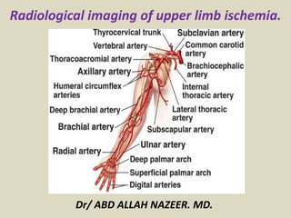
  • 1. Radiological imaging of upper limb ischemia. Dr/ ABD ALLAH NAZEER. MD.
  • 2. Introduction: Upper limb ischemia is an uncommon entity which presents less commonly as compared to lower limb ischaemia, and has a wide range of etiology and controversies in preoperative investigations and management. Less than 2% of patients with upper limb ischaemia presents with rest pain, gangrene or ulcer. Although upper limb loss is less as compared to lower limb, it can result in severe functional impairment and disability if there should be any delay in diagnosis and treatment. The single most common etiology is thromboembolic events secondary to atherosclerosis followed by trauma. Nevertheless, differential diagnoses such as arteritis, thoracic outlet obstruction and aneurysms need to be considered. The signs and symptoms of presentation ranged from nonspecific slight numbness and weakness to pain, pallor and pulselessness. The most common combination of presenting complaints was cold, pulselessness and painful upper limb.
  • 3. Upper extremity occlusive disease is relatively uncommon and accounts for only 5% of all extremity ischemia. Unlike occlusive disease in the lower extremities, which is generally atherosclerotic in origin, the etiology of upper extremity occlusive disease is more varied and can include atherosclerotic disease, arteritis, connective tissue disorders, occupational injuries, and Vasospastic diseases. Etiology Upper extremity occlusive disease can involve the large and small arteries of the arm or a combination. Atherosclerosis is the most common cause of large artery disease and generally involves the subclavian artery, but disease may extend more distally, especially in the setting of diabetes. Patient risk factors are similar to those of other vascular beds. Tobacco use is almost universal. Hypercholesterolemia, hypertension, diabetes and age are also important risk factors. Inflammatory diseases (giant cell and Takayasu’s arteritis) can also involve the large vessels. Small vessel disease due to diabetes can lead to severe complications such as digital ulceration and gangrene.
  • 4. Raynaud’s disease is a Vasospastic disorder that produces a tri-phasic colour change in the hands or less commonly the feet, in response to cold or emotional stimuli. The hallmark of this disorder is pallor, cyanosis and post ischemic hyperemia of a digit or digits, although all three may not always be present. The disorder is classified as primary when no identifiable cause is evident or secondary when it is due to either a more proximal occlusion or other disease process resulting in digital artery vasospasm . Patients whose occupations involve the use of vibrational tools or who use their hands as a hammer (especially the hypothenar eminence) can present with Raynaud’s or frank digital ischemia from either hand-arm vibration syndrome or hypothenar hammer syndrome. Occlusive lesions in the arm and hand secondary to scleroderma, rheumatoid arthritis, systemic lupus and other autoimmune disorders can lead to severe upper extremity ischemia manifesting as pain, ulceration and gangrene. Thromboangiitis obliterans (Buerger’s disease) is an inflammatory disease that can produce pain and ulceration in the fingers and toes. The patient is usually a young man with a long history of tobacco use. Rare causes of upper extremity occlusive disease include iatrogenic injury, trauma, malignancy and frostbite.
  • 5. Common Conditions Associated with Upper Extremity Ischemia: • Thromboembolism • Atheromatous Embolization • Atherosclerosis • Connective Tissue Diseases( • Scleroderma, • Rheumatoid Arthritis, • Sjörgren’s Syndrome, • Systemic Lupus Erythematosus) • Large Artery Vasculitis( • Takayasu’s Disease, • Giant Cell Arteritis) • Small Artery Vasculitis(• Thromboangitis Obliterans (Buerger’s disease). • Blood Dyscrasias • Myeloproliferative Disorders • Thoracic Outlet Syndrome • Latrogenic Injury • Occupational Exposure(• Hand-arm Vibration Syndrome, • Hypothenar Hammer Syndrome). • Vasospasm( • Raynaud’s Disease, • Cocaine Use, • Amphetamine Use).
  • 6. Acute ischemia in the upper extremity constitutes 10–15% of all acute extremity ischemia. The etiology is emboli in 90% of the patients. The reason for this higher rate compared with the leg is that atherosclerosis is less common in arm arteries. Emboli have the same origins as in the lower extremity and usually end up obstructing the brachial artery. Sometimes plaques or an aneurysm in the subclavian or axillary arteries is the primary source of emboli. Embolization to the right arm is more common than to the left due to the vascular anatomy. For the 10% of patients with atherosclerosis and acute thrombosis as the main cause for their arm ischemia, the primary lesions are located in the brachiocephalic trunk or in the subclavian artery. Such pathologies are usually asymptomatic due to well-developed collaterals around the shoulder joint until thrombosis occurs, and they cause either micro- or macroembolization.
  • 7. Clinical Presentation Acute arm ischemia is usually apparent on the basis of the physical examination. The symptoms are often relatively discreet, especially early after onset. The explanation for this is the well developed collateral system circumventing the brachial artery around the elbow, which is the most common site for embolic obstruction. The "six Ps” – pain, pallor, paresthesia, paralysis, pulselessness, poikilothermia– are applicable also for acute arm ischemia, but coldness and color changes are more prominent than for the legs. Accordingly, the most common findings in the physical examination are a cold arm with diminished strength and disturbed hand and finger motor functions. Tingling and numbness are also frequent. The radial pulse is usually absent but is pounding in the upper arm proximal to the obstruction. Gangrene and rest pain appear only when the obstruction is distal to the elbow and affects both of the paired arteries in a finger or in the lower arm. Ischemic signs or symptoms suggesting acute digital artery occlusion in only one or two fingers, imply microembolization.
  • 8. Chronic symptomatic ischemia in the upper extremity is the result of blood flow that is nutritionally inadequate to fulfill metabolic requirements. Symptoms may also result from inappropriate thermoregulatory function. Ischemia may be secondary to inappropriate vessel response when vessels are unable to vasodilate in response to stress or inappropriate vasoconstriction (i.e., vasospastic disease); intima/media injury resulting in aneurysm, embolism, and thrombosis (occlusive disease); or a combination of both (vaso-occlusive disease). Occlusive events may occur secondary to low-flow states in vasospastic disease that precipitate thrombosis development (i.e., secondary occlusion) or secondary to ischemia from occlusive disease that may produce autonomic dysfunction and secondary vasospasm. This cross-over phenomenon creates difficulty in evaluation and management. The goal of this article is to assess contemporary methods of evaluation, classification, and management of upper extremity ischemia.
  • 9. Radiological Imaging: X-Ray to show any evidence of cervical rib. Duplex ultrasound is rapid, can be performed at the bedside, and has near 100% sensitivity for diagnosing complete arterial occlusion. Ankle-brachial index (ABI) will be near zero for patients with acute limb ischemia. CTA has the benefit of rapid availability and high-quality imaging, which allows for precise planning of revascularization. CTA provides imaging of the entire arterial tree from the aortic inflow to the digital level. CTA typically requires 150 mL of iodinated contrast and therefore has to be used with caution in patients with baseline renal insufficiency (glomerular filtration rate <40). For patients with renal insufficiency, aggressive hydration before and after examination with sodium bicarbonate is recommended for CTA or invasive angiography. MRA has a limited role in acute limb ischemia, as the examination can be lengthy (45–60 minutes), is less often available outside regular work hours, and generally has poorer arterial imaging than 64 (or greater) slice CTA. Invasive angiography has the advantage of allowing for simultaneous percutaneous revascularization with both mechanical thrombectomy and thrombolytic therapy.
  • 10.
  • 11.
  • 12. Acute Upper Limb Ischemia with right subclavian and brachial thrombus.
  • 13. Upper limb acute ischemia with recent jugular vein thrombosis.
  • 14. With significant external compression with the probe, a visualized thrombus (blue arrow) is seen within a non-compressible brachial artery
  • 15.
  • 16. The distal (right side subclavian shows no flow.
  • 17. Thrombosis of the brachial artery.
  • 18. Vessels and nerve trauma. Biker’s hammer. (A) Longitudinal and (B) axial colour Doppler US obtained over the palmar aspect of the hypothenar region. Images show a nearly complete thrombosis (black arrowheads) of the superficial branch of the ulnar artery. Some small persistent internal flow signal can be detected (white arrowheads). Note increase in size of the artery and thickening of its wall. HH=hook of the hamate.
  • 19. Left Cervical rib with upper limb chronic ischemia.
  • 20. CT angiography using modern MDCT scanners has evolved into a highly accurate noninvasive diagnostic tool for the evaluation of patients with pathologic abnormalities of the upper extremity arterial system. upper extremity CTA can effectively evaluate for stenosis, occlusion, aneurysm, or embolic events, especially when they affect vessels proximal to the wrist. Atherosclerotic disease affects the upper extremities disproportionately less than the lower extremities. However, proximal involvement of the upper extremities is not infrequent. Risk factors are no different than those for other sites of atherosclerotic involvement and include hypertension, dyslipidemia, diabetes mellitus, age, and a history of smoking . Claudication is the most common presenting symptom, though most patients are asymptomatic because of collateral formation. Blood pressure differences between the upper extremities can frequently be found in these patients. Symptoms are often chronic, with more acute symptoms suggesting acute thrombosis or an embolic event. In the setting of subclavian involvement, upper extremity symptoms may be masked or overshadowed by symptoms related to compromised flow to the ipsilateral vertebral artery due to subclavian steal, particularly syncope and presyncope. In patients with left or right internal mammary bypass grafts, subclavian artery disease can cause myocardial ischemia. Atherosclerotic disease, when identified on imaging, should prompt inquiry into concomitant sites of involvement processes—namely, cardiovascular and cerebrovascular involvement.
  • 21. CT Angiography of the Upper Extremity Arterial System: Axial subvolume maximum intensity projection better outlines stenosis (arrowheads). Axillofemoral bypass (asterisk) is depicted end-on. Volume-rendered image shows axillofemoral bypass graft (arrow) with stenosis proximal and distal to anastomosis in native left axillary artery (arrowheads).
  • 22. Volume-rendered CT angiography shows long occlusion of brachial artery, which was caused by acute thromboembolism.
  • 23. Old woman with acute type A thoracic aortic dissection who presented with acute chest pain and syncope. Physical examination revealed weak right upper extremity pulse. CT angiography of right upper extremity, chest, abdomen, and pelvis was performed to evaluate for aortic dissection and upper extremity arterial pathology. Oblique multiplanar reconstruction image reveals ascending aortic dissection, with intimal flap (arrowheads) covering origins of all supraaortic branch vessels.
  • 24. 49-year-old man in chronic phase of Takayasu disease involving arch and supraaortic vessels. A, Volume-rendered image shows right subclavian artery aneurysm (arrow) and left subclavian artery stent (arrowhead) implanted for steno-occlusive disease. 49-year-old man in chronic phase of Takayasu disease involving arch and supraaortic vessels. B, Axial image shows concentric thickening (arrowheads) of wall of origins of all three supraaortic branches.
  • 25. 64-year-old woman with giant cell arteritis who presented with fever, malaise, and bilateral upper extremity weakness. CT angiography using axial curved multiplanar reconstruction algorithm shows typical irregular wall thickening (arrowheads) of bilateral axillary and subclavian arteries.
  • 26. 29-year-old male heavy smoker with thromboangiitis obliterans who presented with ischemic symptoms of both upper and lower extremities. A, Upper extremity CT angiography (CTA) shows distal ulnar artery occlusion (short thick arrow) and distal radial artery contour irregularity (arrowheads) in right upper extremity. Note typical corkscrew collaterals (long thin arrow). Stenoocclusive disease was also present in left ulnar and interosseous arteries.
  • 27. 50-year-old woman with history of embolic disease to left arm and fibromuscular dysplasia involving bilateral carotid, renal, and external iliac arteries (not shown) who also had involvement of left brachial artery with medial fibrosis, which shows typical “string-of- beads” sign (arrowheads).
  • 28. 17-year-old girl with thoracic outlet syndrome who was evaluated at outside institution for embolic event to left hand. She was found to have aneurysm of left subclavian artery, which was subsequently treated with covered stent. Several months after procedure, patient presented to our institution with heaviness and coolness of left arm and hand. A, Volume- rendered image shows subclavian artery stent, with proximal occlusion (arrowhead) and presence of left cervical rib (single asterisk). First rib is marked with double asterisks. Clavicle was excluded for clarity.
  • 29. 17-year-old girl with thoracic outlet syndrome who was evaluated at outside institution for embolic event to left hand. She was found to have aneurysm of left subclavian artery, which was subsequently treated with covered stent. Several months after procedure, patient presented to our institution with heaviness and coolness of left arm and hand. B, Curved multiplanar reconstruction of left subclavian artery shows completely thrombosed stent (asterisks). Patient underwent subclavian artery vein graft placement and resection of left cervical and first ribs. Volume-rendered image shows vein graft (asterisks) and resection of left cervical and first ribs. Curved multiplanar reconstruction confirms vein graft patency (asterisks).
  • 30. 23-year-old man with arteriovenous malformation in right axilla. B, Maximum intensity projection of upper extremity CT angiography shows malformation (arrowhead), with lateral thoracic artery as main feeder, and early venous drainage, Volume-rendered image shows arteriovenous malformation and anomalous high origin of radial artery (arrowhead), which is normal variant.
  • 31. 17-year-old boy with known diagnosis of Ehlers-Danlos syndrome type 4 who presented to emergency department with extremely painful acute onset swelling of right axilla and Pulseless right upper extremity. A, CT of chest and upper extremity was performed. Unenhanced CT shows large right axillary hematoma (asterisk). B, Maximum intensity projection CT angiography shows extravasation from spontaneously ruptured right axillary artery (arrow). Emergent proximal surgical ligation was performed.
  • 32. Angiogram of the right- upper extremity. (A) Axillary artery occlusion caused by a distal segment. (B) The injured artery and the extensive collateralization.
  • 33. 23-year-old man with dialysis fistula who presented with reduced flows while undergoing hemodialysis and ischemic symptoms in fingers of left hand. B, Subvolume maximum intensity projection (MIP) shows patent arterial anastomosis but diseased radial artery (A) with occlusion distally (arrowheads). There was also stenosis in vein (V) immediately distal to anastomosis (arrow). A, Upper extremity CT angiography volume-rendered view shows dialysis fistula with aneurysm formation (asterisk) on venous side.
  • 34. 23-year-old man with dialysis fistula who presented with reduced flows while undergoing hemodialysis and ischemic symptoms in fingers of left hand. More central subvolume MIP shows critical stenosis of left subclavian vein (arrow).
  • 35. 59-year-old woman who underwent stent placement in left subclavian artery at outside institution presented with recurrent left upper extremity claudication. CT angiography shows stent fracture (arrow) and severe stenosis (arrowheads) due to intimal hyperplasia.
  • 36. Completion angiogram of the right upper limb revealing the axillary– brachial–ulnar axis and the partially-thrombosed aberrant radial artery.
  • 37. Chest roentgenogram demonstrates the clavicular abnormality. Arch aortogram demonstrates a filling defect in the right subclavian artery and occlusion of the axillary artery.
  • 38. Venogram shows occlusion of the right subclavian vein extending to the first rib, with multiple collateral vessels. An angiogram in a 35-year-old woman with right arm ischemia that demonstrates right subclavian artery occlusion from the medial margin of the first rib to the axillary artery at the level of the humeral head.
  • 39. An arteriogram of the right shoulder in neutral position, demonstrating right subclavian stenosis (arrow). An arteriogram of the right upper extremity in abduction, demonstrating increased subclavian artery stenosis and aneurysmal dilation (arrow) in the location of the clavicle and first rib.
  • 40. Thoracic outlet syndrome with stenosis of the left subclavian vein.
  • 41. 73-year-old woman with acute embolus in left axillary artery .
  • 42. Left axillary artery occlusion in a patient with Wegener's granulomatosis .
  • 43. Left brachial artery thrombosis in a patient with Wegener's granulomatosis .
  • 44. Angiogram of upper extremity. The top is in a normal position. The bottom is in a hyperabducted position (arrow indicates area of stenosis).
  • 46. Septic embolization from severe prosthetic aortic valve endocarditis resulted in a 2.5cm brachial artery pseudoaneurysm (circled) that was associated with a pocket of purulence and upper limb ischemia.
  • 47.
  • 48. Angiogram that demonstrates blockage of the ulnar artery.
  • 49. Axial CT angiography source image shows ulnar artery thrombosis with ulnar artery aneurysm (between arrowheads). Ulnar artery reconstruction with right leg saphenous vein autograft was performed, and patient became symptom free. Volume-rendered reconstruction shows occluded ulnar artery (between arrowheads) at Guyon canal.
  • 50. Angiogram of end-stage Raynaud’s disease with extremely poor peripheral circulation. Angiogram that demonstrates an ulnar artery aneurysm.
  • 51. An angiogram demonstrating lack of blood flow to vessels of the hand in Buerger's disease with ischemia to the fingers.
  • 52. Angiogram of a patient with Buerger’s disease that shows decreased circulation of index and thumb. Angiogram that shows decreased circulation in the ulnar digits of the hand.
  • 53. Angiogram of right upper extremity with "string of beads" appearance of the brachial artery (lower left) in fibromuscular dysplasia with chronic ischemia.
  • 54. Conclusions. Upper limb ischaemia remains infrequently seen compared to lower limb ischaemia. Nevertheless, the complications and late effects can result in significant impairment in function. Evaluation of upper limb ischaemia requires careful history taking and clinical examinations. Although angiography remains the gold standard, other non-invasive tools such as duplex US. Prompt surgical intervention results in favorable outcomes.