SlideShare ist ein Scribd-Unternehmen logo
1 von 110
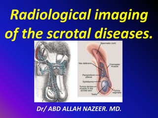
Radiological imaging
of the scrotal diseases.
Dr/ ABD ALLAH NAZEER. MD.
OUTLINE:
Anatomy.
Imaging.
Congenital diseases.
Inflammatory diseases.
Scrotal trauma.
Testicular torsion.
Testicular masses.
Extra-testicular pathology.
Miscellaneous.
Anatomy of the Scrotum
Anatomic structures include:
Testis
Epididymis
Vas deferens
Venous plexus
Testicular artery
Appendix of epididymis (remnant from
embryogenesis)
Appendix of testis (remnant from
embryogenesis)
Anatomy of the Scrotum:
Layers include(Testis: Seminiferous
tubules)
Tunica albuginea
Visceral layer of Tunica vaginalis
Parietal layer of Tunica vaginalis
Fascia
Dartos muscle
Skin
Cryptorchidism (derived from the Greek kryptos, meaning hidden, orchis,
meaning testicle) is the absence of one or both testes from the scrotum. It is a
common birth defect regarding male genitalia. In unique cases,
cryptorchidism can develop later in life, often as late as young adulthood.
About 3% of full-term and 30% of premature infant boys are born with at least
one undescended testis. However, about 80% of cryptorchid testes descend by
the first year of life (the majority within three months), making the true
incidence of cryptorchidism around 1% overall. Cryptorchidism is distinct from
monorchismm, the condition of having only one testicle.
A testis absent from the normal scrotal position can be:
found anywhere along the "path of descent" from high in the posterior
(retroperitoneal) abdomen, just below the kidney, to the inguinal ring; found
in the inguinal canal; ectopic, that is, found to have "wandered" from that
path, usually outside the inguinal canal and sometimes even under the skin of
the thigh, the perineumm, the opposite scrotum, or the femoral canal;
found to be undeveloped (hypoplastic) or severely abnormal (dysgenetic);
found to have vanished (also see anorchia).
Congenital anomalies of the testis.
About two thirds of cases without other abnormalities are
unilateral; one third involve both testes. In 90% of cases an
undescended testis can be felt in the inguinal canal; in a minority the
testis or testes are in the abdomen or nonexistent (truly "hidden").
Undescended testes are associated with reduced fertility, increased
risk of testicular germ cell tumors and psychological problems when
the boy is grown. Undescended testes are also more susceptible to
testicular torsion and infarction and inguinal hernias. "Usually the
testicle will descend into the scrotum without any intervention
during the first year of life, but to reduce these risks, undescended
testes can be brought into the scrotum in infancy by a surgical
procedure called an orchiopexy.
Although cryptorchidism nearly always refers to congenital absence
or maldescent, a testis observed in the scrotum in early infancy can
occasionally "reascend" (move back up) into the inguinal canal. A
testis which can readily move or be moved between the scrotum and
canal is referred to as retractile.
Cryptorchidism with inguinal canal and intra-abdominal testis
Cryptorchidism with undescended testicle in the right inguinal canal
Cryptorchidism. Coronal image shows a nondescended
testis in the right inguinal canal of a child
Axial contrast-enhanced CT image shows fluid collection
with gas in the right scrotum, corresponding to an abscess.
Axial contrast-enhanced CT image shows right inguinal tubular
structures with contrast enhancement corresponding to a varicocele.
Axial contrast-enhanced CT image shows a right fluid
inguino-scrotal collection corresponding to hydrocele.
Spermatic cord lipoma.
Liposarcoma. Axial T1-weighted fat-supressed and contrast-enhanced image
shows enhancement of the solid non-fatty components of the tumor.
Liposarcoma. Coronal T2-weighted (a), axial T2-weighted (b), axial
T1-weighted (c) and T2-weighted fat-supressed images show right
inguino-scrotal mass with heterogeneous signal intensity.
Axial contrast-enhanced CT image shows bilateral inguinal
cystic metastases of epidermoid carcinoma of the penis.
Thank You.

Weitere ähnliche Inhalte

Was ist angesagt?

renal cell carcinoma radiology
renal cell carcinoma radiologyrenal cell carcinoma radiology
renal cell carcinoma radiologydocaashishgupt
 
Ultrasound imaging of Bowel pathology
Ultrasound imaging of Bowel pathologyUltrasound imaging of Bowel pathology
Ultrasound imaging of Bowel pathologyairwave12
 
KEYS OF RADIOLOGY SPOTTERS GIT
KEYS OF RADIOLOGY SPOTTERS GITKEYS OF RADIOLOGY SPOTTERS GIT
KEYS OF RADIOLOGY SPOTTERS GITAnish Choudhary
 
Testicular tumor imaging
Testicular tumor imaging Testicular tumor imaging
Testicular tumor imaging Bom BC
 
Radiological imaging of mediastinal masses
Radiological imaging of mediastinal massesRadiological imaging of mediastinal masses
Radiological imaging of mediastinal massesPankaj Kaira
 
Ultrasound of the abdominal wall hernias
Ultrasound of the abdominal wall herniasUltrasound of the abdominal wall hernias
Ultrasound of the abdominal wall herniasSamir Haffar
 
radiological anatomy of retroperitoneum powerpoint
radiological anatomy of  retroperitoneum powerpointradiological anatomy of  retroperitoneum powerpoint
radiological anatomy of retroperitoneum powerpointDactarAdhikari
 
The Radiology of Malrotation
The Radiology of MalrotationThe Radiology of Malrotation
The Radiology of Malrotationtboulden
 
Radiology Spots PPT- 3 by Dr Chandni Wadhwani
 Radiology Spots PPT- 3 by Dr Chandni Wadhwani Radiology Spots PPT- 3 by Dr Chandni Wadhwani
Radiology Spots PPT- 3 by Dr Chandni WadhwaniChandni Wadhwani
 
Ultrasound of the urinary tract - Renal cysts
Ultrasound of the urinary tract - Renal cystsUltrasound of the urinary tract - Renal cysts
Ultrasound of the urinary tract - Renal cystsSamir Haffar
 
Ultrasound renal stone differential diagnosis .
Ultrasound renal stone differential diagnosis .Ultrasound renal stone differential diagnosis .
Ultrasound renal stone differential diagnosis .AHMED ESAWY
 
Diagnostic Imaging of Pancreatitis
Diagnostic Imaging of PancreatitisDiagnostic Imaging of Pancreatitis
Diagnostic Imaging of PancreatitisMohamed M.A. Zaitoun
 
Ultrasound of the urinary tract - Renal tumors
Ultrasound of the urinary tract - Renal tumorsUltrasound of the urinary tract - Renal tumors
Ultrasound of the urinary tract - Renal tumorsSamir Haffar
 
Mediastinum-RADIOLOGY
Mediastinum-RADIOLOGYMediastinum-RADIOLOGY
Mediastinum-RADIOLOGYNavdeep Shah
 
Cystic liver lesions - An ultrasound perspective
Cystic liver lesions - An ultrasound perspectiveCystic liver lesions - An ultrasound perspective
Cystic liver lesions - An ultrasound perspectiveSamir Haffar
 
Presentation1, ultrasound of the bowel loops and the lymph nodes.
Presentation1, ultrasound of the bowel loops and the lymph nodes.Presentation1, ultrasound of the bowel loops and the lymph nodes.
Presentation1, ultrasound of the bowel loops and the lymph nodes.AbdullahNazeerYassin
 
Radiological anatomy of the male urethra and techniques of imaging
Radiological  anatomy of the male urethra and techniques of imagingRadiological  anatomy of the male urethra and techniques of imaging
Radiological anatomy of the male urethra and techniques of imagingHarrietAimua
 
Diagnostic Imaging of Renal Cystic Diseases
Diagnostic Imaging of Renal Cystic DiseasesDiagnostic Imaging of Renal Cystic Diseases
Diagnostic Imaging of Renal Cystic DiseasesMohamed M.A. Zaitoun
 

Was ist angesagt? (20)

renal cell carcinoma radiology
renal cell carcinoma radiologyrenal cell carcinoma radiology
renal cell carcinoma radiology
 
Ultrasound imaging of Bowel pathology
Ultrasound imaging of Bowel pathologyUltrasound imaging of Bowel pathology
Ultrasound imaging of Bowel pathology
 
KEYS OF RADIOLOGY SPOTTERS GIT
KEYS OF RADIOLOGY SPOTTERS GITKEYS OF RADIOLOGY SPOTTERS GIT
KEYS OF RADIOLOGY SPOTTERS GIT
 
Radiology Spotters
Radiology Spotters Radiology Spotters
Radiology Spotters
 
Testicular tumor imaging
Testicular tumor imaging Testicular tumor imaging
Testicular tumor imaging
 
Radiological imaging of mediastinal masses
Radiological imaging of mediastinal massesRadiological imaging of mediastinal masses
Radiological imaging of mediastinal masses
 
Ultrasound of the abdominal wall hernias
Ultrasound of the abdominal wall herniasUltrasound of the abdominal wall hernias
Ultrasound of the abdominal wall hernias
 
radiological anatomy of retroperitoneum powerpoint
radiological anatomy of  retroperitoneum powerpointradiological anatomy of  retroperitoneum powerpoint
radiological anatomy of retroperitoneum powerpoint
 
The Radiology of Malrotation
The Radiology of MalrotationThe Radiology of Malrotation
The Radiology of Malrotation
 
Radiology Spots PPT- 3 by Dr Chandni Wadhwani
 Radiology Spots PPT- 3 by Dr Chandni Wadhwani Radiology Spots PPT- 3 by Dr Chandni Wadhwani
Radiology Spots PPT- 3 by Dr Chandni Wadhwani
 
Ultrasound of the urinary tract - Renal cysts
Ultrasound of the urinary tract - Renal cystsUltrasound of the urinary tract - Renal cysts
Ultrasound of the urinary tract - Renal cysts
 
Ultrasound renal stone differential diagnosis .
Ultrasound renal stone differential diagnosis .Ultrasound renal stone differential diagnosis .
Ultrasound renal stone differential diagnosis .
 
Diagnostic Imaging of Pancreatitis
Diagnostic Imaging of PancreatitisDiagnostic Imaging of Pancreatitis
Diagnostic Imaging of Pancreatitis
 
Ultrasound of the urinary tract - Renal tumors
Ultrasound of the urinary tract - Renal tumorsUltrasound of the urinary tract - Renal tumors
Ultrasound of the urinary tract - Renal tumors
 
Mediastinum-RADIOLOGY
Mediastinum-RADIOLOGYMediastinum-RADIOLOGY
Mediastinum-RADIOLOGY
 
Cystic liver lesions - An ultrasound perspective
Cystic liver lesions - An ultrasound perspectiveCystic liver lesions - An ultrasound perspective
Cystic liver lesions - An ultrasound perspective
 
Spots with keys (2)
Spots with keys (2)Spots with keys (2)
Spots with keys (2)
 
Presentation1, ultrasound of the bowel loops and the lymph nodes.
Presentation1, ultrasound of the bowel loops and the lymph nodes.Presentation1, ultrasound of the bowel loops and the lymph nodes.
Presentation1, ultrasound of the bowel loops and the lymph nodes.
 
Radiological anatomy of the male urethra and techniques of imaging
Radiological  anatomy of the male urethra and techniques of imagingRadiological  anatomy of the male urethra and techniques of imaging
Radiological anatomy of the male urethra and techniques of imaging
 
Diagnostic Imaging of Renal Cystic Diseases
Diagnostic Imaging of Renal Cystic DiseasesDiagnostic Imaging of Renal Cystic Diseases
Diagnostic Imaging of Renal Cystic Diseases
 

Ähnlich wie Presentation1.pptx, radiological imaging of scrotal diseases.

evaluation of Undescended testes
evaluation of Undescended testesevaluation of Undescended testes
evaluation of Undescended testesVernon Pashi
 
testis presentation.pptx by dr. shahariar hossain
testis presentation.pptx by dr. shahariar hossaintestis presentation.pptx by dr. shahariar hossain
testis presentation.pptx by dr. shahariar hossainshahariarhossainshaw
 
Undescended testis
Undescended testisUndescended testis
Undescended testisGAURAV NAHAR
 
Undescended Testis
Undescended TestisUndescended Testis
Undescended TestisJunish Bagga
 
The Acute Scrotum.pptx
The Acute Scrotum.pptxThe Acute Scrotum.pptx
The Acute Scrotum.pptxJwan AlSofi
 
undescended testis.pptx
undescended testis.pptxundescended testis.pptx
undescended testis.pptxAllenDavid32
 
Presentation1 3 (4).ppt
Presentation1 3 (4).pptPresentation1 3 (4).ppt
Presentation1 3 (4).pptssuser8eb265
 
Rreproduction-and-sexuality-lecture-2
Rreproduction-and-sexuality-lecture-2Rreproduction-and-sexuality-lecture-2
Rreproduction-and-sexuality-lecture-2cjsmann
 
Neurocysticercosis
NeurocysticercosisNeurocysticercosis
Neurocysticercosisdrajay02
 
congenital_anomalies_of_urinary_bladder.ppt
congenital_anomalies_of_urinary_bladder.pptcongenital_anomalies_of_urinary_bladder.ppt
congenital_anomalies_of_urinary_bladder.pptGrashiaBlessy1
 
Anorectal malformations in children
Anorectal malformations in childrenAnorectal malformations in children
Anorectal malformations in childrendhanyav54
 
ULTRASOUND OF OBSTETRICS EMERGENCIES.pptx
ULTRASOUND OF OBSTETRICS EMERGENCIES.pptxULTRASOUND OF OBSTETRICS EMERGENCIES.pptx
ULTRASOUND OF OBSTETRICS EMERGENCIES.pptxArpanUpreti2
 
Endometriosis & Adenomyosis
Endometriosis & AdenomyosisEndometriosis & Adenomyosis
Endometriosis & AdenomyosisBahgat Yassin
 
Undescended Testis- Cryptorchidism
Undescended Testis- CryptorchidismUndescended Testis- Cryptorchidism
Undescended Testis- Cryptorchidismdehdehi
 

Ähnlich wie Presentation1.pptx, radiological imaging of scrotal diseases. (20)

Empty scrotum
Empty scrotumEmpty scrotum
Empty scrotum
 
evaluation of Undescended testes
evaluation of Undescended testesevaluation of Undescended testes
evaluation of Undescended testes
 
testis presentation.pptx by dr. shahariar hossain
testis presentation.pptx by dr. shahariar hossaintestis presentation.pptx by dr. shahariar hossain
testis presentation.pptx by dr. shahariar hossain
 
Undescended testis
Undescended testisUndescended testis
Undescended testis
 
Undescended Testis
Undescended TestisUndescended Testis
Undescended Testis
 
Pediatric Circumcision lecture
Pediatric Circumcision lecturePediatric Circumcision lecture
Pediatric Circumcision lecture
 
ectopic-.pptx
ectopic-.pptxectopic-.pptx
ectopic-.pptx
 
The Acute Scrotum.pptx
The Acute Scrotum.pptxThe Acute Scrotum.pptx
The Acute Scrotum.pptx
 
undescended testis.pptx
undescended testis.pptxundescended testis.pptx
undescended testis.pptx
 
Presentation1 3 (4).ppt
Presentation1 3 (4).pptPresentation1 3 (4).ppt
Presentation1 3 (4).ppt
 
ENDOMETRIOSIS
ENDOMETRIOSIS ENDOMETRIOSIS
ENDOMETRIOSIS
 
Rreproduction-and-sexuality-lecture-2
Rreproduction-and-sexuality-lecture-2Rreproduction-and-sexuality-lecture-2
Rreproduction-and-sexuality-lecture-2
 
Neurocysticercosis
NeurocysticercosisNeurocysticercosis
Neurocysticercosis
 
congenital_anomalies_of_urinary_bladder.ppt
congenital_anomalies_of_urinary_bladder.pptcongenital_anomalies_of_urinary_bladder.ppt
congenital_anomalies_of_urinary_bladder.ppt
 
Uterine malformations
Uterine malformationsUterine malformations
Uterine malformations
 
Anorectal malformations in children
Anorectal malformations in childrenAnorectal malformations in children
Anorectal malformations in children
 
ULTRASOUND OF OBSTETRICS EMERGENCIES.pptx
ULTRASOUND OF OBSTETRICS EMERGENCIES.pptxULTRASOUND OF OBSTETRICS EMERGENCIES.pptx
ULTRASOUND OF OBSTETRICS EMERGENCIES.pptx
 
Cryptochidism
CryptochidismCryptochidism
Cryptochidism
 
Endometriosis & Adenomyosis
Endometriosis & AdenomyosisEndometriosis & Adenomyosis
Endometriosis & Adenomyosis
 
Undescended Testis- Cryptorchidism
Undescended Testis- CryptorchidismUndescended Testis- Cryptorchidism
Undescended Testis- Cryptorchidism
 

Mehr von Abdellah Nazeer

Muculoskeletal Pediatic Imaging..pptx
Muculoskeletal Pediatic Imaging..pptxMuculoskeletal Pediatic Imaging..pptx
Muculoskeletal Pediatic Imaging..pptxAbdellah Nazeer
 
Presentation1, Ultrasound of the bowel loops and the lymph nodes..pptx
Presentation1, Ultrasound of the bowel loops and the lymph nodes..pptxPresentation1, Ultrasound of the bowel loops and the lymph nodes..pptx
Presentation1, Ultrasound of the bowel loops and the lymph nodes..pptxAbdellah Nazeer
 
Presentation1 Short cases MD..pptx
Presentation1 Short cases MD..pptxPresentation1 Short cases MD..pptx
Presentation1 Short cases MD..pptxAbdellah Nazeer
 
Presentation1, MD MCQ Cases..pptx
Presentation1, MD MCQ Cases..pptxPresentation1, MD MCQ Cases..pptx
Presentation1, MD MCQ Cases..pptxAbdellah Nazeer
 
Presentation1, Short Cases Quiz..pptx
Presentation1, Short Cases Quiz..pptxPresentation1, Short Cases Quiz..pptx
Presentation1, Short Cases Quiz..pptxAbdellah Nazeer
 
Presentation1, radiological imaging of lateral hindfoot impingement.
Presentation1, radiological imaging of lateral hindfoot impingement.Presentation1, radiological imaging of lateral hindfoot impingement.
Presentation1, radiological imaging of lateral hindfoot impingement.Abdellah Nazeer
 
Presentation2, radiological anatomy of the liver and spleen.
Presentation2, radiological anatomy of the liver and spleen.Presentation2, radiological anatomy of the liver and spleen.
Presentation2, radiological anatomy of the liver and spleen.Abdellah Nazeer
 
Presentation1, artifacts and pitfalls of the wrist and elbow joints.
Presentation1, artifacts and pitfalls of the wrist and elbow joints.Presentation1, artifacts and pitfalls of the wrist and elbow joints.
Presentation1, artifacts and pitfalls of the wrist and elbow joints.Abdellah Nazeer
 
Presentation1, artifact and pitfalls of the knee, hip and ankle joints.
Presentation1, artifact and pitfalls of the knee, hip and ankle joints.Presentation1, artifact and pitfalls of the knee, hip and ankle joints.
Presentation1, artifact and pitfalls of the knee, hip and ankle joints.Abdellah Nazeer
 
Presentation1, radiological imaging of artifact and pitfalls in shoulder join...
Presentation1, radiological imaging of artifact and pitfalls in shoulder join...Presentation1, radiological imaging of artifact and pitfalls in shoulder join...
Presentation1, radiological imaging of artifact and pitfalls in shoulder join...Abdellah Nazeer
 
Presentation1, radiological imaging of internal abdominal hernia.
Presentation1, radiological imaging of internal abdominal hernia.Presentation1, radiological imaging of internal abdominal hernia.
Presentation1, radiological imaging of internal abdominal hernia.Abdellah Nazeer
 
Presentation11, radiological imaging of ovarian torsion.
Presentation11, radiological imaging of ovarian torsion.Presentation11, radiological imaging of ovarian torsion.
Presentation11, radiological imaging of ovarian torsion.Abdellah Nazeer
 
Presentation1, musculoskeletal anatomy.
Presentation1, musculoskeletal anatomy.Presentation1, musculoskeletal anatomy.
Presentation1, musculoskeletal anatomy.Abdellah Nazeer
 
Presentation1, new mri techniques in the diagnosis and monitoring of multiple...
Presentation1, new mri techniques in the diagnosis and monitoring of multiple...Presentation1, new mri techniques in the diagnosis and monitoring of multiple...
Presentation1, new mri techniques in the diagnosis and monitoring of multiple...Abdellah Nazeer
 
Presentation1, radiological application of diffusion weighted mri in neck mas...
Presentation1, radiological application of diffusion weighted mri in neck mas...Presentation1, radiological application of diffusion weighted mri in neck mas...
Presentation1, radiological application of diffusion weighted mri in neck mas...Abdellah Nazeer
 
Presentation1, radiological application of diffusion weighted images in breas...
Presentation1, radiological application of diffusion weighted images in breas...Presentation1, radiological application of diffusion weighted images in breas...
Presentation1, radiological application of diffusion weighted images in breas...Abdellah Nazeer
 
Presentation1, radiological application of diffusion weighted images in abdom...
Presentation1, radiological application of diffusion weighted images in abdom...Presentation1, radiological application of diffusion weighted images in abdom...
Presentation1, radiological application of diffusion weighted images in abdom...Abdellah Nazeer
 
Presentation1, radiological application of diffusion weighted imges in neuror...
Presentation1, radiological application of diffusion weighted imges in neuror...Presentation1, radiological application of diffusion weighted imges in neuror...
Presentation1, radiological application of diffusion weighted imges in neuror...Abdellah Nazeer
 
Presentation1, mr physics.
Presentation1, mr physics.Presentation1, mr physics.
Presentation1, mr physics.Abdellah Nazeer
 
Presentation1. ct physics.
Presentation1. ct physics.Presentation1. ct physics.
Presentation1. ct physics.Abdellah Nazeer
 

Mehr von Abdellah Nazeer (20)

Muculoskeletal Pediatic Imaging..pptx
Muculoskeletal Pediatic Imaging..pptxMuculoskeletal Pediatic Imaging..pptx
Muculoskeletal Pediatic Imaging..pptx
 
Presentation1, Ultrasound of the bowel loops and the lymph nodes..pptx
Presentation1, Ultrasound of the bowel loops and the lymph nodes..pptxPresentation1, Ultrasound of the bowel loops and the lymph nodes..pptx
Presentation1, Ultrasound of the bowel loops and the lymph nodes..pptx
 
Presentation1 Short cases MD..pptx
Presentation1 Short cases MD..pptxPresentation1 Short cases MD..pptx
Presentation1 Short cases MD..pptx
 
Presentation1, MD MCQ Cases..pptx
Presentation1, MD MCQ Cases..pptxPresentation1, MD MCQ Cases..pptx
Presentation1, MD MCQ Cases..pptx
 
Presentation1, Short Cases Quiz..pptx
Presentation1, Short Cases Quiz..pptxPresentation1, Short Cases Quiz..pptx
Presentation1, Short Cases Quiz..pptx
 
Presentation1, radiological imaging of lateral hindfoot impingement.
Presentation1, radiological imaging of lateral hindfoot impingement.Presentation1, radiological imaging of lateral hindfoot impingement.
Presentation1, radiological imaging of lateral hindfoot impingement.
 
Presentation2, radiological anatomy of the liver and spleen.
Presentation2, radiological anatomy of the liver and spleen.Presentation2, radiological anatomy of the liver and spleen.
Presentation2, radiological anatomy of the liver and spleen.
 
Presentation1, artifacts and pitfalls of the wrist and elbow joints.
Presentation1, artifacts and pitfalls of the wrist and elbow joints.Presentation1, artifacts and pitfalls of the wrist and elbow joints.
Presentation1, artifacts and pitfalls of the wrist and elbow joints.
 
Presentation1, artifact and pitfalls of the knee, hip and ankle joints.
Presentation1, artifact and pitfalls of the knee, hip and ankle joints.Presentation1, artifact and pitfalls of the knee, hip and ankle joints.
Presentation1, artifact and pitfalls of the knee, hip and ankle joints.
 
Presentation1, radiological imaging of artifact and pitfalls in shoulder join...
Presentation1, radiological imaging of artifact and pitfalls in shoulder join...Presentation1, radiological imaging of artifact and pitfalls in shoulder join...
Presentation1, radiological imaging of artifact and pitfalls in shoulder join...
 
Presentation1, radiological imaging of internal abdominal hernia.
Presentation1, radiological imaging of internal abdominal hernia.Presentation1, radiological imaging of internal abdominal hernia.
Presentation1, radiological imaging of internal abdominal hernia.
 
Presentation11, radiological imaging of ovarian torsion.
Presentation11, radiological imaging of ovarian torsion.Presentation11, radiological imaging of ovarian torsion.
Presentation11, radiological imaging of ovarian torsion.
 
Presentation1, musculoskeletal anatomy.
Presentation1, musculoskeletal anatomy.Presentation1, musculoskeletal anatomy.
Presentation1, musculoskeletal anatomy.
 
Presentation1, new mri techniques in the diagnosis and monitoring of multiple...
Presentation1, new mri techniques in the diagnosis and monitoring of multiple...Presentation1, new mri techniques in the diagnosis and monitoring of multiple...
Presentation1, new mri techniques in the diagnosis and monitoring of multiple...
 
Presentation1, radiological application of diffusion weighted mri in neck mas...
Presentation1, radiological application of diffusion weighted mri in neck mas...Presentation1, radiological application of diffusion weighted mri in neck mas...
Presentation1, radiological application of diffusion weighted mri in neck mas...
 
Presentation1, radiological application of diffusion weighted images in breas...
Presentation1, radiological application of diffusion weighted images in breas...Presentation1, radiological application of diffusion weighted images in breas...
Presentation1, radiological application of diffusion weighted images in breas...
 
Presentation1, radiological application of diffusion weighted images in abdom...
Presentation1, radiological application of diffusion weighted images in abdom...Presentation1, radiological application of diffusion weighted images in abdom...
Presentation1, radiological application of diffusion weighted images in abdom...
 
Presentation1, radiological application of diffusion weighted imges in neuror...
Presentation1, radiological application of diffusion weighted imges in neuror...Presentation1, radiological application of diffusion weighted imges in neuror...
Presentation1, radiological application of diffusion weighted imges in neuror...
 
Presentation1, mr physics.
Presentation1, mr physics.Presentation1, mr physics.
Presentation1, mr physics.
 
Presentation1. ct physics.
Presentation1. ct physics.Presentation1. ct physics.
Presentation1. ct physics.
 

Presentation1.pptx, radiological imaging of scrotal diseases.

  • 1. Radiological imaging of the scrotal diseases. Dr/ ABD ALLAH NAZEER. MD.
  • 2. OUTLINE: Anatomy. Imaging. Congenital diseases. Inflammatory diseases. Scrotal trauma. Testicular torsion. Testicular masses. Extra-testicular pathology. Miscellaneous.
  • 3.
  • 4. Anatomy of the Scrotum Anatomic structures include: Testis Epididymis Vas deferens Venous plexus Testicular artery Appendix of epididymis (remnant from embryogenesis) Appendix of testis (remnant from embryogenesis)
  • 5. Anatomy of the Scrotum: Layers include(Testis: Seminiferous tubules) Tunica albuginea Visceral layer of Tunica vaginalis Parietal layer of Tunica vaginalis Fascia Dartos muscle Skin
  • 6.
  • 7.
  • 8.
  • 9. Cryptorchidism (derived from the Greek kryptos, meaning hidden, orchis, meaning testicle) is the absence of one or both testes from the scrotum. It is a common birth defect regarding male genitalia. In unique cases, cryptorchidism can develop later in life, often as late as young adulthood. About 3% of full-term and 30% of premature infant boys are born with at least one undescended testis. However, about 80% of cryptorchid testes descend by the first year of life (the majority within three months), making the true incidence of cryptorchidism around 1% overall. Cryptorchidism is distinct from monorchismm, the condition of having only one testicle. A testis absent from the normal scrotal position can be: found anywhere along the "path of descent" from high in the posterior (retroperitoneal) abdomen, just below the kidney, to the inguinal ring; found in the inguinal canal; ectopic, that is, found to have "wandered" from that path, usually outside the inguinal canal and sometimes even under the skin of the thigh, the perineumm, the opposite scrotum, or the femoral canal; found to be undeveloped (hypoplastic) or severely abnormal (dysgenetic); found to have vanished (also see anorchia). Congenital anomalies of the testis.
  • 10. About two thirds of cases without other abnormalities are unilateral; one third involve both testes. In 90% of cases an undescended testis can be felt in the inguinal canal; in a minority the testis or testes are in the abdomen or nonexistent (truly "hidden"). Undescended testes are associated with reduced fertility, increased risk of testicular germ cell tumors and psychological problems when the boy is grown. Undescended testes are also more susceptible to testicular torsion and infarction and inguinal hernias. "Usually the testicle will descend into the scrotum without any intervention during the first year of life, but to reduce these risks, undescended testes can be brought into the scrotum in infancy by a surgical procedure called an orchiopexy. Although cryptorchidism nearly always refers to congenital absence or maldescent, a testis observed in the scrotum in early infancy can occasionally "reascend" (move back up) into the inguinal canal. A testis which can readily move or be moved between the scrotum and canal is referred to as retractile.
  • 11. Cryptorchidism with inguinal canal and intra-abdominal testis
  • 12. Cryptorchidism with undescended testicle in the right inguinal canal
  • 13. Cryptorchidism. Coronal image shows a nondescended testis in the right inguinal canal of a child
  • 14.
  • 15.
  • 16.
  • 17.
  • 18.
  • 19.
  • 20.
  • 21.
  • 22.
  • 23.
  • 24. Axial contrast-enhanced CT image shows fluid collection with gas in the right scrotum, corresponding to an abscess.
  • 25.
  • 26.
  • 27.
  • 28.
  • 29.
  • 30.
  • 31.
  • 32.
  • 33.
  • 34.
  • 35.
  • 36.
  • 37.
  • 38.
  • 39.
  • 40.
  • 41.
  • 42.
  • 43.
  • 44.
  • 45.
  • 46.
  • 47.
  • 48.
  • 49.
  • 50.
  • 51.
  • 52.
  • 53.
  • 54.
  • 55.
  • 56.
  • 57.
  • 58.
  • 59.
  • 60.
  • 61.
  • 62.
  • 63.
  • 64.
  • 65.
  • 66.
  • 67.
  • 68.
  • 69.
  • 70.
  • 71.
  • 72.
  • 73.
  • 74.
  • 75.
  • 76.
  • 77.
  • 78.
  • 79.
  • 80.
  • 81.
  • 82.
  • 83.
  • 84.
  • 85.
  • 86.
  • 87.
  • 88.
  • 89.
  • 90.
  • 91.
  • 92.
  • 93.
  • 94.
  • 95.
  • 96.
  • 97.
  • 98.
  • 99.
  • 100.
  • 101. Axial contrast-enhanced CT image shows right inguinal tubular structures with contrast enhancement corresponding to a varicocele.
  • 102. Axial contrast-enhanced CT image shows a right fluid inguino-scrotal collection corresponding to hydrocele.
  • 103.
  • 104.
  • 105.
  • 107. Liposarcoma. Axial T1-weighted fat-supressed and contrast-enhanced image shows enhancement of the solid non-fatty components of the tumor.
  • 108. Liposarcoma. Coronal T2-weighted (a), axial T2-weighted (b), axial T1-weighted (c) and T2-weighted fat-supressed images show right inguino-scrotal mass with heterogeneous signal intensity.
  • 109. Axial contrast-enhanced CT image shows bilateral inguinal cystic metastases of epidermoid carcinoma of the penis.