SlideShare ist ein Scribd-Unternehmen logo
1 von 70
Radiological imaging of
diffuse lung disease.
Dr/ ABD ALLAH NAZEER. MD.
Diffuse Interstitial Lung Disease.
Diffuse interstitial lung disease is a group of lung diseases that
affects the connective tissue that forms the support structure of the
air sacs, or alveoli, of the lungs.
During inhalation, the alveoli fill with air. Oxygen within the air
passes through the walls of the alveoli into the blood stream. In
reverse fashion, carbon dioxide passes from the blood into the
alveoli and is exhaled. When affected by an interstitial lung disease,
the tissue supporting the air sacs—called the interstitium—becomes
inflamed and stiff, making it difficult for air sacs to fully expand,
limiting the delivery of oxygen to the body and the removal of
carbon dioxide from the body. As the disease progresses, the tissue
scars and thickens the alveolar walls, further decreasing lung
function.
This inflammation of the interstitium is typically diffuse, meaning it
occurs throughout both lungs rather than being confined to one
area.
The diseases in this group include idiopathic pulmonary fibrosis (IPF),
acute interstitial pneumonia (AIP), cryptogenic organizing pneumonia
(COP) and non-specific interstitial pneumonia (NSIP). Some forms of
interstitial pneumonia are related to the inhalation of cigarette
smoke and occur as a spectrum of injury that includes respiratory
bronchiolitis-interstitial lung disease and desquamative interstitial
pneumonia. Others are associated with multi-systemic diseases such
as rheumatoid arthritis, scleroderma, dermatomyositis and
asbestosis. Occasionally prior drug exposure can result in interstitial
lung disease, such as the anti-cancer drug Bleomycin.
Shortness of breath and a dry cough are the most common symptoms
of diffuse interstitial lung disease. As the disease progresses, weight
loss, muscle and joint pain and fatigue may also occur. At a more
advanced stage, individuals may develop an enlarged heart, an
enlargement of the fingertips called clubbing, and cyanosis—a blue
coloration in the lips, skin and fingernails as a result of reduced
oxygen levels in the blood.
How is diffuse interstitial lung disease evaluated.
To diagnose and determine the cause of interstitial lung disease, a physician will
need to perform a physical examination and order diagnostic tests, including:
Blood tests: These are focused on identifying autoimmune diseases such as
lupus and rheumatoid arthritis which can result in interstitial lung disease as a
result.
Spirometry: This is a test of lung function, in which the patient exhales quickly
and forcefully through a tube connected to a machine that measures how much
air the lungs can hold and how quickly the air moves in and out of the lungs.
Spirometry can help determine if there is an issue with air getting into the lungs
(restriction, such as fibrosis) versus air getting out of the lungs (obstruction,
such as asthma).
Pulse oximetry: This test uses a small device placed on a finger tip to measure
the oxygen saturation of the blood. It shines a specific wavelength of light
though the end of the finger painlessly to measure the amount of oxygen in the
blood.
Chest x-ray : The patterns of lung damage associated with various types of
interstitial lung disease are often identifiable on a chest x-ray. Chest x-rays may
also be used to track the progression of the disease.
CT imaging of the chest : Computed tomography (CT) scanning,
particularly a specific technique known as high resolution CT, is used
to see the fine detail of the interstitium where the disease is occurring.
Based on the imaging appearance, a diagnosis (specifically idiopathic
pulmonary fibrosis) can sometimes be confirmed, thus avoiding the
need for lung biopsy. The CT scan can also help determine the extent
of damage to the lungs and help determine appropriate treatment.
Bronchoscopy and biopsy: In this procedure, a very small sample of
tissue is removed from the lung using a small, flexible tube called a
bronchoscope that is passed through the mouth or nose and into the
lungs.
Surgical biopsy: A surgical biopsy is often needed to obtain a larger
sample tissue than is possible with bronchoscopy. During this
procedure, surgical instruments and a small camera are inserted
through two or three small incisions between the ribs that allow a
physician to see and remove tissue samples from the lungs.
X-Ray chest.
It is important modality for diagnosis of ILDs.
The correlation between the radiographic pattern and the stage of the
disease(clinical or histological) is generally poor.
Review all previous film to assess the rate of changes on disease
activity.
Honeycombing correlated a poor prognosis.
X-Ray limitations:
X-Ray chest is normal in 10-15% of symptomatic patients with
infiltrative lung disease.
The X-Ray sensitivity and a specificity of 80% for detection of DPLD.
The X-Ray can provide a confident diagnosis in 25% of cases.
A diffuse ground glass pattern- early in the disease, progress,- nodules,
linear (reticular) infiltrate, or a combination.
- Infiltrate become coarser and lung volume is lost- honeycomb pattern.
CT pattern of diffuse lung diseases.
Four major patterns (ground-glass opacity, nodular
opacity, reticular opacity, and honeycombing) on thin-
section computed tomographic images were identified.
Idiopathic Pulmonary Fibrosis with subpleural reticular marking, traction
bronchiectasis and honeycombing in a lower lobe distribution.
HRCT cross-sectional view showing a pattern of peripheral reticulation
and honeycomb change that is diagnostic of the presence of UIP.
Idiopathic Pulmonary Fibrosis with subpleural reticular marking, traction
bronchiectasis and honeycombing in a lower lobe distribution.
Idiopathic Pulmonary Fibrosis with subpleural reticular marking, traction
bronchiectasis and honeycombing in a lower lobe distribution.
Hypersensitivity Pneumonitis.
Hypersensitivity Pneumonitis
Drug-related lung disease.
Sarcoidosis HRCT: Hilar and mediastinal lymphadenopathy, beaded
appearance and thickening of brochovascular bundles, nodules along bronchi
Alveolar Proteinosis.
2 cases alveolar proteinosis.
Alveolar proteinosis
Non specific Interstitial Pneumonia.
Desquamative interstitial pneumonia.
Organizing Pneumonia.
Esinophilic granuloma.
Langerhans cell histiocytosis
Lymphomatoid granulomatosis (LYG)
2 cases of bronchoalveolar carcinoma
2 cases of bronchoalveolar carcinoma.
Thank You.

Weitere ähnliche Inhalte

Was ist angesagt?

Collapse- RADIOLOGY
Collapse- RADIOLOGYCollapse- RADIOLOGY
Collapse- RADIOLOGY
Navdeep Shah
 
Presentation1.pptx. interpretation of x ray chest.
Presentation1.pptx. interpretation of x ray chest.Presentation1.pptx. interpretation of x ray chest.
Presentation1.pptx. interpretation of x ray chest.
Abdellah Nazeer
 
Cystic lung diseases
Cystic lung diseasesCystic lung diseases
Cystic lung diseases
Gamal Agmy
 
Presentation1.pptx, radiological imaging of bronchiectasis.
Presentation1.pptx, radiological imaging of bronchiectasis.Presentation1.pptx, radiological imaging of bronchiectasis.
Presentation1.pptx, radiological imaging of bronchiectasis.
Abdellah Nazeer
 
Presentation1.pptx, radiological imaging of pulmonary infection.
Presentation1.pptx, radiological imaging of pulmonary infection.Presentation1.pptx, radiological imaging of pulmonary infection.
Presentation1.pptx, radiological imaging of pulmonary infection.
Abdellah Nazeer
 
Presentation1.pptx radiological imaging of mediastinal masses .
Presentation1.pptx radiological imaging of mediastinal masses .Presentation1.pptx radiological imaging of mediastinal masses .
Presentation1.pptx radiological imaging of mediastinal masses .
Abdellah Nazeer
 
Interactive radiology case presentation
Interactive radiology case presentationInteractive radiology case presentation
Interactive radiology case presentation
Gamal Agmy
 

Was ist angesagt? (20)

Collapse- RADIOLOGY
Collapse- RADIOLOGYCollapse- RADIOLOGY
Collapse- RADIOLOGY
 
Radiological imaging of pleural diseases
Radiological imaging of pleural diseases Radiological imaging of pleural diseases
Radiological imaging of pleural diseases
 
Cystic lung disease
Cystic lung disease   Cystic lung disease
Cystic lung disease
 
Radiological features of Lung cancer Dr. Muhammad Bin Zulfiqar
Radiological features of Lung cancer Dr. Muhammad Bin ZulfiqarRadiological features of Lung cancer Dr. Muhammad Bin Zulfiqar
Radiological features of Lung cancer Dr. Muhammad Bin Zulfiqar
 
Imaging in Skull base
Imaging in Skull baseImaging in Skull base
Imaging in Skull base
 
Solitary pulmonary nodule
Solitary pulmonary noduleSolitary pulmonary nodule
Solitary pulmonary nodule
 
Radiological imaging of pulmonary neoplasms
Radiological imaging of pulmonary neoplasmsRadiological imaging of pulmonary neoplasms
Radiological imaging of pulmonary neoplasms
 
Presentation1.pptx. interpretation of x ray chest.
Presentation1.pptx. interpretation of x ray chest.Presentation1.pptx. interpretation of x ray chest.
Presentation1.pptx. interpretation of x ray chest.
 
Interstitial lung diseases radiology
Interstitial lung diseases radiologyInterstitial lung diseases radiology
Interstitial lung diseases radiology
 
Cystic lung diseases
Cystic lung diseasesCystic lung diseases
Cystic lung diseases
 
Signs in Chest Xray
Signs in Chest Xray Signs in Chest Xray
Signs in Chest Xray
 
Presentation1.pptx, radiological imaging of bronchiectasis.
Presentation1.pptx, radiological imaging of bronchiectasis.Presentation1.pptx, radiological imaging of bronchiectasis.
Presentation1.pptx, radiological imaging of bronchiectasis.
 
Imaging of pulmonary embolism
Imaging of pulmonary embolismImaging of pulmonary embolism
Imaging of pulmonary embolism
 
Presentation1.pptx, radiological imaging of pulmonary infection.
Presentation1.pptx, radiological imaging of pulmonary infection.Presentation1.pptx, radiological imaging of pulmonary infection.
Presentation1.pptx, radiological imaging of pulmonary infection.
 
Presentation1.pptx radiological imaging of mediastinal masses .
Presentation1.pptx radiological imaging of mediastinal masses .Presentation1.pptx radiological imaging of mediastinal masses .
Presentation1.pptx radiological imaging of mediastinal masses .
 
HRCT Chest
HRCT ChestHRCT Chest
HRCT Chest
 
HRCT Nodular pattern
HRCT Nodular pattern HRCT Nodular pattern
HRCT Nodular pattern
 
Lines & mediastinal stripes 01
Lines & mediastinal stripes 01Lines & mediastinal stripes 01
Lines & mediastinal stripes 01
 
Imaging of portal hypertension
Imaging of portal hypertensionImaging of portal hypertension
Imaging of portal hypertension
 
Interactive radiology case presentation
Interactive radiology case presentationInteractive radiology case presentation
Interactive radiology case presentation
 

Andere mochten auch

Presentation1.pptx, radiological imaging of pleural diseases.
Presentation1.pptx, radiological imaging of pleural diseases.Presentation1.pptx, radiological imaging of pleural diseases.
Presentation1.pptx, radiological imaging of pleural diseases.
Abdellah Nazeer
 
Presentation1.pptx, radiological imaging of restrictive lung diseases.
Presentation1.pptx, radiological imaging of restrictive lung diseases.Presentation1.pptx, radiological imaging of restrictive lung diseases.
Presentation1.pptx, radiological imaging of restrictive lung diseases.
Abdellah Nazeer
 
Field Based Application of Automated Image Processing Using Windows Phone Gui...
Field Based Application of Automated Image Processing Using Windows Phone Gui...Field Based Application of Automated Image Processing Using Windows Phone Gui...
Field Based Application of Automated Image Processing Using Windows Phone Gui...
Melvin Marzan
 
Hrct thorax- A-Z
Hrct thorax- A-ZHrct thorax- A-Z
Hrct thorax- A-Z
Nihaal Reddy
 
Presentation1.pptx, radiological imaging of chronic obstructive airway disease.
Presentation1.pptx, radiological imaging of chronic obstructive airway disease.Presentation1.pptx, radiological imaging of chronic obstructive airway disease.
Presentation1.pptx, radiological imaging of chronic obstructive airway disease.
Abdellah Nazeer
 
Presentation1.pptx, chest film reading. lecture 2
Presentation1.pptx, chest film reading. lecture 2Presentation1.pptx, chest film reading. lecture 2
Presentation1.pptx, chest film reading. lecture 2
Abdellah Nazeer
 
Ipf or non ipf interstitial lung diseases
Ipf or non ipf interstitial lung diseasesIpf or non ipf interstitial lung diseases
Ipf or non ipf interstitial lung diseases
Gamal Agmy
 
Presentation1.pptx, chest film reading. lecture 1
Presentation1.pptx, chest film reading. lecture 1Presentation1.pptx, chest film reading. lecture 1
Presentation1.pptx, chest film reading. lecture 1
Abdellah Nazeer
 
Presentation1, radiological imaging of thoracic aortic aneurysm.
Presentation1, radiological imaging of thoracic aortic aneurysm.Presentation1, radiological imaging of thoracic aortic aneurysm.
Presentation1, radiological imaging of thoracic aortic aneurysm.
Abdellah Nazeer
 

Andere mochten auch (20)

Presentation1.pptx, radiological imaging of pleural diseases.
Presentation1.pptx, radiological imaging of pleural diseases.Presentation1.pptx, radiological imaging of pleural diseases.
Presentation1.pptx, radiological imaging of pleural diseases.
 
Presentation1.pptx, radiological imaging of restrictive lung diseases.
Presentation1.pptx, radiological imaging of restrictive lung diseases.Presentation1.pptx, radiological imaging of restrictive lung diseases.
Presentation1.pptx, radiological imaging of restrictive lung diseases.
 
HRCT in Diffuse Lung Diseases - II (Honeycombing, UIP pattern, IPF)
HRCT in Diffuse Lung Diseases - II (Honeycombing, UIP pattern, IPF)HRCT in Diffuse Lung Diseases - II (Honeycombing, UIP pattern, IPF)
HRCT in Diffuse Lung Diseases - II (Honeycombing, UIP pattern, IPF)
 
Approach to interstitial lung disease
Approach to interstitial lung diseaseApproach to interstitial lung disease
Approach to interstitial lung disease
 
Field Based Application of Automated Image Processing Using Windows Phone Gui...
Field Based Application of Automated Image Processing Using Windows Phone Gui...Field Based Application of Automated Image Processing Using Windows Phone Gui...
Field Based Application of Automated Image Processing Using Windows Phone Gui...
 
Hrct iv
Hrct ivHrct iv
Hrct iv
 
Hrct thorax- A-Z
Hrct thorax- A-ZHrct thorax- A-Z
Hrct thorax- A-Z
 
Hrct ii
Hrct iiHrct ii
Hrct ii
 
GreenR: Automatic Plant Disease Diagnosis
GreenR: Automatic Plant Disease DiagnosisGreenR: Automatic Plant Disease Diagnosis
GreenR: Automatic Plant Disease Diagnosis
 
Hrct iii
Hrct iiiHrct iii
Hrct iii
 
Detection of Diseases on Cotton Leaves and its Possible Diagnosis
Detection of Diseases on Cotton Leaves and its Possible DiagnosisDetection of Diseases on Cotton Leaves and its Possible Diagnosis
Detection of Diseases on Cotton Leaves and its Possible Diagnosis
 
Hrct i
Hrct iHrct i
Hrct i
 
Presentation1.pptx, radiological imaging of chronic obstructive airway disease.
Presentation1.pptx, radiological imaging of chronic obstructive airway disease.Presentation1.pptx, radiological imaging of chronic obstructive airway disease.
Presentation1.pptx, radiological imaging of chronic obstructive airway disease.
 
Presentation1.pptx, chest film reading. lecture 2
Presentation1.pptx, chest film reading. lecture 2Presentation1.pptx, chest film reading. lecture 2
Presentation1.pptx, chest film reading. lecture 2
 
Ipf or non ipf interstitial lung diseases
Ipf or non ipf interstitial lung diseasesIpf or non ipf interstitial lung diseases
Ipf or non ipf interstitial lung diseases
 
Coloboma
ColobomaColoboma
Coloboma
 
Presentation1.pptx, chest film reading. lecture 1
Presentation1.pptx, chest film reading. lecture 1Presentation1.pptx, chest film reading. lecture 1
Presentation1.pptx, chest film reading. lecture 1
 
Approach To Diffuse Parenchymal Lung Diseases
Approach To  Diffuse Parenchymal Lung DiseasesApproach To  Diffuse Parenchymal Lung Diseases
Approach To Diffuse Parenchymal Lung Diseases
 
Ct diffuse lung disease
Ct diffuse lung diseaseCt diffuse lung disease
Ct diffuse lung disease
 
Presentation1, radiological imaging of thoracic aortic aneurysm.
Presentation1, radiological imaging of thoracic aortic aneurysm.Presentation1, radiological imaging of thoracic aortic aneurysm.
Presentation1, radiological imaging of thoracic aortic aneurysm.
 

Ähnlich wie Presentation1.pptx, radiological imaging of diffuse lung disease.

Лекция. Абсцесс и гангрена легких. Abscess, lung gangrene англ..ppt
Лекция. Абсцесс и гангрена легких.  Abscess, lung gangrene англ..pptЛекция. Абсцесс и гангрена легких.  Abscess, lung gangrene англ..ppt
Лекция. Абсцесс и гангрена легких. Abscess, lung gangrene англ..ppt
Upasana399630
 
ASSESSMENT, EVALUATION AND TREATMENT OF RESPIRATORY CONDITIONS
ASSESSMENT, EVALUATION AND TREATMENT  OF RESPIRATORY CONDITIONSASSESSMENT, EVALUATION AND TREATMENT  OF RESPIRATORY CONDITIONS
ASSESSMENT, EVALUATION AND TREATMENT OF RESPIRATORY CONDITIONS
Peace Samuel
 
presentation1-140523194630-phpapp01.pptx
presentation1-140523194630-phpapp01.pptxpresentation1-140523194630-phpapp01.pptx
presentation1-140523194630-phpapp01.pptx
Oluseyi7
 
Presentation1.pptx, radiological imaging of copd.
Presentation1.pptx, radiological imaging of copd.Presentation1.pptx, radiological imaging of copd.
Presentation1.pptx, radiological imaging of copd.
Abdellah Nazeer
 
DOC-20221119-WA0000..pptx
DOC-20221119-WA0000..pptxDOC-20221119-WA0000..pptx
DOC-20221119-WA0000..pptx
bhmMhmm
 

Ähnlich wie Presentation1.pptx, radiological imaging of diffuse lung disease. (20)

Interstitial lung diseases
Interstitial lung diseasesInterstitial lung diseases
Interstitial lung diseases
 
Лекция. Абсцесс и гангрена легких. Abscess, lung gangrene англ..ppt
Лекция. Абсцесс и гангрена легких.  Abscess, lung gangrene англ..pptЛекция. Абсцесс и гангрена легких.  Abscess, lung gangrene англ..ppt
Лекция. Абсцесс и гангрена легких. Abscess, lung gangrene англ..ppt
 
Radiology& Imaging in rehabilitation.pdf
Radiology& Imaging in rehabilitation.pdfRadiology& Imaging in rehabilitation.pdf
Radiology& Imaging in rehabilitation.pdf
 
Lung Failure And AORTIC INSUFFICIENCY.pptx
Lung Failure And AORTIC INSUFFICIENCY.pptxLung Failure And AORTIC INSUFFICIENCY.pptx
Lung Failure And AORTIC INSUFFICIENCY.pptx
 
Chest x ray
Chest x rayChest x ray
Chest x ray
 
CHRONIC OBUSTRUCTIVE PULMONARY DISEASE POWER POINT.pptx
CHRONIC OBUSTRUCTIVE PULMONARY DISEASE POWER POINT.pptxCHRONIC OBUSTRUCTIVE PULMONARY DISEASE POWER POINT.pptx
CHRONIC OBUSTRUCTIVE PULMONARY DISEASE POWER POINT.pptx
 
Mahra Nourbakhsh's Lecture for Pathology Assistants, Respiratory System
Mahra Nourbakhsh's Lecture for Pathology Assistants, Respiratory SystemMahra Nourbakhsh's Lecture for Pathology Assistants, Respiratory System
Mahra Nourbakhsh's Lecture for Pathology Assistants, Respiratory System
 
Complex clinical imaging of radiological system
Complex clinical imaging of radiological system Complex clinical imaging of radiological system
Complex clinical imaging of radiological system
 
Biology pro (autosaved)
Biology pro (autosaved)Biology pro (autosaved)
Biology pro (autosaved)
 
PLEURAL EFFUSION.pptx
PLEURAL EFFUSION.pptxPLEURAL EFFUSION.pptx
PLEURAL EFFUSION.pptx
 
ASSESSMENT, EVALUATION AND TREATMENT OF RESPIRATORY CONDITIONS
ASSESSMENT, EVALUATION AND TREATMENT  OF RESPIRATORY CONDITIONSASSESSMENT, EVALUATION AND TREATMENT  OF RESPIRATORY CONDITIONS
ASSESSMENT, EVALUATION AND TREATMENT OF RESPIRATORY CONDITIONS
 
presentation1-140523194630-phpapp01.pptx
presentation1-140523194630-phpapp01.pptxpresentation1-140523194630-phpapp01.pptx
presentation1-140523194630-phpapp01.pptx
 
Presentation1.pptx, radiological imaging of copd.
Presentation1.pptx, radiological imaging of copd.Presentation1.pptx, radiological imaging of copd.
Presentation1.pptx, radiological imaging of copd.
 
DOC-20221119-WA0000..pptx
DOC-20221119-WA0000..pptxDOC-20221119-WA0000..pptx
DOC-20221119-WA0000..pptx
 
Pneumonia
PneumoniaPneumonia
Pneumonia
 
Pneumonia summary
Pneumonia summaryPneumonia summary
Pneumonia summary
 
Common suppurative diseases of lung- Bronchiectasis...!
Common suppurative diseases of lung- Bronchiectasis...!Common suppurative diseases of lung- Bronchiectasis...!
Common suppurative diseases of lung- Bronchiectasis...!
 
Lung abscess pdf
Lung abscess pdfLung abscess pdf
Lung abscess pdf
 
Extrapleural Pneumonectomy
Extrapleural PneumonectomyExtrapleural Pneumonectomy
Extrapleural Pneumonectomy
 
Suppurative lung diseases
Suppurative lung diseasesSuppurative lung diseases
Suppurative lung diseases
 

Mehr von Abdellah Nazeer

Presentation1, radiological imaging of lateral hindfoot impingement.
Presentation1, radiological imaging of lateral hindfoot impingement.Presentation1, radiological imaging of lateral hindfoot impingement.
Presentation1, radiological imaging of lateral hindfoot impingement.
Abdellah Nazeer
 
Presentation2, radiological anatomy of the liver and spleen.
Presentation2, radiological anatomy of the liver and spleen.Presentation2, radiological anatomy of the liver and spleen.
Presentation2, radiological anatomy of the liver and spleen.
Abdellah Nazeer
 
Presentation1, artifacts and pitfalls of the wrist and elbow joints.
Presentation1, artifacts and pitfalls of the wrist and elbow joints.Presentation1, artifacts and pitfalls of the wrist and elbow joints.
Presentation1, artifacts and pitfalls of the wrist and elbow joints.
Abdellah Nazeer
 
Presentation1, artifact and pitfalls of the knee, hip and ankle joints.
Presentation1, artifact and pitfalls of the knee, hip and ankle joints.Presentation1, artifact and pitfalls of the knee, hip and ankle joints.
Presentation1, artifact and pitfalls of the knee, hip and ankle joints.
Abdellah Nazeer
 
Presentation1, radiological imaging of artifact and pitfalls in shoulder join...
Presentation1, radiological imaging of artifact and pitfalls in shoulder join...Presentation1, radiological imaging of artifact and pitfalls in shoulder join...
Presentation1, radiological imaging of artifact and pitfalls in shoulder join...
Abdellah Nazeer
 
Presentation1, radiological imaging of internal abdominal hernia.
Presentation1, radiological imaging of internal abdominal hernia.Presentation1, radiological imaging of internal abdominal hernia.
Presentation1, radiological imaging of internal abdominal hernia.
Abdellah Nazeer
 
Presentation11, radiological imaging of ovarian torsion.
Presentation11, radiological imaging of ovarian torsion.Presentation11, radiological imaging of ovarian torsion.
Presentation11, radiological imaging of ovarian torsion.
Abdellah Nazeer
 
Presentation1, new mri techniques in the diagnosis and monitoring of multiple...
Presentation1, new mri techniques in the diagnosis and monitoring of multiple...Presentation1, new mri techniques in the diagnosis and monitoring of multiple...
Presentation1, new mri techniques in the diagnosis and monitoring of multiple...
Abdellah Nazeer
 
Presentation1, radiological application of diffusion weighted mri in neck mas...
Presentation1, radiological application of diffusion weighted mri in neck mas...Presentation1, radiological application of diffusion weighted mri in neck mas...
Presentation1, radiological application of diffusion weighted mri in neck mas...
Abdellah Nazeer
 
Presentation1, radiological application of diffusion weighted images in breas...
Presentation1, radiological application of diffusion weighted images in breas...Presentation1, radiological application of diffusion weighted images in breas...
Presentation1, radiological application of diffusion weighted images in breas...
Abdellah Nazeer
 
Presentation1, radiological application of diffusion weighted images in abdom...
Presentation1, radiological application of diffusion weighted images in abdom...Presentation1, radiological application of diffusion weighted images in abdom...
Presentation1, radiological application of diffusion weighted images in abdom...
Abdellah Nazeer
 
Presentation1, radiological application of diffusion weighted imges in neuror...
Presentation1, radiological application of diffusion weighted imges in neuror...Presentation1, radiological application of diffusion weighted imges in neuror...
Presentation1, radiological application of diffusion weighted imges in neuror...
Abdellah Nazeer
 

Mehr von Abdellah Nazeer (20)

Muculoskeletal Pediatic Imaging..pptx
Muculoskeletal Pediatic Imaging..pptxMuculoskeletal Pediatic Imaging..pptx
Muculoskeletal Pediatic Imaging..pptx
 
Presentation1, Ultrasound of the bowel loops and the lymph nodes..pptx
Presentation1, Ultrasound of the bowel loops and the lymph nodes..pptxPresentation1, Ultrasound of the bowel loops and the lymph nodes..pptx
Presentation1, Ultrasound of the bowel loops and the lymph nodes..pptx
 
Presentation1 Short cases MD..pptx
Presentation1 Short cases MD..pptxPresentation1 Short cases MD..pptx
Presentation1 Short cases MD..pptx
 
Presentation1, MD MCQ Cases..pptx
Presentation1, MD MCQ Cases..pptxPresentation1, MD MCQ Cases..pptx
Presentation1, MD MCQ Cases..pptx
 
Presentation1, Short Cases Quiz..pptx
Presentation1, Short Cases Quiz..pptxPresentation1, Short Cases Quiz..pptx
Presentation1, Short Cases Quiz..pptx
 
Presentation1, radiological imaging of lateral hindfoot impingement.
Presentation1, radiological imaging of lateral hindfoot impingement.Presentation1, radiological imaging of lateral hindfoot impingement.
Presentation1, radiological imaging of lateral hindfoot impingement.
 
Presentation2, radiological anatomy of the liver and spleen.
Presentation2, radiological anatomy of the liver and spleen.Presentation2, radiological anatomy of the liver and spleen.
Presentation2, radiological anatomy of the liver and spleen.
 
Presentation1, artifacts and pitfalls of the wrist and elbow joints.
Presentation1, artifacts and pitfalls of the wrist and elbow joints.Presentation1, artifacts and pitfalls of the wrist and elbow joints.
Presentation1, artifacts and pitfalls of the wrist and elbow joints.
 
Presentation1, artifact and pitfalls of the knee, hip and ankle joints.
Presentation1, artifact and pitfalls of the knee, hip and ankle joints.Presentation1, artifact and pitfalls of the knee, hip and ankle joints.
Presentation1, artifact and pitfalls of the knee, hip and ankle joints.
 
Presentation1, radiological imaging of artifact and pitfalls in shoulder join...
Presentation1, radiological imaging of artifact and pitfalls in shoulder join...Presentation1, radiological imaging of artifact and pitfalls in shoulder join...
Presentation1, radiological imaging of artifact and pitfalls in shoulder join...
 
Presentation1, radiological imaging of internal abdominal hernia.
Presentation1, radiological imaging of internal abdominal hernia.Presentation1, radiological imaging of internal abdominal hernia.
Presentation1, radiological imaging of internal abdominal hernia.
 
Presentation11, radiological imaging of ovarian torsion.
Presentation11, radiological imaging of ovarian torsion.Presentation11, radiological imaging of ovarian torsion.
Presentation11, radiological imaging of ovarian torsion.
 
Presentation1, musculoskeletal anatomy.
Presentation1, musculoskeletal anatomy.Presentation1, musculoskeletal anatomy.
Presentation1, musculoskeletal anatomy.
 
Presentation1, new mri techniques in the diagnosis and monitoring of multiple...
Presentation1, new mri techniques in the diagnosis and monitoring of multiple...Presentation1, new mri techniques in the diagnosis and monitoring of multiple...
Presentation1, new mri techniques in the diagnosis and monitoring of multiple...
 
Presentation1, radiological application of diffusion weighted mri in neck mas...
Presentation1, radiological application of diffusion weighted mri in neck mas...Presentation1, radiological application of diffusion weighted mri in neck mas...
Presentation1, radiological application of diffusion weighted mri in neck mas...
 
Presentation1, radiological application of diffusion weighted images in breas...
Presentation1, radiological application of diffusion weighted images in breas...Presentation1, radiological application of diffusion weighted images in breas...
Presentation1, radiological application of diffusion weighted images in breas...
 
Presentation1, radiological application of diffusion weighted images in abdom...
Presentation1, radiological application of diffusion weighted images in abdom...Presentation1, radiological application of diffusion weighted images in abdom...
Presentation1, radiological application of diffusion weighted images in abdom...
 
Presentation1, radiological application of diffusion weighted imges in neuror...
Presentation1, radiological application of diffusion weighted imges in neuror...Presentation1, radiological application of diffusion weighted imges in neuror...
Presentation1, radiological application of diffusion weighted imges in neuror...
 
Presentation1, mr physics.
Presentation1, mr physics.Presentation1, mr physics.
Presentation1, mr physics.
 
Presentation1. ct physics.
Presentation1. ct physics.Presentation1. ct physics.
Presentation1. ct physics.
 

KĂźrzlich hochgeladen

VIP Service Call Girls Sindhi Colony 📳 7877925207 For 18+ VIP Call Girl At Th...
VIP Service Call Girls Sindhi Colony 📳 7877925207 For 18+ VIP Call Girl At Th...VIP Service Call Girls Sindhi Colony 📳 7877925207 For 18+ VIP Call Girl At Th...
VIP Service Call Girls Sindhi Colony 📳 7877925207 For 18+ VIP Call Girl At Th...
jageshsingh5554
 
College Call Girls in Haridwar 9667172968 Short 4000 Night 10000 Best call gi...
College Call Girls in Haridwar 9667172968 Short 4000 Night 10000 Best call gi...College Call Girls in Haridwar 9667172968 Short 4000 Night 10000 Best call gi...
College Call Girls in Haridwar 9667172968 Short 4000 Night 10000 Best call gi...
perfect solution
 
Russian Escorts Girls Nehru Place ZINATHI 🔝9711199012 ☪ 24/7 Call Girls Delhi
Russian Escorts Girls  Nehru Place ZINATHI 🔝9711199012 ☪ 24/7 Call Girls DelhiRussian Escorts Girls  Nehru Place ZINATHI 🔝9711199012 ☪ 24/7 Call Girls Delhi
Russian Escorts Girls Nehru Place ZINATHI 🔝9711199012 ☪ 24/7 Call Girls Delhi
AlinaDevecerski
 

KĂźrzlich hochgeladen (20)

(Low Rate RASHMI ) Rate Of Call Girls Jaipur ❣ 8445551418 ❣ Elite Models & Ce...
(Low Rate RASHMI ) Rate Of Call Girls Jaipur ❣ 8445551418 ❣ Elite Models & Ce...(Low Rate RASHMI ) Rate Of Call Girls Jaipur ❣ 8445551418 ❣ Elite Models & Ce...
(Low Rate RASHMI ) Rate Of Call Girls Jaipur ❣ 8445551418 ❣ Elite Models & Ce...
 
Top Quality Call Girl Service Kalyanpur 6378878445 Available Call Girls Any Time
Top Quality Call Girl Service Kalyanpur 6378878445 Available Call Girls Any TimeTop Quality Call Girl Service Kalyanpur 6378878445 Available Call Girls Any Time
Top Quality Call Girl Service Kalyanpur 6378878445 Available Call Girls Any Time
 
Call Girls Gwalior Just Call 8617370543 Top Class Call Girl Service Available
Call Girls Gwalior Just Call 8617370543 Top Class Call Girl Service AvailableCall Girls Gwalior Just Call 8617370543 Top Class Call Girl Service Available
Call Girls Gwalior Just Call 8617370543 Top Class Call Girl Service Available
 
Top Rated Bangalore Call Girls Richmond Circle ⟟ 9332606886 ⟟ Call Me For Ge...
Top Rated Bangalore Call Girls Richmond Circle ⟟  9332606886 ⟟ Call Me For Ge...Top Rated Bangalore Call Girls Richmond Circle ⟟  9332606886 ⟟ Call Me For Ge...
Top Rated Bangalore Call Girls Richmond Circle ⟟ 9332606886 ⟟ Call Me For Ge...
 
Call Girls Bareilly Just Call 8250077686 Top Class Call Girl Service Available
Call Girls Bareilly Just Call 8250077686 Top Class Call Girl Service AvailableCall Girls Bareilly Just Call 8250077686 Top Class Call Girl Service Available
Call Girls Bareilly Just Call 8250077686 Top Class Call Girl Service Available
 
Call Girls Haridwar Just Call 8250077686 Top Class Call Girl Service Available
Call Girls Haridwar Just Call 8250077686 Top Class Call Girl Service AvailableCall Girls Haridwar Just Call 8250077686 Top Class Call Girl Service Available
Call Girls Haridwar Just Call 8250077686 Top Class Call Girl Service Available
 
VIP Call Girls Indore Kirti 💚😋 9256729539 🚀 Indore Escorts
VIP Call Girls Indore Kirti 💚😋  9256729539 🚀 Indore EscortsVIP Call Girls Indore Kirti 💚😋  9256729539 🚀 Indore Escorts
VIP Call Girls Indore Kirti 💚😋 9256729539 🚀 Indore Escorts
 
Top Rated Bangalore Call Girls Ramamurthy Nagar ⟟ 9332606886 ⟟ Call Me For G...
Top Rated Bangalore Call Girls Ramamurthy Nagar ⟟  9332606886 ⟟ Call Me For G...Top Rated Bangalore Call Girls Ramamurthy Nagar ⟟  9332606886 ⟟ Call Me For G...
Top Rated Bangalore Call Girls Ramamurthy Nagar ⟟ 9332606886 ⟟ Call Me For G...
 
Call Girls Dehradun Just Call 9907093804 Top Class Call Girl Service Available
Call Girls Dehradun Just Call 9907093804 Top Class Call Girl Service AvailableCall Girls Dehradun Just Call 9907093804 Top Class Call Girl Service Available
Call Girls Dehradun Just Call 9907093804 Top Class Call Girl Service Available
 
Call Girls Coimbatore Just Call 9907093804 Top Class Call Girl Service Available
Call Girls Coimbatore Just Call 9907093804 Top Class Call Girl Service AvailableCall Girls Coimbatore Just Call 9907093804 Top Class Call Girl Service Available
Call Girls Coimbatore Just Call 9907093804 Top Class Call Girl Service Available
 
The Most Attractive Hyderabad Call Girls Kothapet 𖠋 6297143586 𖠋 Will You Mis...
The Most Attractive Hyderabad Call Girls Kothapet 𖠋 6297143586 𖠋 Will You Mis...The Most Attractive Hyderabad Call Girls Kothapet 𖠋 6297143586 𖠋 Will You Mis...
The Most Attractive Hyderabad Call Girls Kothapet 𖠋 6297143586 𖠋 Will You Mis...
 
Call Girls Visakhapatnam Just Call 9907093804 Top Class Call Girl Service Ava...
Call Girls Visakhapatnam Just Call 9907093804 Top Class Call Girl Service Ava...Call Girls Visakhapatnam Just Call 9907093804 Top Class Call Girl Service Ava...
Call Girls Visakhapatnam Just Call 9907093804 Top Class Call Girl Service Ava...
 
All Time Service Available Call Girls Marine Drive 📳 9820252231 For 18+ VIP C...
All Time Service Available Call Girls Marine Drive 📳 9820252231 For 18+ VIP C...All Time Service Available Call Girls Marine Drive 📳 9820252231 For 18+ VIP C...
All Time Service Available Call Girls Marine Drive 📳 9820252231 For 18+ VIP C...
 
Call Girls Bangalore Just Call 8250077686 Top Class Call Girl Service Available
Call Girls Bangalore Just Call 8250077686 Top Class Call Girl Service AvailableCall Girls Bangalore Just Call 8250077686 Top Class Call Girl Service Available
Call Girls Bangalore Just Call 8250077686 Top Class Call Girl Service Available
 
VIP Hyderabad Call Girls Bahadurpally 7877925207 ₹5000 To 25K With AC Room 💚😋
VIP Hyderabad Call Girls Bahadurpally 7877925207 ₹5000 To 25K With AC Room 💚😋VIP Hyderabad Call Girls Bahadurpally 7877925207 ₹5000 To 25K With AC Room 💚😋
VIP Hyderabad Call Girls Bahadurpally 7877925207 ₹5000 To 25K With AC Room 💚😋
 
Night 7k to 12k Chennai City Center Call Girls 👉👉 7427069034⭐⭐ 100% Genuine E...
Night 7k to 12k Chennai City Center Call Girls 👉👉 7427069034⭐⭐ 100% Genuine E...Night 7k to 12k Chennai City Center Call Girls 👉👉 7427069034⭐⭐ 100% Genuine E...
Night 7k to 12k Chennai City Center Call Girls 👉👉 7427069034⭐⭐ 100% Genuine E...
 
VIP Service Call Girls Sindhi Colony 📳 7877925207 For 18+ VIP Call Girl At Th...
VIP Service Call Girls Sindhi Colony 📳 7877925207 For 18+ VIP Call Girl At Th...VIP Service Call Girls Sindhi Colony 📳 7877925207 For 18+ VIP Call Girl At Th...
VIP Service Call Girls Sindhi Colony 📳 7877925207 For 18+ VIP Call Girl At Th...
 
Night 7k to 12k Navi Mumbai Call Girl Photo 👉 BOOK NOW 9833363713 👈 ♀️ night ...
Night 7k to 12k Navi Mumbai Call Girl Photo 👉 BOOK NOW 9833363713 👈 ♀️ night ...Night 7k to 12k Navi Mumbai Call Girl Photo 👉 BOOK NOW 9833363713 👈 ♀️ night ...
Night 7k to 12k Navi Mumbai Call Girl Photo 👉 BOOK NOW 9833363713 👈 ♀️ night ...
 
College Call Girls in Haridwar 9667172968 Short 4000 Night 10000 Best call gi...
College Call Girls in Haridwar 9667172968 Short 4000 Night 10000 Best call gi...College Call Girls in Haridwar 9667172968 Short 4000 Night 10000 Best call gi...
College Call Girls in Haridwar 9667172968 Short 4000 Night 10000 Best call gi...
 
Russian Escorts Girls Nehru Place ZINATHI 🔝9711199012 ☪ 24/7 Call Girls Delhi
Russian Escorts Girls  Nehru Place ZINATHI 🔝9711199012 ☪ 24/7 Call Girls DelhiRussian Escorts Girls  Nehru Place ZINATHI 🔝9711199012 ☪ 24/7 Call Girls Delhi
Russian Escorts Girls Nehru Place ZINATHI 🔝9711199012 ☪ 24/7 Call Girls Delhi
 

Presentation1.pptx, radiological imaging of diffuse lung disease.

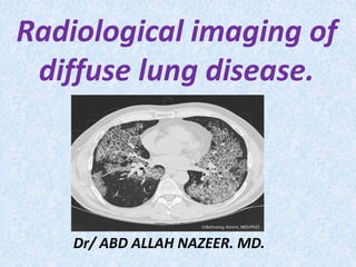
  • 1. Radiological imaging of diffuse lung disease. Dr/ ABD ALLAH NAZEER. MD.
  • 2. Diffuse Interstitial Lung Disease. Diffuse interstitial lung disease is a group of lung diseases that affects the connective tissue that forms the support structure of the air sacs, or alveoli, of the lungs. During inhalation, the alveoli fill with air. Oxygen within the air passes through the walls of the alveoli into the blood stream. In reverse fashion, carbon dioxide passes from the blood into the alveoli and is exhaled. When affected by an interstitial lung disease, the tissue supporting the air sacs—called the interstitium—becomes inflamed and stiff, making it difficult for air sacs to fully expand, limiting the delivery of oxygen to the body and the removal of carbon dioxide from the body. As the disease progresses, the tissue scars and thickens the alveolar walls, further decreasing lung function. This inflammation of the interstitium is typically diffuse, meaning it occurs throughout both lungs rather than being confined to one area.
  • 3. The diseases in this group include idiopathic pulmonary fibrosis (IPF), acute interstitial pneumonia (AIP), cryptogenic organizing pneumonia (COP) and non-specific interstitial pneumonia (NSIP). Some forms of interstitial pneumonia are related to the inhalation of cigarette smoke and occur as a spectrum of injury that includes respiratory bronchiolitis-interstitial lung disease and desquamative interstitial pneumonia. Others are associated with multi-systemic diseases such as rheumatoid arthritis, scleroderma, dermatomyositis and asbestosis. Occasionally prior drug exposure can result in interstitial lung disease, such as the anti-cancer drug Bleomycin. Shortness of breath and a dry cough are the most common symptoms of diffuse interstitial lung disease. As the disease progresses, weight loss, muscle and joint pain and fatigue may also occur. At a more advanced stage, individuals may develop an enlarged heart, an enlargement of the fingertips called clubbing, and cyanosis—a blue coloration in the lips, skin and fingernails as a result of reduced oxygen levels in the blood.
  • 4.
  • 5.
  • 6.
  • 7.
  • 8.
  • 9. How is diffuse interstitial lung disease evaluated. To diagnose and determine the cause of interstitial lung disease, a physician will need to perform a physical examination and order diagnostic tests, including: Blood tests: These are focused on identifying autoimmune diseases such as lupus and rheumatoid arthritis which can result in interstitial lung disease as a result. Spirometry: This is a test of lung function, in which the patient exhales quickly and forcefully through a tube connected to a machine that measures how much air the lungs can hold and how quickly the air moves in and out of the lungs. Spirometry can help determine if there is an issue with air getting into the lungs (restriction, such as fibrosis) versus air getting out of the lungs (obstruction, such as asthma). Pulse oximetry: This test uses a small device placed on a finger tip to measure the oxygen saturation of the blood. It shines a specific wavelength of light though the end of the finger painlessly to measure the amount of oxygen in the blood. Chest x-ray : The patterns of lung damage associated with various types of interstitial lung disease are often identifiable on a chest x-ray. Chest x-rays may also be used to track the progression of the disease.
  • 10. CT imaging of the chest : Computed tomography (CT) scanning, particularly a specific technique known as high resolution CT, is used to see the fine detail of the interstitium where the disease is occurring. Based on the imaging appearance, a diagnosis (specifically idiopathic pulmonary fibrosis) can sometimes be confirmed, thus avoiding the need for lung biopsy. The CT scan can also help determine the extent of damage to the lungs and help determine appropriate treatment. Bronchoscopy and biopsy: In this procedure, a very small sample of tissue is removed from the lung using a small, flexible tube called a bronchoscope that is passed through the mouth or nose and into the lungs. Surgical biopsy: A surgical biopsy is often needed to obtain a larger sample tissue than is possible with bronchoscopy. During this procedure, surgical instruments and a small camera are inserted through two or three small incisions between the ribs that allow a physician to see and remove tissue samples from the lungs.
  • 11.
  • 12.
  • 13.
  • 14.
  • 15.
  • 16. X-Ray chest. It is important modality for diagnosis of ILDs. The correlation between the radiographic pattern and the stage of the disease(clinical or histological) is generally poor. Review all previous film to assess the rate of changes on disease activity. Honeycombing correlated a poor prognosis. X-Ray limitations: X-Ray chest is normal in 10-15% of symptomatic patients with infiltrative lung disease. The X-Ray sensitivity and a specificity of 80% for detection of DPLD. The X-Ray can provide a confident diagnosis in 25% of cases. A diffuse ground glass pattern- early in the disease, progress,- nodules, linear (reticular) infiltrate, or a combination. - Infiltrate become coarser and lung volume is lost- honeycomb pattern.
  • 17.
  • 18.
  • 19.
  • 20.
  • 21. CT pattern of diffuse lung diseases. Four major patterns (ground-glass opacity, nodular opacity, reticular opacity, and honeycombing) on thin- section computed tomographic images were identified.
  • 22.
  • 23.
  • 24.
  • 25.
  • 26. Idiopathic Pulmonary Fibrosis with subpleural reticular marking, traction bronchiectasis and honeycombing in a lower lobe distribution.
  • 27. HRCT cross-sectional view showing a pattern of peripheral reticulation and honeycomb change that is diagnostic of the presence of UIP.
  • 28. Idiopathic Pulmonary Fibrosis with subpleural reticular marking, traction bronchiectasis and honeycombing in a lower lobe distribution.
  • 29. Idiopathic Pulmonary Fibrosis with subpleural reticular marking, traction bronchiectasis and honeycombing in a lower lobe distribution.
  • 34.
  • 35.
  • 36.
  • 37.
  • 38.
  • 39.
  • 40.
  • 41.
  • 42.
  • 43.
  • 44. Sarcoidosis HRCT: Hilar and mediastinal lymphadenopathy, beaded appearance and thickening of brochovascular bundles, nodules along bronchi
  • 45.
  • 46.
  • 48.
  • 49. 2 cases alveolar proteinosis.
  • 51.
  • 52.
  • 53.
  • 54.
  • 55.
  • 56.
  • 58.
  • 63.
  • 65.
  • 68. 2 cases of bronchoalveolar carcinoma
  • 69. 2 cases of bronchoalveolar carcinoma.