SlideShare ist ein Scribd-Unternehmen logo
1 von 50
Functional MRI of brain neoplasm. 
Dr/ ABD ALLAH NAZEER. MD.
Functional magnetic resonance imaging (fMRI) is increasingly 
used in the work-up of patients at the preoperative stage to assess the 
relationship between the functionally eloquent cortex and brain pathology. Inter-individual 
normal variations of anatomy render such assessment unreliable based 
on structural imaging alone despite the definition of clear anatomical landmarks. 
This is even more of an issue when normal anatomy is obscured by a tumour mass 
effect or when functional anatomy is altered due to cortical plasticity. 
fMRI has seen a rapid evolution from its first human application in 1991 to an 
essential tool in the exploration of human brain function, most prominently in the 
scientific arena. 
In 2007, new Current Procedure Terminology (CPT) codes were developed for fMRI 
by the American Medical Association, signifying the transition of fMRI to a 
valuable tool in a clinical setting. 
Recent major advances in clinical fMRI make its acquisition, image processing, and 
even integration of its findings for neuronavigational purposes relatively easy. 
However, the technique is not without limitations and validation issues which are 
easily forgotten when color activation maps become readily available at the single 
click of a button. The theoretical background, the validity in brain tumour 
patients, and several considerations of fMRI are addressed.
fMRI Background: 
Blood Oxygenation Level-Dependent (BOLD) fMRI is the most commonly used 
functional MR neuroimaging technique. BOLD fMRI takes advantage of the tight 
link between local neuronal activity and blood flow, called neurovascular coupling 
due to neurovascular coupling, blood flow and volume increase locally with an 
increase of neuronal activity. This leads to an increase in oxygenated blood that is 
disproportionate to the increased need of oxygen for neuronal activity. As a result, 
there is a relative decrease of paramagnetic deoxygenated hemoglobin which in 
turn leads to an increase of MR signal in those areas of the brain that are active. 
Such signal changes are small and relative, which means that many 
measurements need to be made, typically during an alternation of active and 
baseline conditions in a task that aims to activate the functional brain region of 
interest. Furthermore, the signal changes occur at a delay after and are more 
prolonged than the neuronal activity, defined by the hemodynamic response 
function. A statistical model is created to assess the correlation of the measured 
signal changes with the task, taking the hemodynamic response function into 
account. The resulting statistical map is threshold at a certain p or T value and 
overlaid in color on a high-resolution anatomical image which is acquired 
separately. This is the typical color “activation” map produced by an fMRI image 
processing software, which is merely a combination of anatomical and statistical 
information very indirectly representing neuronal activation.
Task-Based fMRI 
For clinical application, almost exclusively task-based fMRI is used. During 
the performance of a task by the subject in the scanner, rapid imaging of 
the brain is performed. Typically, the entire brain is scanned at intervals of 
3–5 s for a duration of about 5 min so that > 100 measurements are made 
per task. The task consists of active and baseline conditions, which 
commonly alternate in blocks of 20–40 s. Such so-called blocked 
paradigms are statistically robust, since a lot of signal is acquired for each 
condition, but they are restrained because they do not leave much room 
for unexpected or short stimuli. In an event-related task design, individual 
stimuli, each representing a specific condition, are presented in random 
order and rapid succession. Such a task design offers the possibility to 
present unexpected stimuli as well as many different condition. Rendering 
it very flexible, but statistically less robust since the signal acquired per 
condition is generally low. For clinical application, blocked designs are 
generally well-suited and preferable.
Clinical Pre-Surgical fMRI Studies: 
The aim of neurosurgery in brain tumour patients is maximum tumour resection, while 
at the same time minimizing the risk of new functional deficits post-operatively. For 
optimal results, the relationship between the tumour margins and eloquent brain 
regions needs to be established as accurately as possible. The gold standard for such 
assessment is intraoperative ECM, which has in fact been shown to significantly modify 
long-term survival in low-grade glioma patients. However, intraoperative ECM is 
invasive, requires experience and expertise of the neurosurgical team, increases 
surgery duration, and requires awake and active participation, collaboration, and 
motivation of the patient. Additionally, only a limited number of tasks can be tested. 
Functional MRI may be used to make a risk assessment preoperatively, which is of 
particular value in young low-grade glioma patients, to plan and guide the 
neurosurgical approach, shorten surgery duration, and obtain prognostic information 
prior to surgery. For fMRI to be used in such a setting, both high sensitivity and high 
specificity are required. High sensitivity for eloquent brain regions is needed to reduce 
the false negative rate so that no eloquent cortex is missed and no functional deficit is 
induced by surgery. At high specificity the false positive rate is low, which means that 
the visualized areas of activation relate to truly eloquent or critical brain regions. At 
low specificity non-critical brain regions are also visualized, inducing the risk that such 
areas are avoided at surgery and are subsequently exposed to a less extensive 
resection than would have been possible.
Motor Function: 
Motor cortex assessment has been validated in a multitude of studies 
that generally report a good correlation between fMRI and 
intraoperative ECM. Reported sensitivities for localization of the primary 
motor cortex range from 88–100 % . Specificities are also high, ranging 
from 87–100 % . Such high reliability may be contributed to by the robust 
activation that is seen with simple motor tasks that can be easily 
performed by the majority of patients. Also the functional anatomical 
stability of the sensorimotor area, at both the macroscopic and 
microscopic levels, probably contributes to the reliability of fMRI of 
motor function . Spatial accuracy of fMRI motor cortex localization is 
found to be within the range of 1–2 cm. Yetkin et al reported that all 
intraoperative ECM and fMRI sites of activation were within 2 cm, while 
87 % were within 1 cm. Importantly though, reliability seems to be 
decreased with high tumour grades, as demonstrated by Bizzi et al. In 
benign and malignant brain mass lesions in or near the primary motor 
cortex, overall sensitivity was 88 %, but only 65 % in grade-IV gliomas.
Language Function: 
In contrast to the high validity shown for fMRI of motor function, results 
from language function validation studies are controversial, varying from 
100 % sensitivity for fMRI to identify all critical language areas to as low 
as 22 %. Reported specificity is even more variable, ranging from 0– 100 %. 
Validation studies of fMRI of language function in brain tumour patients 
are relatively scarce, are generally performed in small patient populations, 
and suffer from differences in the validation methods used among the 
studies, disparities of brain lesions, and the variety of the language tasks 
performed preoperatively and during intraoperative ECM. The mapping of 
language function is also more complex than that of the sensorimotor 
cortex due to the lack of consistent surface landmarks and substantial 
inter-individual variability. The language cortical network seems to consist 
of critical regions, essential for language processing, and participating but 
non-critical areas, which may be resected without inducing a permanent 
language deficit. These areas cannot be reliably distinguished with fMRI, 
resulting in low specificity of fMRI compared with intraoperative ECM.
Other Functions: 
The visual cortex has been a frequent topic of study since the early days of 
fMRI, due to its relatively strong BOLD response and easy implementation 
of stimulus paradigms. Presurgical mapping of the primary visual cortex 
has been described and may be indicated when the normal anatomy is 
severely distorted by the tumour and/or when the brain structure of 
interest is located deep inside the brain and cannot be assessed by ECM. 
Commonly used stimulus paradigms are flashing lights presented with 
light-proof goggles and reversing black-and-white checkerboards . 
Another function that may be assessed with fMRI is visuospatial attention, 
failure of which results in spatial neglect. 
This condition arises with damage of the temporoparietal or frontal 
cortex, the thalamus or the basal ganglia, generally of the right 
hemisphere. It is an invalidating condition, in which patients behave as 
if the left part of the world does not exist. Functional localization may be 
assessed with fMRI using a line bisection task, in which patients are asked 
to bisect 20-cm horizontal lines, which has been used successfully during 
ECM.
Critical Issues: 
There are several limitations of fMRI that need to be considered when using 
the technique for pre-surgical assessment of brain tumour patients. These 
include issues that are inherent to the technique, such as spatial and 
geometric uncertainty, tumour effects on the BOLD signal, inter- and intra-individual 
variability, lack of discrimination between essential and modulating 
brain regions, and lack of information on the underlying white matter. The 
imaging sequence used for BOLD fMRI is particularly sensitive to 
postoperative effects, such as metallic implants and surgical staples, air 
underneath the skull flap, and blood products, as well. This means that 
additional care needs to be taken in patients who have had previous surgery, 
biopsy, or hemorrhage. Small regions of hemosiderin deposition may not be 
visible on conventional imaging, but will show large artifacts in the BOLD 
fMRI data. Such artifacts are obscured on the fMRI activation color maps, 
which would simply show decreased or no activation in the artefactual area. 
It is therefore crucial that the raw data are scrutinized for such artifacts in 
every patient. Other issues with pre-surgical fMRI, that may be resolved in 
the future, are the lack of standardization of tasks and image processing 
techniques.
Four resting state networks in a normal subject. Also shown are corresponding time courses of 
the independent component coefficients.(Colors quantify the z-value of correlation of the time 
series with the corresponding ICA coefficient. Either positive or negative z-values are shown.)
Activation patterns in response to visual stimuli. Note the lack of 
activation in the central visual cortex when using projection screen.
Visual area locations. A, Parasagittal anatomical image from one subject 
indicating the slice selection (perpendicular to the calcarine sulcus) for the 
retinotopy measurements and the control conditions. B–D, Visual areas V1, V2, 
V3, V3A, and V4v depicted on in-plane anatomies from a similar location (near the 
middle of the 8 in-planes) in three control subjects. E–G, Visual areas depicted on 
in-plane anatomies from a similar location in three dyslexic subjects.
Location of area MT1. A, Parasagittal anatomical image from one subject indicating the 
slice selection (parallel to the calcarine sulcus) for the moving dots and the test conditions. 
The blue lines were slices that contained the MT1 region of interest (ROI) in this subject. B– 
D, Brain activity in one slice containing the MT1 ROI from three control subjects. Reddish 
voxels show regions with greater response to moving versus stationary dot patterns. 
Images were chosen to optimally show MT1 in the right hemisphere (arrows), although 
activity from left hemisphere MT1 and V1 are also present in some cases. The image in B is 
from the same brain as the sagittal image in A and corresponds to the most inferior of the 
three blue slices. The MT1 ROI was defined by outlining (dotted line in B) the strongest 
area of activity that was approximately lateral to the junction between the calcarine sulcus 
and the parieto-occipital sulcus, and beyond the retinotopically organized visual areas. 
E–G, Slices with MT1 ROIs in three dyslexic subjects.
This image is in 
radiological format 
and shows an 
auditory language 
map averaged over 
40 subjects. 
There is bilateral 
primary auditory 
cortex (yellow 
arrows), left 
Wernicke’s (green 
arrows) and Broca’s 
(red arrows), and 
SMA (orange arrows). 
In this auditory task, 
the subject hears 
phrases, such as “The 
funny guys at the 
circus” and thinks 
in his/her mind of a 
word that fits.
Typical pattern of language activation in Reverse Word Reading Task
Figure 4. Functional reorganization of motor area was observed in the patients 
with short term and long term spinal cord injury (sci). 
Figure 4a. Control. 
Figure 4b. Two years after sci. 
Figure 4c. More than 10 years after sci.
Localization of primary motor cortices in 28 years old patients with brain tumor: 
Note that the posterior and medial displacement of motor cortices by the mass lesion
Localization of language cortices in 28 years old patients with brain tumor: 
Note that the strong left lateralization of language activities.
Localization of 
primary motor 
cortices in 28 years 
old patients with 
brain tumor in 3 views
fMRI in preoperative planning. 
3a. Preoperative planning for brain biopsy in a patient with a lesion (arrow) in 
the left frontal lobe (Broca’s area). Significant bilateral language activation 
was observed in this patient. 
3b. fMRI was used to determine the language area and its relation to the tumor 
for a patient with glioblastoma (arrow).
Patient K, male, 37 years old, with glioblastoma in the left temporoparietal 
area,activation from visual task, R - right hemisphere; L - left hemisphere.
Patient P, male, 28 years old, with glioblastoma in the left temporal lobe, activation 
from visual and auditory tasks, R - right hemisphere; L - left hemisphere, WA – 
activation in Wernicke's area. Red - activation elicited by visual task, blue - activation 
elicited by auditory task, purple - activation that coincided in two tasks.
Patient KO, female, 53 years old, with glioblastoma in the left parietal area, 
activation from visual task (red), R - right hemisphere; L - left hemisphere.
Preoperative (A) activity showing the focus 
of activity in 4 patients obtained from the 
comparison of the visuomotor conditional 
blocks of trials with the motor control 
blocks of trials and postoperative (B) 
anatomical images. A: Functional MR 
images show the focus of activity within 
the rostral part of the premotor cortex in 
the lateral view (in all patients) and in the 
top view (in Case 2) of the cortical surface 
rendering in standard stereotactic space of 
the left hemisphere of each patient. The 
gray shaded area of the cortical surface 
shows the tumor location. Each blue cross 
indicates the location of the preoperative 
activity increase in the PMdr. The color 
scale indicates the t value range. B: 
Magnetic resonance images show the 
cortical surfaces as renderings (in standard 
stereotactic space) of the left hemisphere of 
each patient based on the postoperative 
MR image. The deformation of the cortical 
surface shows the extent of the tumor 
resection. The data demonstrate that, in 
each patient, the region involved in the 
visuomotor conditional function was not 
removed during the tumor resection.
Images showing the 
preoperative activity 
obtained from the 
comparison of the 
motor control with the 
visual control block of 
trials. The cortical 
surfaces are renderings 
(in standard stereotactic 
space) of the left 
hemisphere of each 
patient. The horizontal 
yellow line indicates the 
dorsoventral level of the 
horizontal sections 
illustrated in the images 
on the right. The 
increases in activity 
correspond to the 
location of the primary 
hand motor region in 
the precentral 
knob of each patient,
Tumor heterogeneity Recognition Evaluation on the High Grade Glioma of 
the BraTS dataset: Qualitative Example Case BRATS HG0001 a) Gadolinium T1 MRI 
Original Image b) FLAIR MRI Original Image c) Overlap between Gadolinium T1 MRI 
original image and the Ground Truth masks d) Overlap between Gadolinium T1 MRI 
original image and the automatic recognized masks.(Red points are Edema and Cyan 
point are the Active Tumor
Tumor heterogeneity Recognition Evaluation on the High Grade Glioma of 
the BraTS dataset: Qualitative Example Case BRATS HG0001 a) Gadolinium T1 MRI 
Original Image b) FLAIR MRI Original Image c) Overlap between Gadolinium T1 MRI 
original image and the Ground Truth masks d) Overlap between Gadolinium T1 MRI 
original image and the automatic recognized masks.(Red points are Edema and Cyan 
point are the Active Tumor)
18-year old boy with a glioma grade 3 (arrows) near the left rolandic area, demonstrating 
the importance of fMRI in planning surgery and avoiding postoperative neurological 
deficits. fMRI using a visuomotor paradigm showed functional activation less than 2 cm 
posterior to the tumour and partial resection was proposed (A,B). Partial resection was 
performed using intra-operative cortical stimulation, and followed by application of gene 
therapy in the tumour remnant posteriorly. Postoperatively, no motor deficit was present.
26-year old man with right frontal glioma demonstrating the role of fMRI in 
deciding on operability. This man was left-handed which increased chances that 
language areas are not confined solely to the left hemisphere. fMRI using a verb 
generation paradigm showed that the language areas Broca (B) and Wernicke (W) 
were strongly lateralized to the left hemisphere because no activation was seen in 
the right hemisphere. Complete resection of the right frontal glioma (T) was 
performed and the patient did not have any language deficit postoperatively.
59-year old woman with arteriovenous malformation (AVM) in the right parieto-occipital 
region, demonstrating risk for postoperative deficit because activation areas are less 
than 2 cm of the lesion. fMRI using a visuomotor paradigm showed visual activation 
areas (green) medially and caudally relative to the AVM (circle), at less than 1 cm 
distance. After endovascular approach with partial embolisation, complete resection was 
performed surgically. Postoperatively, a left lower quadrant anopsy was present.
(Top row) Acquired axial CT and anatomical MRI slices from the AVM patient. 
(Bottom row left) fMRI activation map fused with the anatomical MRI data 
showing the Broadmann-17 functional structure and (lower right) corresponding 
color-coded (red = left-right; green= anterior-posterior; blue = superior-inferior). 
The FA map was used for calculating the tractography data.
Recurrent left parietal lobe 
anaplastic astrocytoma in 37- 
year-old right-handed woman. 
Surgery was not initially planned 
because of presumed 
involvement of receptive speech 
area. Left inferior and middle 
frontal gyral activation (yellow 
arrows) is consistent with 
dominant expressive speech area 
and is located at anterior border 
of more cephalad component of 
lesion. Left superior and middle 
temporal gyral activation (green 
arrows) is consistent with 
dominant receptive speech area 
and abuts inferior border of 
temporal component of lesion, 
with superior temporal gyral 
activation component lying 
anteroinferior to lesion.
Preoperative fMRI in patients during contralesional movements. Preoperative anatomic and 
functional MRI in Patients 1, 5, and 8 (left column) showed large tumoral infiltration of the 
supplementary motor area (SMA) (asterisk), associated with under activity in the SMA in the 
pathologic hemisphere, and over activity in the healthy SMA resulting in negative magnitude 
laterality indices (LI) (0.40, 0.12, 0.06). Recovery began within the first 3 days and lasted 
between 30 and 60 days. In Patients 6, 9, and 11 (right column), tumoral infiltration (asterisk) 
was lower, and LI remained positive (0.08, 0.19, 0.12). Arrows indicate predominant SMA 
activation. Recovery began after 7 to 21 days, and lasted between 90 and 120 days.
Postoperative fMRI. Anatomic and functional MRI in one representative control 
and in Patients 4, 6, and 12 during ipsilesional (IL) and contralesional (CL) 
movements after surgery. In controls and in patients during IL movements, 
supplementary motor area (SMA) (arrow) and premotor cortex (PMC) (arrowhead) 
activation predominated in the hemisphere contralateral to the moving hand. In 
patients during CL movements, SMA activation was located in the healthy 
hemisphere and PMC activation became more important in the ipsilateral healthy 
hemisphere. The resection cavity is indicated by the asterisk on axial planes.
Axial TIWI in neurologic format show left megalencephaly with an enlarged dysmorphic 
left cerebral hemisphere. There is poly microgyria as well as heterotopia. During this 
auditory task, there was only activation in the right primary auditory cortex (yellow arrow) 
rather the expected bilateral activation. (Color version of figure is available online.)
Conclusions. 
Functional MRI is a valuable tool in the pre-surgical assessment of 
brain tumour patients, but needs to be used with care. 
Interpretation of the results requires a lot of experience and may be 
difficult. Knowledge of functional brain anatomy is a first 
requirement for risk evaluation and to determine which 
fMRI tasks need to be performed. The shortcomings of fMRI in a 
clinical setting as described above need to explicitly be taken into 
account in every patient. In our institution, fMRI, combined with DTI 
tractography, is used to aid neurosurgical planning but 
intraoperative ECM is always used for confirmation when activation 
is shown in the proximity of the brain tumour or if activation is 
atypical. Most importantly, the absence of fMRI activation does not 
exclude the presence of functional neuronal tissue, not even within 
infiltrative tumours.
Thank You.

Weitere ähnliche Inhalte

Was ist angesagt?

Presentation1.pptx, diffusion tensor imaging of white matter tract in cerebra...
Presentation1.pptx, diffusion tensor imaging of white matter tract in cerebra...Presentation1.pptx, diffusion tensor imaging of white matter tract in cerebra...
Presentation1.pptx, diffusion tensor imaging of white matter tract in cerebra...Abdellah Nazeer
 
MRI Procedure of Brain
MRI Procedure of BrainMRI Procedure of Brain
MRI Procedure of BrainSudil Paudyal
 
MRI spectroscopy- Its Application, Principle & Techniques
MRI spectroscopy- Its Application, Principle & Techniques MRI spectroscopy- Its Application, Principle & Techniques
MRI spectroscopy- Its Application, Principle & Techniques Nitish Virmani
 
MR spectroscopy
MR spectroscopyMR spectroscopy
MR spectroscopyairwave12
 
Presentation1.pptx, radiological anatomy of the brain.
Presentation1.pptx, radiological anatomy of the brain.Presentation1.pptx, radiological anatomy of the brain.
Presentation1.pptx, radiological anatomy of the brain.Abdellah Nazeer
 
BASICS IN NEUROIMAGING: CT, MRI AND PET
BASICS IN NEUROIMAGING: CT, MRI AND PETBASICS IN NEUROIMAGING: CT, MRI AND PET
BASICS IN NEUROIMAGING: CT, MRI AND PETPramod Krishnan
 
Radiologic anatomy of the cranial nerves
Radiologic anatomy of the cranial nervesRadiologic anatomy of the cranial nerves
Radiologic anatomy of the cranial nerveshazem youssef
 
Magnetic Resonance Diffusion
Magnetic Resonance DiffusionMagnetic Resonance Diffusion
Magnetic Resonance DiffusionRahman Ud Din
 
Diffusion weighted imaging
Diffusion  weighted imagingDiffusion  weighted imaging
Diffusion weighted imagingSharath Raj
 
Mri shoulder joint with common pathologies
Mri shoulder joint with common pathologiesMri shoulder joint with common pathologies
Mri shoulder joint with common pathologiesGobardhan Thapa
 

Was ist angesagt? (20)

Presentation1.pptx, diffusion tensor imaging of white matter tract in cerebra...
Presentation1.pptx, diffusion tensor imaging of white matter tract in cerebra...Presentation1.pptx, diffusion tensor imaging of white matter tract in cerebra...
Presentation1.pptx, diffusion tensor imaging of white matter tract in cerebra...
 
Ppt mri brain
Ppt mri brainPpt mri brain
Ppt mri brain
 
Fmri overview
Fmri overviewFmri overview
Fmri overview
 
MRI Procedure of Brain
MRI Procedure of BrainMRI Procedure of Brain
MRI Procedure of Brain
 
MRI spectroscopy- Its Application, Principle & Techniques
MRI spectroscopy- Its Application, Principle & Techniques MRI spectroscopy- Its Application, Principle & Techniques
MRI spectroscopy- Its Application, Principle & Techniques
 
Radiological anatomy of the brain
Radiological anatomy of the brainRadiological anatomy of the brain
Radiological anatomy of the brain
 
MR spectroscopy
MR spectroscopyMR spectroscopy
MR spectroscopy
 
Presentation1.pptx, radiological anatomy of the brain.
Presentation1.pptx, radiological anatomy of the brain.Presentation1.pptx, radiological anatomy of the brain.
Presentation1.pptx, radiological anatomy of the brain.
 
Basics Of MRI
Basics Of MRIBasics Of MRI
Basics Of MRI
 
FMRI
FMRIFMRI
FMRI
 
MRI Tractography IMAGING
MRI Tractography IMAGINGMRI Tractography IMAGING
MRI Tractography IMAGING
 
BASICS IN NEUROIMAGING: CT, MRI AND PET
BASICS IN NEUROIMAGING: CT, MRI AND PETBASICS IN NEUROIMAGING: CT, MRI AND PET
BASICS IN NEUROIMAGING: CT, MRI AND PET
 
Radiologic anatomy of the cranial nerves
Radiologic anatomy of the cranial nervesRadiologic anatomy of the cranial nerves
Radiologic anatomy of the cranial nerves
 
Advance fMRI (Fast fMRI)
Advance fMRI (Fast fMRI)Advance fMRI (Fast fMRI)
Advance fMRI (Fast fMRI)
 
Magnetic Resonance Diffusion
Magnetic Resonance DiffusionMagnetic Resonance Diffusion
Magnetic Resonance Diffusion
 
Brain anatomy on ct & amp; mri
Brain anatomy on ct & amp; mriBrain anatomy on ct & amp; mri
Brain anatomy on ct & amp; mri
 
Diffusion weighted imaging
Diffusion  weighted imagingDiffusion  weighted imaging
Diffusion weighted imaging
 
Mri shoulder joint with common pathologies
Mri shoulder joint with common pathologiesMri shoulder joint with common pathologies
Mri shoulder joint with common pathologies
 
MRI artifacts
MRI artifactsMRI artifacts
MRI artifacts
 
MRI of Brain: Basics
MRI of Brain: BasicsMRI of Brain: Basics
MRI of Brain: Basics
 

Andere mochten auch

fMRI Brain Imaging
fMRI Brain ImagingfMRI Brain Imaging
fMRI Brain ImagingMerrett
 
fMRI terms: HRF and BOLD
fMRI terms: HRF and BOLDfMRI terms: HRF and BOLD
fMRI terms: HRF and BOLDRussell James
 
Amthor BMES Presentation Final
Amthor BMES Presentation FinalAmthor BMES Presentation Final
Amthor BMES Presentation FinalMatthew Gray
 
Cosmo-not: a brief look at methods of analysis in functional MRI and in diffu...
Cosmo-not: a brief look at methods of analysis in functional MRI and in diffu...Cosmo-not: a brief look at methods of analysis in functional MRI and in diffu...
Cosmo-not: a brief look at methods of analysis in functional MRI and in diffu...CosmoAIMS Bassett
 
Intro to Transcranial Direct Curent Stimulation (tDCS)
Intro to Transcranial Direct Curent Stimulation (tDCS)Intro to Transcranial Direct Curent Stimulation (tDCS)
Intro to Transcranial Direct Curent Stimulation (tDCS)Daniel Stevenson
 
Class 1 f_mri_intro
Class 1 f_mri_introClass 1 f_mri_intro
Class 1 f_mri_intro景淳 許
 
A (quick) introduction to Magnetic Resonance Imagery preprocessing and analysis
A (quick) introduction to Magnetic Resonance Imagery preprocessing and analysisA (quick) introduction to Magnetic Resonance Imagery preprocessing and analysis
A (quick) introduction to Magnetic Resonance Imagery preprocessing and analysisStephen Larroque
 
Magnetic resonance imaging
Magnetic resonance imagingMagnetic resonance imaging
Magnetic resonance imagingAlbein Vivek
 
Neuroradiology in ms
Neuroradiology in msNeuroradiology in ms
Neuroradiology in msAmr Hassan
 

Andere mochten auch (20)

Introduction to fMRI
Introduction to fMRIIntroduction to fMRI
Introduction to fMRI
 
Fetal MRI
Fetal MRIFetal MRI
Fetal MRI
 
Fetal MRI
Fetal MRIFetal MRI
Fetal MRI
 
fMRI Brain Imaging
fMRI Brain ImagingfMRI Brain Imaging
fMRI Brain Imaging
 
fMRI Study Design
fMRI Study DesignfMRI Study Design
fMRI Study Design
 
fMRI terms: HRF and BOLD
fMRI terms: HRF and BOLDfMRI terms: HRF and BOLD
fMRI terms: HRF and BOLD
 
Tensor de difusão e tratografia
Tensor de difusão e tratografiaTensor de difusão e tratografia
Tensor de difusão e tratografia
 
Copy of NCSU Workshop - DTI.1
Copy of NCSU Workshop - DTI.1Copy of NCSU Workshop - DTI.1
Copy of NCSU Workshop - DTI.1
 
Amthor BMES Presentation Final
Amthor BMES Presentation FinalAmthor BMES Presentation Final
Amthor BMES Presentation Final
 
Cosmo-not: a brief look at methods of analysis in functional MRI and in diffu...
Cosmo-not: a brief look at methods of analysis in functional MRI and in diffu...Cosmo-not: a brief look at methods of analysis in functional MRI and in diffu...
Cosmo-not: a brief look at methods of analysis in functional MRI and in diffu...
 
Dti basics
Dti basicsDti basics
Dti basics
 
Brief Introduction to MRI
Brief Introduction to MRIBrief Introduction to MRI
Brief Introduction to MRI
 
An introduction to MRI
An introduction to MRIAn introduction to MRI
An introduction to MRI
 
Intro to Transcranial Direct Curent Stimulation (tDCS)
Intro to Transcranial Direct Curent Stimulation (tDCS)Intro to Transcranial Direct Curent Stimulation (tDCS)
Intro to Transcranial Direct Curent Stimulation (tDCS)
 
Class 1 f_mri_intro
Class 1 f_mri_introClass 1 f_mri_intro
Class 1 f_mri_intro
 
A (quick) introduction to Magnetic Resonance Imagery preprocessing and analysis
A (quick) introduction to Magnetic Resonance Imagery preprocessing and analysisA (quick) introduction to Magnetic Resonance Imagery preprocessing and analysis
A (quick) introduction to Magnetic Resonance Imagery preprocessing and analysis
 
Magnetic resonance imaging
Magnetic resonance imagingMagnetic resonance imaging
Magnetic resonance imaging
 
Mri final ppt
Mri final pptMri final ppt
Mri final ppt
 
MRI Brain
MRI BrainMRI Brain
MRI Brain
 
Neuroradiology in ms
Neuroradiology in msNeuroradiology in ms
Neuroradiology in ms
 

Ähnlich wie Presentation1.pptx, f mri of brain neoplasm

Seminar on Brain Mapping techniques.pptx
Seminar on Brain Mapping techniques.pptxSeminar on Brain Mapping techniques.pptx
Seminar on Brain Mapping techniques.pptxSoumen Kanjilal
 
Final Progress Report- Farzana
Final Progress Report- FarzanaFinal Progress Report- Farzana
Final Progress Report- FarzanaFarzana Ali
 
Positron-emission tomography studies of cross-modality inhibition in selectiv...
Positron-emission tomography studies of cross-modality inhibition in selectiv...Positron-emission tomography studies of cross-modality inhibition in selectiv...
Positron-emission tomography studies of cross-modality inhibition in selectiv...Dr Brendan O'Sullivan
 
Kohut Literature Review fMRI corrections
Kohut Literature Review fMRI correctionsKohut Literature Review fMRI corrections
Kohut Literature Review fMRI correctionsJulie Kohut
 
Brain Abnormality Categorization
Brain Abnormality CategorizationBrain Abnormality Categorization
Brain Abnormality CategorizationIRJET Journal
 
Anxiety In Adolescent Rhesus Monkeys
Anxiety In Adolescent Rhesus MonkeysAnxiety In Adolescent Rhesus Monkeys
Anxiety In Adolescent Rhesus MonkeysVeronica Smith
 
Proton magnetic resonance spectroscopy
Proton magnetic resonance spectroscopyProton magnetic resonance spectroscopy
Proton magnetic resonance spectroscopyhazem youssef
 
Neuroradiology in dementia
Neuroradiology in dementiaNeuroradiology in dementia
Neuroradiology in dementiaNeurologyKota
 
Magnetoencephalography an emerging biological marker for neurodegenerative an...
Magnetoencephalography an emerging biological marker for neurodegenerative an...Magnetoencephalography an emerging biological marker for neurodegenerative an...
Magnetoencephalography an emerging biological marker for neurodegenerative an...Adonis Sfera, MD
 
Emerging MRI and metabolic neuroimaging techniques in mild traumatic brain in...
Emerging MRI and metabolic neuroimaging techniques in mild traumatic brain in...Emerging MRI and metabolic neuroimaging techniques in mild traumatic brain in...
Emerging MRI and metabolic neuroimaging techniques in mild traumatic brain in...IntesarAldweri
 
PauloLMelo_Report_2nd_lab_rotation_ICNAS_IBILI
PauloLMelo_Report_2nd_lab_rotation_ICNAS_IBILIPauloLMelo_Report_2nd_lab_rotation_ICNAS_IBILI
PauloLMelo_Report_2nd_lab_rotation_ICNAS_IBILIPaulo Melo
 
Tumor Detection Based On Symmetry Information
Tumor Detection Based On Symmetry InformationTumor Detection Based On Symmetry Information
Tumor Detection Based On Symmetry InformationIJERA Editor
 
Evaluation of visual function with opaque media
Evaluation of visual function with opaque media Evaluation of visual function with opaque media
Evaluation of visual function with opaque media Kunal Shinde
 
Computer Aided Therapeutic of Alzheimer’s Disease Eulogizing Pattern Classifi...
Computer Aided Therapeutic of Alzheimer’s Disease Eulogizing Pattern Classifi...Computer Aided Therapeutic of Alzheimer’s Disease Eulogizing Pattern Classifi...
Computer Aided Therapeutic of Alzheimer’s Disease Eulogizing Pattern Classifi...DR.P.S.JAGADEESH KUMAR
 

Ähnlich wie Presentation1.pptx, f mri of brain neoplasm (20)

Epilepsy surgery
Epilepsy surgeryEpilepsy surgery
Epilepsy surgery
 
Seminar on Brain Mapping techniques.pptx
Seminar on Brain Mapping techniques.pptxSeminar on Brain Mapping techniques.pptx
Seminar on Brain Mapping techniques.pptx
 
Final Progress Report- Farzana
Final Progress Report- FarzanaFinal Progress Report- Farzana
Final Progress Report- Farzana
 
Functional MRI in Neuroradio
Functional  MRI in NeuroradioFunctional  MRI in Neuroradio
Functional MRI in Neuroradio
 
Positron-emission tomography studies of cross-modality inhibition in selectiv...
Positron-emission tomography studies of cross-modality inhibition in selectiv...Positron-emission tomography studies of cross-modality inhibition in selectiv...
Positron-emission tomography studies of cross-modality inhibition in selectiv...
 
Kohut Literature Review fMRI corrections
Kohut Literature Review fMRI correctionsKohut Literature Review fMRI corrections
Kohut Literature Review fMRI corrections
 
Gadgets in neuro
Gadgets in neuroGadgets in neuro
Gadgets in neuro
 
Brain Abnormality Categorization
Brain Abnormality CategorizationBrain Abnormality Categorization
Brain Abnormality Categorization
 
Anxiety In Adolescent Rhesus Monkeys
Anxiety In Adolescent Rhesus MonkeysAnxiety In Adolescent Rhesus Monkeys
Anxiety In Adolescent Rhesus Monkeys
 
Proton magnetic resonance spectroscopy
Proton magnetic resonance spectroscopyProton magnetic resonance spectroscopy
Proton magnetic resonance spectroscopy
 
Neuroradiology in dementia
Neuroradiology in dementiaNeuroradiology in dementia
Neuroradiology in dementia
 
Magnetoencephalography an emerging biological marker for neurodegenerative an...
Magnetoencephalography an emerging biological marker for neurodegenerative an...Magnetoencephalography an emerging biological marker for neurodegenerative an...
Magnetoencephalography an emerging biological marker for neurodegenerative an...
 
Facial nerve tests
Facial nerve testsFacial nerve tests
Facial nerve tests
 
Emerging MRI and metabolic neuroimaging techniques in mild traumatic brain in...
Emerging MRI and metabolic neuroimaging techniques in mild traumatic brain in...Emerging MRI and metabolic neuroimaging techniques in mild traumatic brain in...
Emerging MRI and metabolic neuroimaging techniques in mild traumatic brain in...
 
USP.ppt
USP.pptUSP.ppt
USP.ppt
 
PauloLMelo_Report_2nd_lab_rotation_ICNAS_IBILI
PauloLMelo_Report_2nd_lab_rotation_ICNAS_IBILIPauloLMelo_Report_2nd_lab_rotation_ICNAS_IBILI
PauloLMelo_Report_2nd_lab_rotation_ICNAS_IBILI
 
IJET-V2I6P20
IJET-V2I6P20IJET-V2I6P20
IJET-V2I6P20
 
Tumor Detection Based On Symmetry Information
Tumor Detection Based On Symmetry InformationTumor Detection Based On Symmetry Information
Tumor Detection Based On Symmetry Information
 
Evaluation of visual function with opaque media
Evaluation of visual function with opaque media Evaluation of visual function with opaque media
Evaluation of visual function with opaque media
 
Computer Aided Therapeutic of Alzheimer’s Disease Eulogizing Pattern Classifi...
Computer Aided Therapeutic of Alzheimer’s Disease Eulogizing Pattern Classifi...Computer Aided Therapeutic of Alzheimer’s Disease Eulogizing Pattern Classifi...
Computer Aided Therapeutic of Alzheimer’s Disease Eulogizing Pattern Classifi...
 

Mehr von Abdellah Nazeer

Muculoskeletal Pediatic Imaging..pptx
Muculoskeletal Pediatic Imaging..pptxMuculoskeletal Pediatic Imaging..pptx
Muculoskeletal Pediatic Imaging..pptxAbdellah Nazeer
 
Presentation1, Ultrasound of the bowel loops and the lymph nodes..pptx
Presentation1, Ultrasound of the bowel loops and the lymph nodes..pptxPresentation1, Ultrasound of the bowel loops and the lymph nodes..pptx
Presentation1, Ultrasound of the bowel loops and the lymph nodes..pptxAbdellah Nazeer
 
Presentation1 Short cases MD..pptx
Presentation1 Short cases MD..pptxPresentation1 Short cases MD..pptx
Presentation1 Short cases MD..pptxAbdellah Nazeer
 
Presentation1, MD MCQ Cases..pptx
Presentation1, MD MCQ Cases..pptxPresentation1, MD MCQ Cases..pptx
Presentation1, MD MCQ Cases..pptxAbdellah Nazeer
 
Presentation1, Short Cases Quiz..pptx
Presentation1, Short Cases Quiz..pptxPresentation1, Short Cases Quiz..pptx
Presentation1, Short Cases Quiz..pptxAbdellah Nazeer
 
Presentation1, radiological imaging of lateral hindfoot impingement.
Presentation1, radiological imaging of lateral hindfoot impingement.Presentation1, radiological imaging of lateral hindfoot impingement.
Presentation1, radiological imaging of lateral hindfoot impingement.Abdellah Nazeer
 
Presentation2, radiological anatomy of the liver and spleen.
Presentation2, radiological anatomy of the liver and spleen.Presentation2, radiological anatomy of the liver and spleen.
Presentation2, radiological anatomy of the liver and spleen.Abdellah Nazeer
 
Presentation1, artifacts and pitfalls of the wrist and elbow joints.
Presentation1, artifacts and pitfalls of the wrist and elbow joints.Presentation1, artifacts and pitfalls of the wrist and elbow joints.
Presentation1, artifacts and pitfalls of the wrist and elbow joints.Abdellah Nazeer
 
Presentation1, artifact and pitfalls of the knee, hip and ankle joints.
Presentation1, artifact and pitfalls of the knee, hip and ankle joints.Presentation1, artifact and pitfalls of the knee, hip and ankle joints.
Presentation1, artifact and pitfalls of the knee, hip and ankle joints.Abdellah Nazeer
 
Presentation1, radiological imaging of artifact and pitfalls in shoulder join...
Presentation1, radiological imaging of artifact and pitfalls in shoulder join...Presentation1, radiological imaging of artifact and pitfalls in shoulder join...
Presentation1, radiological imaging of artifact and pitfalls in shoulder join...Abdellah Nazeer
 
Presentation1, radiological imaging of internal abdominal hernia.
Presentation1, radiological imaging of internal abdominal hernia.Presentation1, radiological imaging of internal abdominal hernia.
Presentation1, radiological imaging of internal abdominal hernia.Abdellah Nazeer
 
Presentation11, radiological imaging of ovarian torsion.
Presentation11, radiological imaging of ovarian torsion.Presentation11, radiological imaging of ovarian torsion.
Presentation11, radiological imaging of ovarian torsion.Abdellah Nazeer
 
Presentation1, musculoskeletal anatomy.
Presentation1, musculoskeletal anatomy.Presentation1, musculoskeletal anatomy.
Presentation1, musculoskeletal anatomy.Abdellah Nazeer
 
Presentation1, new mri techniques in the diagnosis and monitoring of multiple...
Presentation1, new mri techniques in the diagnosis and monitoring of multiple...Presentation1, new mri techniques in the diagnosis and monitoring of multiple...
Presentation1, new mri techniques in the diagnosis and monitoring of multiple...Abdellah Nazeer
 
Presentation1, radiological application of diffusion weighted mri in neck mas...
Presentation1, radiological application of diffusion weighted mri in neck mas...Presentation1, radiological application of diffusion weighted mri in neck mas...
Presentation1, radiological application of diffusion weighted mri in neck mas...Abdellah Nazeer
 
Presentation1, radiological application of diffusion weighted images in breas...
Presentation1, radiological application of diffusion weighted images in breas...Presentation1, radiological application of diffusion weighted images in breas...
Presentation1, radiological application of diffusion weighted images in breas...Abdellah Nazeer
 
Presentation1, radiological application of diffusion weighted images in abdom...
Presentation1, radiological application of diffusion weighted images in abdom...Presentation1, radiological application of diffusion weighted images in abdom...
Presentation1, radiological application of diffusion weighted images in abdom...Abdellah Nazeer
 
Presentation1, radiological application of diffusion weighted imges in neuror...
Presentation1, radiological application of diffusion weighted imges in neuror...Presentation1, radiological application of diffusion weighted imges in neuror...
Presentation1, radiological application of diffusion weighted imges in neuror...Abdellah Nazeer
 
Presentation1, mr physics.
Presentation1, mr physics.Presentation1, mr physics.
Presentation1, mr physics.Abdellah Nazeer
 
Presentation1. ct physics.
Presentation1. ct physics.Presentation1. ct physics.
Presentation1. ct physics.Abdellah Nazeer
 

Mehr von Abdellah Nazeer (20)

Muculoskeletal Pediatic Imaging..pptx
Muculoskeletal Pediatic Imaging..pptxMuculoskeletal Pediatic Imaging..pptx
Muculoskeletal Pediatic Imaging..pptx
 
Presentation1, Ultrasound of the bowel loops and the lymph nodes..pptx
Presentation1, Ultrasound of the bowel loops and the lymph nodes..pptxPresentation1, Ultrasound of the bowel loops and the lymph nodes..pptx
Presentation1, Ultrasound of the bowel loops and the lymph nodes..pptx
 
Presentation1 Short cases MD..pptx
Presentation1 Short cases MD..pptxPresentation1 Short cases MD..pptx
Presentation1 Short cases MD..pptx
 
Presentation1, MD MCQ Cases..pptx
Presentation1, MD MCQ Cases..pptxPresentation1, MD MCQ Cases..pptx
Presentation1, MD MCQ Cases..pptx
 
Presentation1, Short Cases Quiz..pptx
Presentation1, Short Cases Quiz..pptxPresentation1, Short Cases Quiz..pptx
Presentation1, Short Cases Quiz..pptx
 
Presentation1, radiological imaging of lateral hindfoot impingement.
Presentation1, radiological imaging of lateral hindfoot impingement.Presentation1, radiological imaging of lateral hindfoot impingement.
Presentation1, radiological imaging of lateral hindfoot impingement.
 
Presentation2, radiological anatomy of the liver and spleen.
Presentation2, radiological anatomy of the liver and spleen.Presentation2, radiological anatomy of the liver and spleen.
Presentation2, radiological anatomy of the liver and spleen.
 
Presentation1, artifacts and pitfalls of the wrist and elbow joints.
Presentation1, artifacts and pitfalls of the wrist and elbow joints.Presentation1, artifacts and pitfalls of the wrist and elbow joints.
Presentation1, artifacts and pitfalls of the wrist and elbow joints.
 
Presentation1, artifact and pitfalls of the knee, hip and ankle joints.
Presentation1, artifact and pitfalls of the knee, hip and ankle joints.Presentation1, artifact and pitfalls of the knee, hip and ankle joints.
Presentation1, artifact and pitfalls of the knee, hip and ankle joints.
 
Presentation1, radiological imaging of artifact and pitfalls in shoulder join...
Presentation1, radiological imaging of artifact and pitfalls in shoulder join...Presentation1, radiological imaging of artifact and pitfalls in shoulder join...
Presentation1, radiological imaging of artifact and pitfalls in shoulder join...
 
Presentation1, radiological imaging of internal abdominal hernia.
Presentation1, radiological imaging of internal abdominal hernia.Presentation1, radiological imaging of internal abdominal hernia.
Presentation1, radiological imaging of internal abdominal hernia.
 
Presentation11, radiological imaging of ovarian torsion.
Presentation11, radiological imaging of ovarian torsion.Presentation11, radiological imaging of ovarian torsion.
Presentation11, radiological imaging of ovarian torsion.
 
Presentation1, musculoskeletal anatomy.
Presentation1, musculoskeletal anatomy.Presentation1, musculoskeletal anatomy.
Presentation1, musculoskeletal anatomy.
 
Presentation1, new mri techniques in the diagnosis and monitoring of multiple...
Presentation1, new mri techniques in the diagnosis and monitoring of multiple...Presentation1, new mri techniques in the diagnosis and monitoring of multiple...
Presentation1, new mri techniques in the diagnosis and monitoring of multiple...
 
Presentation1, radiological application of diffusion weighted mri in neck mas...
Presentation1, radiological application of diffusion weighted mri in neck mas...Presentation1, radiological application of diffusion weighted mri in neck mas...
Presentation1, radiological application of diffusion weighted mri in neck mas...
 
Presentation1, radiological application of diffusion weighted images in breas...
Presentation1, radiological application of diffusion weighted images in breas...Presentation1, radiological application of diffusion weighted images in breas...
Presentation1, radiological application of diffusion weighted images in breas...
 
Presentation1, radiological application of diffusion weighted images in abdom...
Presentation1, radiological application of diffusion weighted images in abdom...Presentation1, radiological application of diffusion weighted images in abdom...
Presentation1, radiological application of diffusion weighted images in abdom...
 
Presentation1, radiological application of diffusion weighted imges in neuror...
Presentation1, radiological application of diffusion weighted imges in neuror...Presentation1, radiological application of diffusion weighted imges in neuror...
Presentation1, radiological application of diffusion weighted imges in neuror...
 
Presentation1, mr physics.
Presentation1, mr physics.Presentation1, mr physics.
Presentation1, mr physics.
 
Presentation1. ct physics.
Presentation1. ct physics.Presentation1. ct physics.
Presentation1. ct physics.
 

Presentation1.pptx, f mri of brain neoplasm

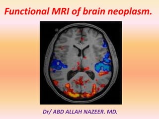
  • 1. Functional MRI of brain neoplasm. Dr/ ABD ALLAH NAZEER. MD.
  • 2. Functional magnetic resonance imaging (fMRI) is increasingly used in the work-up of patients at the preoperative stage to assess the relationship between the functionally eloquent cortex and brain pathology. Inter-individual normal variations of anatomy render such assessment unreliable based on structural imaging alone despite the definition of clear anatomical landmarks. This is even more of an issue when normal anatomy is obscured by a tumour mass effect or when functional anatomy is altered due to cortical plasticity. fMRI has seen a rapid evolution from its first human application in 1991 to an essential tool in the exploration of human brain function, most prominently in the scientific arena. In 2007, new Current Procedure Terminology (CPT) codes were developed for fMRI by the American Medical Association, signifying the transition of fMRI to a valuable tool in a clinical setting. Recent major advances in clinical fMRI make its acquisition, image processing, and even integration of its findings for neuronavigational purposes relatively easy. However, the technique is not without limitations and validation issues which are easily forgotten when color activation maps become readily available at the single click of a button. The theoretical background, the validity in brain tumour patients, and several considerations of fMRI are addressed.
  • 3. fMRI Background: Blood Oxygenation Level-Dependent (BOLD) fMRI is the most commonly used functional MR neuroimaging technique. BOLD fMRI takes advantage of the tight link between local neuronal activity and blood flow, called neurovascular coupling due to neurovascular coupling, blood flow and volume increase locally with an increase of neuronal activity. This leads to an increase in oxygenated blood that is disproportionate to the increased need of oxygen for neuronal activity. As a result, there is a relative decrease of paramagnetic deoxygenated hemoglobin which in turn leads to an increase of MR signal in those areas of the brain that are active. Such signal changes are small and relative, which means that many measurements need to be made, typically during an alternation of active and baseline conditions in a task that aims to activate the functional brain region of interest. Furthermore, the signal changes occur at a delay after and are more prolonged than the neuronal activity, defined by the hemodynamic response function. A statistical model is created to assess the correlation of the measured signal changes with the task, taking the hemodynamic response function into account. The resulting statistical map is threshold at a certain p or T value and overlaid in color on a high-resolution anatomical image which is acquired separately. This is the typical color “activation” map produced by an fMRI image processing software, which is merely a combination of anatomical and statistical information very indirectly representing neuronal activation.
  • 4. Task-Based fMRI For clinical application, almost exclusively task-based fMRI is used. During the performance of a task by the subject in the scanner, rapid imaging of the brain is performed. Typically, the entire brain is scanned at intervals of 3–5 s for a duration of about 5 min so that > 100 measurements are made per task. The task consists of active and baseline conditions, which commonly alternate in blocks of 20–40 s. Such so-called blocked paradigms are statistically robust, since a lot of signal is acquired for each condition, but they are restrained because they do not leave much room for unexpected or short stimuli. In an event-related task design, individual stimuli, each representing a specific condition, are presented in random order and rapid succession. Such a task design offers the possibility to present unexpected stimuli as well as many different condition. Rendering it very flexible, but statistically less robust since the signal acquired per condition is generally low. For clinical application, blocked designs are generally well-suited and preferable.
  • 5. Clinical Pre-Surgical fMRI Studies: The aim of neurosurgery in brain tumour patients is maximum tumour resection, while at the same time minimizing the risk of new functional deficits post-operatively. For optimal results, the relationship between the tumour margins and eloquent brain regions needs to be established as accurately as possible. The gold standard for such assessment is intraoperative ECM, which has in fact been shown to significantly modify long-term survival in low-grade glioma patients. However, intraoperative ECM is invasive, requires experience and expertise of the neurosurgical team, increases surgery duration, and requires awake and active participation, collaboration, and motivation of the patient. Additionally, only a limited number of tasks can be tested. Functional MRI may be used to make a risk assessment preoperatively, which is of particular value in young low-grade glioma patients, to plan and guide the neurosurgical approach, shorten surgery duration, and obtain prognostic information prior to surgery. For fMRI to be used in such a setting, both high sensitivity and high specificity are required. High sensitivity for eloquent brain regions is needed to reduce the false negative rate so that no eloquent cortex is missed and no functional deficit is induced by surgery. At high specificity the false positive rate is low, which means that the visualized areas of activation relate to truly eloquent or critical brain regions. At low specificity non-critical brain regions are also visualized, inducing the risk that such areas are avoided at surgery and are subsequently exposed to a less extensive resection than would have been possible.
  • 6. Motor Function: Motor cortex assessment has been validated in a multitude of studies that generally report a good correlation between fMRI and intraoperative ECM. Reported sensitivities for localization of the primary motor cortex range from 88–100 % . Specificities are also high, ranging from 87–100 % . Such high reliability may be contributed to by the robust activation that is seen with simple motor tasks that can be easily performed by the majority of patients. Also the functional anatomical stability of the sensorimotor area, at both the macroscopic and microscopic levels, probably contributes to the reliability of fMRI of motor function . Spatial accuracy of fMRI motor cortex localization is found to be within the range of 1–2 cm. Yetkin et al reported that all intraoperative ECM and fMRI sites of activation were within 2 cm, while 87 % were within 1 cm. Importantly though, reliability seems to be decreased with high tumour grades, as demonstrated by Bizzi et al. In benign and malignant brain mass lesions in or near the primary motor cortex, overall sensitivity was 88 %, but only 65 % in grade-IV gliomas.
  • 7. Language Function: In contrast to the high validity shown for fMRI of motor function, results from language function validation studies are controversial, varying from 100 % sensitivity for fMRI to identify all critical language areas to as low as 22 %. Reported specificity is even more variable, ranging from 0– 100 %. Validation studies of fMRI of language function in brain tumour patients are relatively scarce, are generally performed in small patient populations, and suffer from differences in the validation methods used among the studies, disparities of brain lesions, and the variety of the language tasks performed preoperatively and during intraoperative ECM. The mapping of language function is also more complex than that of the sensorimotor cortex due to the lack of consistent surface landmarks and substantial inter-individual variability. The language cortical network seems to consist of critical regions, essential for language processing, and participating but non-critical areas, which may be resected without inducing a permanent language deficit. These areas cannot be reliably distinguished with fMRI, resulting in low specificity of fMRI compared with intraoperative ECM.
  • 8. Other Functions: The visual cortex has been a frequent topic of study since the early days of fMRI, due to its relatively strong BOLD response and easy implementation of stimulus paradigms. Presurgical mapping of the primary visual cortex has been described and may be indicated when the normal anatomy is severely distorted by the tumour and/or when the brain structure of interest is located deep inside the brain and cannot be assessed by ECM. Commonly used stimulus paradigms are flashing lights presented with light-proof goggles and reversing black-and-white checkerboards . Another function that may be assessed with fMRI is visuospatial attention, failure of which results in spatial neglect. This condition arises with damage of the temporoparietal or frontal cortex, the thalamus or the basal ganglia, generally of the right hemisphere. It is an invalidating condition, in which patients behave as if the left part of the world does not exist. Functional localization may be assessed with fMRI using a line bisection task, in which patients are asked to bisect 20-cm horizontal lines, which has been used successfully during ECM.
  • 9. Critical Issues: There are several limitations of fMRI that need to be considered when using the technique for pre-surgical assessment of brain tumour patients. These include issues that are inherent to the technique, such as spatial and geometric uncertainty, tumour effects on the BOLD signal, inter- and intra-individual variability, lack of discrimination between essential and modulating brain regions, and lack of information on the underlying white matter. The imaging sequence used for BOLD fMRI is particularly sensitive to postoperative effects, such as metallic implants and surgical staples, air underneath the skull flap, and blood products, as well. This means that additional care needs to be taken in patients who have had previous surgery, biopsy, or hemorrhage. Small regions of hemosiderin deposition may not be visible on conventional imaging, but will show large artifacts in the BOLD fMRI data. Such artifacts are obscured on the fMRI activation color maps, which would simply show decreased or no activation in the artefactual area. It is therefore crucial that the raw data are scrutinized for such artifacts in every patient. Other issues with pre-surgical fMRI, that may be resolved in the future, are the lack of standardization of tasks and image processing techniques.
  • 10. Four resting state networks in a normal subject. Also shown are corresponding time courses of the independent component coefficients.(Colors quantify the z-value of correlation of the time series with the corresponding ICA coefficient. Either positive or negative z-values are shown.)
  • 11. Activation patterns in response to visual stimuli. Note the lack of activation in the central visual cortex when using projection screen.
  • 12. Visual area locations. A, Parasagittal anatomical image from one subject indicating the slice selection (perpendicular to the calcarine sulcus) for the retinotopy measurements and the control conditions. B–D, Visual areas V1, V2, V3, V3A, and V4v depicted on in-plane anatomies from a similar location (near the middle of the 8 in-planes) in three control subjects. E–G, Visual areas depicted on in-plane anatomies from a similar location in three dyslexic subjects.
  • 13. Location of area MT1. A, Parasagittal anatomical image from one subject indicating the slice selection (parallel to the calcarine sulcus) for the moving dots and the test conditions. The blue lines were slices that contained the MT1 region of interest (ROI) in this subject. B– D, Brain activity in one slice containing the MT1 ROI from three control subjects. Reddish voxels show regions with greater response to moving versus stationary dot patterns. Images were chosen to optimally show MT1 in the right hemisphere (arrows), although activity from left hemisphere MT1 and V1 are also present in some cases. The image in B is from the same brain as the sagittal image in A and corresponds to the most inferior of the three blue slices. The MT1 ROI was defined by outlining (dotted line in B) the strongest area of activity that was approximately lateral to the junction between the calcarine sulcus and the parieto-occipital sulcus, and beyond the retinotopically organized visual areas. E–G, Slices with MT1 ROIs in three dyslexic subjects.
  • 14. This image is in radiological format and shows an auditory language map averaged over 40 subjects. There is bilateral primary auditory cortex (yellow arrows), left Wernicke’s (green arrows) and Broca’s (red arrows), and SMA (orange arrows). In this auditory task, the subject hears phrases, such as “The funny guys at the circus” and thinks in his/her mind of a word that fits.
  • 15. Typical pattern of language activation in Reverse Word Reading Task
  • 16. Figure 4. Functional reorganization of motor area was observed in the patients with short term and long term spinal cord injury (sci). Figure 4a. Control. Figure 4b. Two years after sci. Figure 4c. More than 10 years after sci.
  • 17.
  • 18.
  • 19.
  • 20. Localization of primary motor cortices in 28 years old patients with brain tumor: Note that the posterior and medial displacement of motor cortices by the mass lesion
  • 21. Localization of language cortices in 28 years old patients with brain tumor: Note that the strong left lateralization of language activities.
  • 22.
  • 23. Localization of primary motor cortices in 28 years old patients with brain tumor in 3 views
  • 24.
  • 25. fMRI in preoperative planning. 3a. Preoperative planning for brain biopsy in a patient with a lesion (arrow) in the left frontal lobe (Broca’s area). Significant bilateral language activation was observed in this patient. 3b. fMRI was used to determine the language area and its relation to the tumor for a patient with glioblastoma (arrow).
  • 26.
  • 27.
  • 28.
  • 29.
  • 30.
  • 31.
  • 32. Patient K, male, 37 years old, with glioblastoma in the left temporoparietal area,activation from visual task, R - right hemisphere; L - left hemisphere.
  • 33. Patient P, male, 28 years old, with glioblastoma in the left temporal lobe, activation from visual and auditory tasks, R - right hemisphere; L - left hemisphere, WA – activation in Wernicke's area. Red - activation elicited by visual task, blue - activation elicited by auditory task, purple - activation that coincided in two tasks.
  • 34. Patient KO, female, 53 years old, with glioblastoma in the left parietal area, activation from visual task (red), R - right hemisphere; L - left hemisphere.
  • 35. Preoperative (A) activity showing the focus of activity in 4 patients obtained from the comparison of the visuomotor conditional blocks of trials with the motor control blocks of trials and postoperative (B) anatomical images. A: Functional MR images show the focus of activity within the rostral part of the premotor cortex in the lateral view (in all patients) and in the top view (in Case 2) of the cortical surface rendering in standard stereotactic space of the left hemisphere of each patient. The gray shaded area of the cortical surface shows the tumor location. Each blue cross indicates the location of the preoperative activity increase in the PMdr. The color scale indicates the t value range. B: Magnetic resonance images show the cortical surfaces as renderings (in standard stereotactic space) of the left hemisphere of each patient based on the postoperative MR image. The deformation of the cortical surface shows the extent of the tumor resection. The data demonstrate that, in each patient, the region involved in the visuomotor conditional function was not removed during the tumor resection.
  • 36. Images showing the preoperative activity obtained from the comparison of the motor control with the visual control block of trials. The cortical surfaces are renderings (in standard stereotactic space) of the left hemisphere of each patient. The horizontal yellow line indicates the dorsoventral level of the horizontal sections illustrated in the images on the right. The increases in activity correspond to the location of the primary hand motor region in the precentral knob of each patient,
  • 37. Tumor heterogeneity Recognition Evaluation on the High Grade Glioma of the BraTS dataset: Qualitative Example Case BRATS HG0001 a) Gadolinium T1 MRI Original Image b) FLAIR MRI Original Image c) Overlap between Gadolinium T1 MRI original image and the Ground Truth masks d) Overlap between Gadolinium T1 MRI original image and the automatic recognized masks.(Red points are Edema and Cyan point are the Active Tumor
  • 38. Tumor heterogeneity Recognition Evaluation on the High Grade Glioma of the BraTS dataset: Qualitative Example Case BRATS HG0001 a) Gadolinium T1 MRI Original Image b) FLAIR MRI Original Image c) Overlap between Gadolinium T1 MRI original image and the Ground Truth masks d) Overlap between Gadolinium T1 MRI original image and the automatic recognized masks.(Red points are Edema and Cyan point are the Active Tumor)
  • 39.
  • 40.
  • 41. 18-year old boy with a glioma grade 3 (arrows) near the left rolandic area, demonstrating the importance of fMRI in planning surgery and avoiding postoperative neurological deficits. fMRI using a visuomotor paradigm showed functional activation less than 2 cm posterior to the tumour and partial resection was proposed (A,B). Partial resection was performed using intra-operative cortical stimulation, and followed by application of gene therapy in the tumour remnant posteriorly. Postoperatively, no motor deficit was present.
  • 42. 26-year old man with right frontal glioma demonstrating the role of fMRI in deciding on operability. This man was left-handed which increased chances that language areas are not confined solely to the left hemisphere. fMRI using a verb generation paradigm showed that the language areas Broca (B) and Wernicke (W) were strongly lateralized to the left hemisphere because no activation was seen in the right hemisphere. Complete resection of the right frontal glioma (T) was performed and the patient did not have any language deficit postoperatively.
  • 43. 59-year old woman with arteriovenous malformation (AVM) in the right parieto-occipital region, demonstrating risk for postoperative deficit because activation areas are less than 2 cm of the lesion. fMRI using a visuomotor paradigm showed visual activation areas (green) medially and caudally relative to the AVM (circle), at less than 1 cm distance. After endovascular approach with partial embolisation, complete resection was performed surgically. Postoperatively, a left lower quadrant anopsy was present.
  • 44. (Top row) Acquired axial CT and anatomical MRI slices from the AVM patient. (Bottom row left) fMRI activation map fused with the anatomical MRI data showing the Broadmann-17 functional structure and (lower right) corresponding color-coded (red = left-right; green= anterior-posterior; blue = superior-inferior). The FA map was used for calculating the tractography data.
  • 45. Recurrent left parietal lobe anaplastic astrocytoma in 37- year-old right-handed woman. Surgery was not initially planned because of presumed involvement of receptive speech area. Left inferior and middle frontal gyral activation (yellow arrows) is consistent with dominant expressive speech area and is located at anterior border of more cephalad component of lesion. Left superior and middle temporal gyral activation (green arrows) is consistent with dominant receptive speech area and abuts inferior border of temporal component of lesion, with superior temporal gyral activation component lying anteroinferior to lesion.
  • 46. Preoperative fMRI in patients during contralesional movements. Preoperative anatomic and functional MRI in Patients 1, 5, and 8 (left column) showed large tumoral infiltration of the supplementary motor area (SMA) (asterisk), associated with under activity in the SMA in the pathologic hemisphere, and over activity in the healthy SMA resulting in negative magnitude laterality indices (LI) (0.40, 0.12, 0.06). Recovery began within the first 3 days and lasted between 30 and 60 days. In Patients 6, 9, and 11 (right column), tumoral infiltration (asterisk) was lower, and LI remained positive (0.08, 0.19, 0.12). Arrows indicate predominant SMA activation. Recovery began after 7 to 21 days, and lasted between 90 and 120 days.
  • 47. Postoperative fMRI. Anatomic and functional MRI in one representative control and in Patients 4, 6, and 12 during ipsilesional (IL) and contralesional (CL) movements after surgery. In controls and in patients during IL movements, supplementary motor area (SMA) (arrow) and premotor cortex (PMC) (arrowhead) activation predominated in the hemisphere contralateral to the moving hand. In patients during CL movements, SMA activation was located in the healthy hemisphere and PMC activation became more important in the ipsilateral healthy hemisphere. The resection cavity is indicated by the asterisk on axial planes.
  • 48. Axial TIWI in neurologic format show left megalencephaly with an enlarged dysmorphic left cerebral hemisphere. There is poly microgyria as well as heterotopia. During this auditory task, there was only activation in the right primary auditory cortex (yellow arrow) rather the expected bilateral activation. (Color version of figure is available online.)
  • 49. Conclusions. Functional MRI is a valuable tool in the pre-surgical assessment of brain tumour patients, but needs to be used with care. Interpretation of the results requires a lot of experience and may be difficult. Knowledge of functional brain anatomy is a first requirement for risk evaluation and to determine which fMRI tasks need to be performed. The shortcomings of fMRI in a clinical setting as described above need to explicitly be taken into account in every patient. In our institution, fMRI, combined with DTI tractography, is used to aid neurosurgical planning but intraoperative ECM is always used for confirmation when activation is shown in the proximity of the brain tumour or if activation is atypical. Most importantly, the absence of fMRI activation does not exclude the presence of functional neuronal tissue, not even within infiltrative tumours.