SlideShare ist ein Scribd-Unternehmen logo
1 von 45
Dr/ ABD ALLAH NAZEER. MD.
Radiological Imaging of thoracic aortic aneurysm.
Thoracic aortic aneurysms are relatively uncommon compared
to abdominal aortic aneurysms. There is a wide range of causes, and the
ascending aorta is most commonly affected. CTA and MRA are the
modalities of choice to image this condition.
Terminology
The term aneurysm is used when the axial diameter of the thoracic
aorta is > 5 cm and when it measures 4-5 cm the term dilatation is used.
Epidemiology
Most commonly occur in 50 to 60-year-old age group and incidence is
estimated at ~7.5 per 100,000 patient years. There is a male
predominance (M:F=3:1).
Clinical presentation
Thoracic aneurysms are often identified incidentally on imaging of the
chest. A symptomatic presentation may be due to mass effect on airway or
esophagus. Alternatively, they may present due to a complication,
including rupture, aorto-bronchial or aorto-oesophageal fistulae.
Pseudoaneurysms of the thoracic aorta are usually the result of significant
thoracic trauma, both penetrating and blunt, and carry a very high
mortality, with 80-90% of patients dying before reaching hospital
Pathology
Location
Aneurysmal dilatation can affect any
part of the thoracic aorta. Relative
frequencies are (with some involving
more than one segment):
aortic root/ascending aorta: 60%
aortic arch: 10%
descending aorta: 40%
thoracoabdominal segment: 10%
Etiology
Thoracic aortic aneurysms can be divided pathologically according to their relationship
to the aortic wall:
true aneurysm
atherosclerotic aneurysms (most common)
inflammatory/aortitis
rheumatoid arthritis (RA)
ankylosing spondylitis
Takayasu arteritis
giant cell arteritis (GCA)
syphilis
connective tissue disease
cystic medial necrosis
Marfan disease
Ehlers-Danlos syndrome
Loeys-Dietz syndrome
bicuspid aortic valve
false aneurysm
trauma
post surgery
mycotic aneurysms
Associations
intracranial cerebral aneurysms (~10% prevalence).
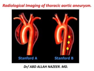
Stanford classification:
The Acute Aortic Syndrome (AAS) is classified according to Stanford.
Stanford Type A lesions involve the ascending aorta and aortic arch
and may or may not involve the descending aorta(60-70%).
Stanford Type B lesions involve the thoracic aorta distal to the left
subclavian artery(30-40%).
The Stanford classification has replaced the DeBakey classification
(type I= ascending, arch and descending aorta: type II= only
ascending aorta: type III= only descending aorta).
DeBakey system divides dissection into three (3) types:
DeBakey I – originates at the ascending aorta and involves at least the aortic arch and may extend to
the descending aorta
DeBakey II – involves the ascending aorta only
DeBakey types I and II are equivalent to Stanford type A
DeBakey III – begins in the descending aorta usually distal to the origin of the subclavian artery and is
equivalent to Stanford type B. It is further classified to type a and b.
- IIIa refers to dissections that originate distal to the left
subclavian artery but extend proximally or distally, mostly above
the diaphragm
- IIIb is similar dissection that originate distal to the left
subclavian artery however extends below the diaphragm
Radiographic features
The location and shape of thoracic aortic aneurysms are variable. An
aortic aneurysm, as aneurysms elsewhere, can be described as saccular or
fusiform. In the case of fusiform dilatation, the term aneurysm should be
applied when the diameter is >4 cm.
Plain radiograph
The thoracic aorta can usually be seen on both frontal and lateral chest
radiographs, and aneurysms are often obvious. However, it is difficult to
assess size accurately (due to magnification effects and often poor
visualization on the side of the artery).
Additionally, mediastinal masses may mimic aortic aneurysms.
Mural calcification is seen both in atherosclerotic disease as well as
various causes of aortitis.
Ultrasound
Unlike abdominal aneurysms that can usually be readily assessed and
monitored with ultrasound, thoracic aortic aneurysms are encased in bone
and air making transthoracic ultrasound of no use.
Transoesophageal echocardiography can visualize much of the descending
aorta, but due to its invasive nature is not routinely used.
CT
CTA is the work-horse of aneurysm assessment able to rapidly image the relevant vascular
territory with high resolution. It can visualise both the sac and the lumen and detect
potential complications.
Typically aneurysms appear as dilatations of the lumen. The walls may be thin or thickened
by the presence of a mural thrombus (circumferential or more frequently eccentric).
Calcified atherosclerotic disease is often identified not only in the wall of an aneurysm but
adjacent arteries.
If rupture or leak has occurred hematoma/fluid may be seen adjacent to the aorta, in the
left pleural cavity or the pericardium.
See main article: reporting tips for aortic aneurysms.
MRI
MRI has the advantage of not requiring ionizing radiation or large volumes of iodinated
contrast 2. This is particularly advantageous in young patients with connective tissue
disorders. However, there are limitations in patients with pacemakers, and those with
reduced renal function (see nephrogenic systemic fibrosis)
Acquisitions capable of being reformatted in three dimensions are essential to allow for
accurate luminal measurement.
Digital subtraction angiography (DSA)
Although angiography has long been considered the gold standard for vascular imaging, it
has largely been superseded by CTA and MRA, which can obtain 3D volumetric data, and
able to assess the extraluminal soft tissues.
Angiography is however used during endovascular repair.
TAA. This huge aneurysm of the aortic arch and descending thoracic
aorta was an incidental finding on this chest radiograph
Syphilitic aneurysm Interval evolution of a Syphilitic Aortic Aneurysm (arrows)
involving the thoracic aorta as seen on these PA chest x rays taken two years apart.
Aortic Aneurysm Dissection.
Chest X-ray (postero-anterior projection) showing that the
descending thoracic aorta is extremely dilated and tortuous.
A transthoracic echocardiogram, in a parasternal long-axis view, demonstrating a
dilated aortic root (4.4 cm) and ascending aorta (4.2 cm). Whereas the aortic root is
well visualized, the ascending aorta is less so, as is often the case with transthoracic
imaging. RV indicates right ventricle; LV, left ventricle; and LA, left atrium.
Parasternal long-axis view by transthoracic echocardiography. (A)
Annuloaortic ectasia with pyriform morphology. (B) Ascending aorta
aneurysm located in the upper part of the sinotubular junction.
Pear shaped proximal ascending thoracic aorta in a patient with Marfan syndrome.
Transoesophageal echocardiography (long-axis view). This illustrates
an ascending aortic aneurysm, with dilation at the level of the sinuses
(onion bulb appearance) in a patient with Marfan syndrome.
Aortography of an ascending aortic aneurysm in
a patient with an idiopathic aortic aneurysm.
Dilation is predominating at the aortic sinuses
Marfan syndrome and annuloaortic ectasia in a 40-year-old man. Contrast-enhanced CT
scan (a) and three-dimensional VR image (b) show a pear-shaped aorta that tapers to a
normal aortic arch, a finding characteristic of Marfan syndrome and annuloaortic ectasia.
Ascending aortic aneurysm and bicuspid aortic valve in a 40-year-old woman. (a, b) Contrast-
enhanced CT scan (a) and VR image (b) show an ascending aortic aneurysm. (c) Oblique axial
image through the plane of the aortic valve shows the bicuspid nature of the valve.
Takayasu arteritis in a 35-year-old woman. (a, b) Contrast-enhanced CT scans obtained
at the level of the ascending (a) and distal descending (b) aorta show diffuse aortic
wall thickening and an ascending aortic aneurysm. (c) VR image shows multiple areas
of stenosis and aneurysm formation involving the aorta and its branches.
Contrast-enhanced axial (a) and oblique sagittal reformatted (b) CT images obtained in a 65-year-old
man show a type B aortic dissection with a partially thrombosed false lumen and a descending TAA.
Syphilitic aneurysm Contrast enhanced CT in a 57 year old male with
chest pain demonstrating concentric wall thickening (arrows) of the
ascending aorta and aortic arch in a patient with syphilitic aortitis.
Mycotic aneurysm Scout chest image and axial contrast enhanced CT thorax in this patient with
disseminated Staphylococcus infection in an IV drug abuser, demonstrating a large mycotic
aneurysm of the brachiocephalic artery (arrows) with circumferential aortic wall thickening.
Ascending aortic aneurysm.
Aortic dilation—(a) sagittal oblique reconstructed CT in a 41-year-old male shows dilated
ascending aorta (4.6 cm) not having reached the size required for a diagnosis of aneurysm.
(b) Axial CT image shows the dilated ascending aorta but normal descending thoracic aorta.
Fusiform ascending aortic aneurysm—sagittal reconstructed CT image in a 47-year-old male shows a
smooth, fusiform aneurysm of the ascending aorta, which tapers to normal caliber at the proximal arch.
A, Standard axial image from a contrast-enhanced CT scan showing what appears to be an oval-shaped descending thoracic
aortic aneurysm, appearing to measure as much as 8.0×5.2 cm in diameter (arrows). B, Three-dimensional reconstruction in a
left anterior oblique view of same CT scan demonstrating that the descending aorta is tortuous and was consequently cut off-
axis (dotted arrow) on axial CT image. The true maximal diameter of this aortic segment was only 5.6 cm (solid arrow).
Infectious aortitis s/p aortic graft repair Contrast enhanced CT demonstrating hypo enhancing
concentric wall thickening (arrows) of the left brachiocephalic artery (A), aortic arch (B), and
the descending aorta (C,D) in a patient with infectious aortitis s/p aortic graft repair
MR angiogram demonstrating a 4.7-cm ascending thoracic aortic aneurysm.
Contrast-enhanced CT scan demonstrating a 7.5×8.3-cm ascending
thoracic aortic aneurysm. A indicates ascending; D, descending.
Ascending thoracic aortic aneurysm.
Mycotic aneurysm. A: Axial contrast enhanced magnetic resonance angiography (MRA) shows large proximal
descending aortic aneurysm with eccentric thrombus and invasion of adjacent thoracic vertebral body (arrow); B:
Sagittal short tau inversion recovery shows large descending aortic aneurysm (white arrow) with high signal within
the wall posteriorly (red arrow) indicating acute inflammation; C: Sagittal contrast enhanced MRA maximum intensity
projection reconstruction shows proximal descending aortic mycotic aneurysm (arrow) extending posteriorly.
Aortic pseudoaneurysm after bypass graft. A: Sagittal contrast enhanced magnetic resonance
angiography (MRA) maximum intensity projection reconstruction shows proximal anastomotic
pseudoaneurysm (arrow) of the aorta post ascending to descending aortic bypass graft after prior
type B aortic dissection; B: Axial contrast enhanced MRA shows proximal anastomotic
pseudoaneurysm (arrow) compressing the main pulmonary artery posteriorly; C: Three-dimensional
volume rendered MRA shows proximal aortic anastomotic pseudoaneurysm (arrow).
Non-contrast and B) gadolinium-enhanced MRA thin MIP images of a 42-year-old
male with history of bicuspid aortic valve. The ascending aorta is aneurismal,
measuring up to 4.6cm, and is well depicted with the non-contrast MRA technique.
Preoperative angiography in a 52 year-old man with a thoracic aortic descending
aneurysm (A). Computed tomography scans after successful stent-graft exclusion of
the aneurysm demonstrate continued shrinkage of the periprothetic thrombus mass (1-4).
Syphilitic aneurysm Thoracic aortography demonstrating the syphilitic fusiform dilatation of
the ascending (arrowheads) and saccular aneurysm of the descending thoracic aorta (arrows).
CT Scan and Ascending aortogram showing ascending aortic aneurysm.
Thank You.

Weitere ähnliche Inhalte

Was ist angesagt?

Chest xray for evaluation of cardiovascular system
Chest xray for evaluation of cardiovascular systemChest xray for evaluation of cardiovascular system
Chest xray for evaluation of cardiovascular systemPRAVEEN GUPTA
 
Presentation1, interpretation of x ray of the abdomen.
Presentation1, interpretation of x ray of the abdomen.Presentation1, interpretation of x ray of the abdomen.
Presentation1, interpretation of x ray of the abdomen.Abdellah Nazeer
 
KEYS OF RADIOLOGY SPOTTERS GIT
KEYS OF RADIOLOGY SPOTTERS GITKEYS OF RADIOLOGY SPOTTERS GIT
KEYS OF RADIOLOGY SPOTTERS GITAnish Choudhary
 
The Radiology of Malrotation
The Radiology of MalrotationThe Radiology of Malrotation
The Radiology of Malrotationtboulden
 
Presentation1, ultrasound examination of the chest.
Presentation1, ultrasound examination of the chest.Presentation1, ultrasound examination of the chest.
Presentation1, ultrasound examination of the chest.Abdellah Nazeer
 
Radiographic Presentation of Congenital Heart Disease
Radiographic Presentation of Congenital Heart DiseaseRadiographic Presentation of Congenital Heart Disease
Radiographic Presentation of Congenital Heart DiseaseTarique Ajij
 
Collapse- RADIOLOGY
Collapse- RADIOLOGYCollapse- RADIOLOGY
Collapse- RADIOLOGYNavdeep Shah
 
Paediatric chest imaging
Paediatric chest imagingPaediatric chest imaging
Paediatric chest imagingSidra Afzal
 
Presentation1.pptx, radiological imaging of pleural diseases.
Presentation1.pptx, radiological imaging of pleural diseases.Presentation1.pptx, radiological imaging of pleural diseases.
Presentation1.pptx, radiological imaging of pleural diseases.Abdellah Nazeer
 
Presentation1.pptx. interpretation of x ray chest.
Presentation1.pptx. interpretation of x ray chest.Presentation1.pptx. interpretation of x ray chest.
Presentation1.pptx. interpretation of x ray chest.Abdellah Nazeer
 
Presentation1, radiological imaging of scimitar syndrome
Presentation1, radiological imaging of scimitar syndromePresentation1, radiological imaging of scimitar syndrome
Presentation1, radiological imaging of scimitar syndromeAbdellah Nazeer
 
Doppler ultrasound of A-V access for hemodialysis
Doppler ultrasound of A-V access for hemodialysisDoppler ultrasound of A-V access for hemodialysis
Doppler ultrasound of A-V access for hemodialysisSamir Haffar
 
Radiology of Pulmonary Hypertension
Radiology of Pulmonary HypertensionRadiology of Pulmonary Hypertension
Radiology of Pulmonary HypertensionHatlan Al Hatlan
 
Doppler ultrasound of carotid arteries
Doppler ultrasound of carotid arteriesDoppler ultrasound of carotid arteries
Doppler ultrasound of carotid arteriesSamir Haffar
 

Was ist angesagt? (20)

Chest xray for evaluation of cardiovascular system
Chest xray for evaluation of cardiovascular systemChest xray for evaluation of cardiovascular system
Chest xray for evaluation of cardiovascular system
 
CT Angiography Lower Limb
CT Angiography Lower LimbCT Angiography Lower Limb
CT Angiography Lower Limb
 
Rib notching
Rib notching Rib notching
Rib notching
 
Presentation1, interpretation of x ray of the abdomen.
Presentation1, interpretation of x ray of the abdomen.Presentation1, interpretation of x ray of the abdomen.
Presentation1, interpretation of x ray of the abdomen.
 
KEYS OF RADIOLOGY SPOTTERS GIT
KEYS OF RADIOLOGY SPOTTERS GITKEYS OF RADIOLOGY SPOTTERS GIT
KEYS OF RADIOLOGY SPOTTERS GIT
 
Chest x ray
Chest x rayChest x ray
Chest x ray
 
Mediastinal mass
Mediastinal massMediastinal mass
Mediastinal mass
 
Pleural effusion (dr. mahesh)
Pleural effusion (dr. mahesh)Pleural effusion (dr. mahesh)
Pleural effusion (dr. mahesh)
 
The Radiology of Malrotation
The Radiology of MalrotationThe Radiology of Malrotation
The Radiology of Malrotation
 
Presentation1, ultrasound examination of the chest.
Presentation1, ultrasound examination of the chest.Presentation1, ultrasound examination of the chest.
Presentation1, ultrasound examination of the chest.
 
Radiographic Presentation of Congenital Heart Disease
Radiographic Presentation of Congenital Heart DiseaseRadiographic Presentation of Congenital Heart Disease
Radiographic Presentation of Congenital Heart Disease
 
Collapse- RADIOLOGY
Collapse- RADIOLOGYCollapse- RADIOLOGY
Collapse- RADIOLOGY
 
Paediatric chest imaging
Paediatric chest imagingPaediatric chest imaging
Paediatric chest imaging
 
Presentation1.pptx, radiological imaging of pleural diseases.
Presentation1.pptx, radiological imaging of pleural diseases.Presentation1.pptx, radiological imaging of pleural diseases.
Presentation1.pptx, radiological imaging of pleural diseases.
 
Presentation1.pptx. interpretation of x ray chest.
Presentation1.pptx. interpretation of x ray chest.Presentation1.pptx. interpretation of x ray chest.
Presentation1.pptx. interpretation of x ray chest.
 
Presentation1, radiological imaging of scimitar syndrome
Presentation1, radiological imaging of scimitar syndromePresentation1, radiological imaging of scimitar syndrome
Presentation1, radiological imaging of scimitar syndrome
 
Doppler ultrasound of A-V access for hemodialysis
Doppler ultrasound of A-V access for hemodialysisDoppler ultrasound of A-V access for hemodialysis
Doppler ultrasound of A-V access for hemodialysis
 
Radiology of Pulmonary Hypertension
Radiology of Pulmonary HypertensionRadiology of Pulmonary Hypertension
Radiology of Pulmonary Hypertension
 
Doppler ultrasound of carotid arteries
Doppler ultrasound of carotid arteriesDoppler ultrasound of carotid arteries
Doppler ultrasound of carotid arteries
 
Gastric carcinoma radiology ppt
Gastric carcinoma radiology  ppt Gastric carcinoma radiology  ppt
Gastric carcinoma radiology ppt
 

Ähnlich wie Presentation1, radiological imaging of thoracic aortic aneurysm.

Imaging of aortic pathologies
Imaging of aortic pathologies Imaging of aortic pathologies
Imaging of aortic pathologies Pankaj Kaira
 
CT Imaging for Acute Aortic Syndrome
CT Imaging for Acute Aortic SyndromeCT Imaging for Acute Aortic Syndrome
CT Imaging for Acute Aortic SyndromeSun Yai-Cheng
 
Aortic aneurysm imaging
Aortic aneurysm imagingAortic aneurysm imaging
Aortic aneurysm imagingSanal Kumar
 
Imaging of Aortic Dissection
Imaging of Aortic DissectionImaging of Aortic Dissection
Imaging of Aortic DissectionSakher Alkhaderi
 
Acquired heart disease II
Acquired heart disease IIAcquired heart disease II
Acquired heart disease IIabeerahameed1
 
Emergency cadiopulmonary imaging
Emergency cadiopulmonary imagingEmergency cadiopulmonary imaging
Emergency cadiopulmonary imagingAmir Mahmoud
 
Spectrum Of Ct Findings In Rupture And Impendinging Rupture Of AAA
Spectrum Of Ct Findings In Rupture And Impendinging Rupture Of AAASpectrum Of Ct Findings In Rupture And Impendinging Rupture Of AAA
Spectrum Of Ct Findings In Rupture And Impendinging Rupture Of AAAXiu Srithammasit
 
Role of MDCT MULTISCLICE in coronary artery part 5 (non atherosclerotic coron...
Role of MDCT MULTISCLICE in coronary artery part 5 (non atherosclerotic coron...Role of MDCT MULTISCLICE in coronary artery part 5 (non atherosclerotic coron...
Role of MDCT MULTISCLICE in coronary artery part 5 (non atherosclerotic coron...AHMED ESAWY
 
Ct chest fundamentals
Ct chest fundamentalsCt chest fundamentals
Ct chest fundamentalsDr Emad efat
 
Thoracic Ultrasound For The Respiratory System In Critically Ill Patients
Thoracic Ultrasound For The Respiratory System In Critically Ill PatientsThoracic Ultrasound For The Respiratory System In Critically Ill Patients
Thoracic Ultrasound For The Respiratory System In Critically Ill PatientsBassel Ericsoussi, MD
 
thorax med.pptx
thorax med.pptxthorax med.pptx
thorax med.pptxDRVISHAL12
 
Radiological imaging of mediastinal masses
Radiological imaging of mediastinal massesRadiological imaging of mediastinal masses
Radiological imaging of mediastinal massesPankaj Kaira
 
Computed tomography Chest Fundamentals update
Computed tomography Chest Fundamentals updateComputed tomography Chest Fundamentals update
Computed tomography Chest Fundamentals updateDr Emad efat
 

Ähnlich wie Presentation1, radiological imaging of thoracic aortic aneurysm. (20)

Imaging of aortic pathologies
Imaging of aortic pathologies Imaging of aortic pathologies
Imaging of aortic pathologies
 
Thoracic Outlet Syndrome
Thoracic Outlet SyndromeThoracic Outlet Syndrome
Thoracic Outlet Syndrome
 
CT Imaging for Acute Aortic Syndrome
CT Imaging for Acute Aortic SyndromeCT Imaging for Acute Aortic Syndrome
CT Imaging for Acute Aortic Syndrome
 
Aortic aneurysm imaging
Aortic aneurysm imagingAortic aneurysm imaging
Aortic aneurysm imaging
 
Imaging of Aortic Dissection
Imaging of Aortic DissectionImaging of Aortic Dissection
Imaging of Aortic Dissection
 
Acquired heart disease II
Acquired heart disease IIAcquired heart disease II
Acquired heart disease II
 
Emergency cadiopulmonary imaging
Emergency cadiopulmonary imagingEmergency cadiopulmonary imaging
Emergency cadiopulmonary imaging
 
Echocardiography of CHD in Adults
Echocardiography of CHD in AdultsEchocardiography of CHD in Adults
Echocardiography of CHD in Adults
 
Aortic aneurysm
Aortic aneurysmAortic aneurysm
Aortic aneurysm
 
Spectrum Of Ct Findings In Rupture And Impendinging Rupture Of AAA
Spectrum Of Ct Findings In Rupture And Impendinging Rupture Of AAASpectrum Of Ct Findings In Rupture And Impendinging Rupture Of AAA
Spectrum Of Ct Findings In Rupture And Impendinging Rupture Of AAA
 
Role of MDCT MULTISCLICE in coronary artery part 5 (non atherosclerotic coron...
Role of MDCT MULTISCLICE in coronary artery part 5 (non atherosclerotic coron...Role of MDCT MULTISCLICE in coronary artery part 5 (non atherosclerotic coron...
Role of MDCT MULTISCLICE in coronary artery part 5 (non atherosclerotic coron...
 
USG of Aorta and Coeliac axis
USG of Aorta and Coeliac axisUSG of Aorta and Coeliac axis
USG of Aorta and Coeliac axis
 
Aortic dissection
Aortic dissectionAortic dissection
Aortic dissection
 
Aortic Dissection.pptx
Aortic Dissection.pptxAortic Dissection.pptx
Aortic Dissection.pptx
 
Ct chest fundamentals
Ct chest fundamentalsCt chest fundamentals
Ct chest fundamentals
 
Thoracic Ultrasound For The Respiratory System In Critically Ill Patients
Thoracic Ultrasound For The Respiratory System In Critically Ill PatientsThoracic Ultrasound For The Respiratory System In Critically Ill Patients
Thoracic Ultrasound For The Respiratory System In Critically Ill Patients
 
thorax med.pptx
thorax med.pptxthorax med.pptx
thorax med.pptx
 
RADIOLOGY THORAX
RADIOLOGY THORAXRADIOLOGY THORAX
RADIOLOGY THORAX
 
Radiological imaging of mediastinal masses
Radiological imaging of mediastinal massesRadiological imaging of mediastinal masses
Radiological imaging of mediastinal masses
 
Computed tomography Chest Fundamentals update
Computed tomography Chest Fundamentals updateComputed tomography Chest Fundamentals update
Computed tomography Chest Fundamentals update
 

Mehr von Abdellah Nazeer

Muculoskeletal Pediatic Imaging..pptx
Muculoskeletal Pediatic Imaging..pptxMuculoskeletal Pediatic Imaging..pptx
Muculoskeletal Pediatic Imaging..pptxAbdellah Nazeer
 
Presentation1, Ultrasound of the bowel loops and the lymph nodes..pptx
Presentation1, Ultrasound of the bowel loops and the lymph nodes..pptxPresentation1, Ultrasound of the bowel loops and the lymph nodes..pptx
Presentation1, Ultrasound of the bowel loops and the lymph nodes..pptxAbdellah Nazeer
 
Presentation1 Short cases MD..pptx
Presentation1 Short cases MD..pptxPresentation1 Short cases MD..pptx
Presentation1 Short cases MD..pptxAbdellah Nazeer
 
Presentation1, MD MCQ Cases..pptx
Presentation1, MD MCQ Cases..pptxPresentation1, MD MCQ Cases..pptx
Presentation1, MD MCQ Cases..pptxAbdellah Nazeer
 
Presentation1, Short Cases Quiz..pptx
Presentation1, Short Cases Quiz..pptxPresentation1, Short Cases Quiz..pptx
Presentation1, Short Cases Quiz..pptxAbdellah Nazeer
 
Presentation1, radiological imaging of lateral hindfoot impingement.
Presentation1, radiological imaging of lateral hindfoot impingement.Presentation1, radiological imaging of lateral hindfoot impingement.
Presentation1, radiological imaging of lateral hindfoot impingement.Abdellah Nazeer
 
Presentation2, radiological anatomy of the liver and spleen.
Presentation2, radiological anatomy of the liver and spleen.Presentation2, radiological anatomy of the liver and spleen.
Presentation2, radiological anatomy of the liver and spleen.Abdellah Nazeer
 
Presentation1, artifacts and pitfalls of the wrist and elbow joints.
Presentation1, artifacts and pitfalls of the wrist and elbow joints.Presentation1, artifacts and pitfalls of the wrist and elbow joints.
Presentation1, artifacts and pitfalls of the wrist and elbow joints.Abdellah Nazeer
 
Presentation1, artifact and pitfalls of the knee, hip and ankle joints.
Presentation1, artifact and pitfalls of the knee, hip and ankle joints.Presentation1, artifact and pitfalls of the knee, hip and ankle joints.
Presentation1, artifact and pitfalls of the knee, hip and ankle joints.Abdellah Nazeer
 
Presentation1, radiological imaging of artifact and pitfalls in shoulder join...
Presentation1, radiological imaging of artifact and pitfalls in shoulder join...Presentation1, radiological imaging of artifact and pitfalls in shoulder join...
Presentation1, radiological imaging of artifact and pitfalls in shoulder join...Abdellah Nazeer
 
Presentation1, radiological imaging of internal abdominal hernia.
Presentation1, radiological imaging of internal abdominal hernia.Presentation1, radiological imaging of internal abdominal hernia.
Presentation1, radiological imaging of internal abdominal hernia.Abdellah Nazeer
 
Presentation11, radiological imaging of ovarian torsion.
Presentation11, radiological imaging of ovarian torsion.Presentation11, radiological imaging of ovarian torsion.
Presentation11, radiological imaging of ovarian torsion.Abdellah Nazeer
 
Presentation1, musculoskeletal anatomy.
Presentation1, musculoskeletal anatomy.Presentation1, musculoskeletal anatomy.
Presentation1, musculoskeletal anatomy.Abdellah Nazeer
 
Presentation1, new mri techniques in the diagnosis and monitoring of multiple...
Presentation1, new mri techniques in the diagnosis and monitoring of multiple...Presentation1, new mri techniques in the diagnosis and monitoring of multiple...
Presentation1, new mri techniques in the diagnosis and monitoring of multiple...Abdellah Nazeer
 
Presentation1, radiological application of diffusion weighted mri in neck mas...
Presentation1, radiological application of diffusion weighted mri in neck mas...Presentation1, radiological application of diffusion weighted mri in neck mas...
Presentation1, radiological application of diffusion weighted mri in neck mas...Abdellah Nazeer
 
Presentation1, radiological application of diffusion weighted images in breas...
Presentation1, radiological application of diffusion weighted images in breas...Presentation1, radiological application of diffusion weighted images in breas...
Presentation1, radiological application of diffusion weighted images in breas...Abdellah Nazeer
 
Presentation1, radiological application of diffusion weighted images in abdom...
Presentation1, radiological application of diffusion weighted images in abdom...Presentation1, radiological application of diffusion weighted images in abdom...
Presentation1, radiological application of diffusion weighted images in abdom...Abdellah Nazeer
 
Presentation1, radiological application of diffusion weighted imges in neuror...
Presentation1, radiological application of diffusion weighted imges in neuror...Presentation1, radiological application of diffusion weighted imges in neuror...
Presentation1, radiological application of diffusion weighted imges in neuror...Abdellah Nazeer
 
Presentation1, mr physics.
Presentation1, mr physics.Presentation1, mr physics.
Presentation1, mr physics.Abdellah Nazeer
 
Presentation1. ct physics.
Presentation1. ct physics.Presentation1. ct physics.
Presentation1. ct physics.Abdellah Nazeer
 

Mehr von Abdellah Nazeer (20)

Muculoskeletal Pediatic Imaging..pptx
Muculoskeletal Pediatic Imaging..pptxMuculoskeletal Pediatic Imaging..pptx
Muculoskeletal Pediatic Imaging..pptx
 
Presentation1, Ultrasound of the bowel loops and the lymph nodes..pptx
Presentation1, Ultrasound of the bowel loops and the lymph nodes..pptxPresentation1, Ultrasound of the bowel loops and the lymph nodes..pptx
Presentation1, Ultrasound of the bowel loops and the lymph nodes..pptx
 
Presentation1 Short cases MD..pptx
Presentation1 Short cases MD..pptxPresentation1 Short cases MD..pptx
Presentation1 Short cases MD..pptx
 
Presentation1, MD MCQ Cases..pptx
Presentation1, MD MCQ Cases..pptxPresentation1, MD MCQ Cases..pptx
Presentation1, MD MCQ Cases..pptx
 
Presentation1, Short Cases Quiz..pptx
Presentation1, Short Cases Quiz..pptxPresentation1, Short Cases Quiz..pptx
Presentation1, Short Cases Quiz..pptx
 
Presentation1, radiological imaging of lateral hindfoot impingement.
Presentation1, radiological imaging of lateral hindfoot impingement.Presentation1, radiological imaging of lateral hindfoot impingement.
Presentation1, radiological imaging of lateral hindfoot impingement.
 
Presentation2, radiological anatomy of the liver and spleen.
Presentation2, radiological anatomy of the liver and spleen.Presentation2, radiological anatomy of the liver and spleen.
Presentation2, radiological anatomy of the liver and spleen.
 
Presentation1, artifacts and pitfalls of the wrist and elbow joints.
Presentation1, artifacts and pitfalls of the wrist and elbow joints.Presentation1, artifacts and pitfalls of the wrist and elbow joints.
Presentation1, artifacts and pitfalls of the wrist and elbow joints.
 
Presentation1, artifact and pitfalls of the knee, hip and ankle joints.
Presentation1, artifact and pitfalls of the knee, hip and ankle joints.Presentation1, artifact and pitfalls of the knee, hip and ankle joints.
Presentation1, artifact and pitfalls of the knee, hip and ankle joints.
 
Presentation1, radiological imaging of artifact and pitfalls in shoulder join...
Presentation1, radiological imaging of artifact and pitfalls in shoulder join...Presentation1, radiological imaging of artifact and pitfalls in shoulder join...
Presentation1, radiological imaging of artifact and pitfalls in shoulder join...
 
Presentation1, radiological imaging of internal abdominal hernia.
Presentation1, radiological imaging of internal abdominal hernia.Presentation1, radiological imaging of internal abdominal hernia.
Presentation1, radiological imaging of internal abdominal hernia.
 
Presentation11, radiological imaging of ovarian torsion.
Presentation11, radiological imaging of ovarian torsion.Presentation11, radiological imaging of ovarian torsion.
Presentation11, radiological imaging of ovarian torsion.
 
Presentation1, musculoskeletal anatomy.
Presentation1, musculoskeletal anatomy.Presentation1, musculoskeletal anatomy.
Presentation1, musculoskeletal anatomy.
 
Presentation1, new mri techniques in the diagnosis and monitoring of multiple...
Presentation1, new mri techniques in the diagnosis and monitoring of multiple...Presentation1, new mri techniques in the diagnosis and monitoring of multiple...
Presentation1, new mri techniques in the diagnosis and monitoring of multiple...
 
Presentation1, radiological application of diffusion weighted mri in neck mas...
Presentation1, radiological application of diffusion weighted mri in neck mas...Presentation1, radiological application of diffusion weighted mri in neck mas...
Presentation1, radiological application of diffusion weighted mri in neck mas...
 
Presentation1, radiological application of diffusion weighted images in breas...
Presentation1, radiological application of diffusion weighted images in breas...Presentation1, radiological application of diffusion weighted images in breas...
Presentation1, radiological application of diffusion weighted images in breas...
 
Presentation1, radiological application of diffusion weighted images in abdom...
Presentation1, radiological application of diffusion weighted images in abdom...Presentation1, radiological application of diffusion weighted images in abdom...
Presentation1, radiological application of diffusion weighted images in abdom...
 
Presentation1, radiological application of diffusion weighted imges in neuror...
Presentation1, radiological application of diffusion weighted imges in neuror...Presentation1, radiological application of diffusion weighted imges in neuror...
Presentation1, radiological application of diffusion weighted imges in neuror...
 
Presentation1, mr physics.
Presentation1, mr physics.Presentation1, mr physics.
Presentation1, mr physics.
 
Presentation1. ct physics.
Presentation1. ct physics.Presentation1. ct physics.
Presentation1. ct physics.
 

Kürzlich hochgeladen

Call 8250092165 Patna Call Girls ₹4.5k Cash Payment With Room Delivery
Call 8250092165 Patna Call Girls ₹4.5k Cash Payment With Room DeliveryCall 8250092165 Patna Call Girls ₹4.5k Cash Payment With Room Delivery
Call 8250092165 Patna Call Girls ₹4.5k Cash Payment With Room DeliveryJyoti singh
 
Circulatory Shock, types and stages, compensatory mechanisms
Circulatory Shock, types and stages, compensatory mechanismsCirculatory Shock, types and stages, compensatory mechanisms
Circulatory Shock, types and stages, compensatory mechanismsMedicoseAcademics
 
💚Reliable Call Girls Chandigarh 💯Niamh 📲🔝8868886958🔝Call Girl In Chandigarh N...
💚Reliable Call Girls Chandigarh 💯Niamh 📲🔝8868886958🔝Call Girl In Chandigarh N...💚Reliable Call Girls Chandigarh 💯Niamh 📲🔝8868886958🔝Call Girl In Chandigarh N...
💚Reliable Call Girls Chandigarh 💯Niamh 📲🔝8868886958🔝Call Girl In Chandigarh N...Sheetaleventcompany
 
💚Call Girls In Amritsar 💯Anvi 📲🔝8725944379🔝Amritsar Call Girl No💰Advance Cash...
💚Call Girls In Amritsar 💯Anvi 📲🔝8725944379🔝Amritsar Call Girl No💰Advance Cash...💚Call Girls In Amritsar 💯Anvi 📲🔝8725944379🔝Amritsar Call Girl No💰Advance Cash...
💚Call Girls In Amritsar 💯Anvi 📲🔝8725944379🔝Amritsar Call Girl No💰Advance Cash...Sheetaleventcompany
 
ANATOMY AND PHYSIOLOGY OF RESPIRATORY SYSTEM.pptx
ANATOMY AND PHYSIOLOGY OF RESPIRATORY SYSTEM.pptxANATOMY AND PHYSIOLOGY OF RESPIRATORY SYSTEM.pptx
ANATOMY AND PHYSIOLOGY OF RESPIRATORY SYSTEM.pptxSwetaba Besh
 
Pune Call Girl Service 📞9xx000xx09📞Just Call Divya📲 Call Girl In Pune No💰Adva...
Pune Call Girl Service 📞9xx000xx09📞Just Call Divya📲 Call Girl In Pune No💰Adva...Pune Call Girl Service 📞9xx000xx09📞Just Call Divya📲 Call Girl In Pune No💰Adva...
Pune Call Girl Service 📞9xx000xx09📞Just Call Divya📲 Call Girl In Pune No💰Adva...Sheetaleventcompany
 
(RIYA)🎄Airhostess Call Girl Jaipur Call Now 8445551418 Premium Collection Of ...
(RIYA)🎄Airhostess Call Girl Jaipur Call Now 8445551418 Premium Collection Of ...(RIYA)🎄Airhostess Call Girl Jaipur Call Now 8445551418 Premium Collection Of ...
(RIYA)🎄Airhostess Call Girl Jaipur Call Now 8445551418 Premium Collection Of ...TanyaAhuja34
 
Goa Call Girl Service 📞9xx000xx09📞Just Call Divya📲 Call Girl In Goa No💰Advanc...
Goa Call Girl Service 📞9xx000xx09📞Just Call Divya📲 Call Girl In Goa No💰Advanc...Goa Call Girl Service 📞9xx000xx09📞Just Call Divya📲 Call Girl In Goa No💰Advanc...
Goa Call Girl Service 📞9xx000xx09📞Just Call Divya📲 Call Girl In Goa No💰Advanc...Sheetaleventcompany
 
Chennai ❣️ Call Girl 6378878445 Call Girls in Chennai Escort service book now
Chennai ❣️ Call Girl 6378878445 Call Girls in Chennai Escort service book nowChennai ❣️ Call Girl 6378878445 Call Girls in Chennai Escort service book now
Chennai ❣️ Call Girl 6378878445 Call Girls in Chennai Escort service book nowtanudubay92
 
Call Girls Shahdol Just Call 8250077686 Top Class Call Girl Service Available
Call Girls Shahdol Just Call 8250077686 Top Class Call Girl Service AvailableCall Girls Shahdol Just Call 8250077686 Top Class Call Girl Service Available
Call Girls Shahdol Just Call 8250077686 Top Class Call Girl Service AvailableDipal Arora
 
Cara Menggugurkan Kandungan Dengan Cepat Selesai Dalam 24 Jam Secara Alami Bu...
Cara Menggugurkan Kandungan Dengan Cepat Selesai Dalam 24 Jam Secara Alami Bu...Cara Menggugurkan Kandungan Dengan Cepat Selesai Dalam 24 Jam Secara Alami Bu...
Cara Menggugurkan Kandungan Dengan Cepat Selesai Dalam 24 Jam Secara Alami Bu...Cara Menggugurkan Kandungan 087776558899
 
❤️Call Girl Service In Chandigarh☎️9814379184☎️ Call Girl in Chandigarh☎️ Cha...
❤️Call Girl Service In Chandigarh☎️9814379184☎️ Call Girl in Chandigarh☎️ Cha...❤️Call Girl Service In Chandigarh☎️9814379184☎️ Call Girl in Chandigarh☎️ Cha...
❤️Call Girl Service In Chandigarh☎️9814379184☎️ Call Girl in Chandigarh☎️ Cha...Sheetaleventcompany
 
💚Chandigarh Call Girls Service 💯Piya 📲🔝8868886958🔝Call Girls In Chandigarh No...
💚Chandigarh Call Girls Service 💯Piya 📲🔝8868886958🔝Call Girls In Chandigarh No...💚Chandigarh Call Girls Service 💯Piya 📲🔝8868886958🔝Call Girls In Chandigarh No...
💚Chandigarh Call Girls Service 💯Piya 📲🔝8868886958🔝Call Girls In Chandigarh No...Sheetaleventcompany
 
Gastric Cancer: Сlinical Implementation of Artificial Intelligence, Synergeti...
Gastric Cancer: Сlinical Implementation of Artificial Intelligence, Synergeti...Gastric Cancer: Сlinical Implementation of Artificial Intelligence, Synergeti...
Gastric Cancer: Сlinical Implementation of Artificial Intelligence, Synergeti...Oleg Kshivets
 
Difference Between Skeletal Smooth and Cardiac Muscles
Difference Between Skeletal Smooth and Cardiac MusclesDifference Between Skeletal Smooth and Cardiac Muscles
Difference Between Skeletal Smooth and Cardiac MusclesMedicoseAcademics
 
Call girls Service Phullen / 9332606886 Genuine Call girls with real Photos a...
Call girls Service Phullen / 9332606886 Genuine Call girls with real Photos a...Call girls Service Phullen / 9332606886 Genuine Call girls with real Photos a...
Call girls Service Phullen / 9332606886 Genuine Call girls with real Photos a...call girls hydrabad
 
Gorgeous Call Girls Dehradun {8854095900} ❤️VVIP ROCKY Call Girls in Dehradun...
Gorgeous Call Girls Dehradun {8854095900} ❤️VVIP ROCKY Call Girls in Dehradun...Gorgeous Call Girls Dehradun {8854095900} ❤️VVIP ROCKY Call Girls in Dehradun...
Gorgeous Call Girls Dehradun {8854095900} ❤️VVIP ROCKY Call Girls in Dehradun...Sheetaleventcompany
 
Call Girl in Chennai | Whatsapp No 📞 7427069034 📞 VIP Escorts Service Availab...
Call Girl in Chennai | Whatsapp No 📞 7427069034 📞 VIP Escorts Service Availab...Call Girl in Chennai | Whatsapp No 📞 7427069034 📞 VIP Escorts Service Availab...
Call Girl in Chennai | Whatsapp No 📞 7427069034 📞 VIP Escorts Service Availab...amritaverma53
 
Exclusive Call Girls Bangalore {7304373326} ❤️VVIP POOJA Call Girls in Bangal...
Exclusive Call Girls Bangalore {7304373326} ❤️VVIP POOJA Call Girls in Bangal...Exclusive Call Girls Bangalore {7304373326} ❤️VVIP POOJA Call Girls in Bangal...
Exclusive Call Girls Bangalore {7304373326} ❤️VVIP POOJA Call Girls in Bangal...Sheetaleventcompany
 
ANATOMY AND PHYSIOLOGY OF REPRODUCTIVE SYSTEM.pptx
ANATOMY AND PHYSIOLOGY OF REPRODUCTIVE SYSTEM.pptxANATOMY AND PHYSIOLOGY OF REPRODUCTIVE SYSTEM.pptx
ANATOMY AND PHYSIOLOGY OF REPRODUCTIVE SYSTEM.pptxSwetaba Besh
 

Kürzlich hochgeladen (20)

Call 8250092165 Patna Call Girls ₹4.5k Cash Payment With Room Delivery
Call 8250092165 Patna Call Girls ₹4.5k Cash Payment With Room DeliveryCall 8250092165 Patna Call Girls ₹4.5k Cash Payment With Room Delivery
Call 8250092165 Patna Call Girls ₹4.5k Cash Payment With Room Delivery
 
Circulatory Shock, types and stages, compensatory mechanisms
Circulatory Shock, types and stages, compensatory mechanismsCirculatory Shock, types and stages, compensatory mechanisms
Circulatory Shock, types and stages, compensatory mechanisms
 
💚Reliable Call Girls Chandigarh 💯Niamh 📲🔝8868886958🔝Call Girl In Chandigarh N...
💚Reliable Call Girls Chandigarh 💯Niamh 📲🔝8868886958🔝Call Girl In Chandigarh N...💚Reliable Call Girls Chandigarh 💯Niamh 📲🔝8868886958🔝Call Girl In Chandigarh N...
💚Reliable Call Girls Chandigarh 💯Niamh 📲🔝8868886958🔝Call Girl In Chandigarh N...
 
💚Call Girls In Amritsar 💯Anvi 📲🔝8725944379🔝Amritsar Call Girl No💰Advance Cash...
💚Call Girls In Amritsar 💯Anvi 📲🔝8725944379🔝Amritsar Call Girl No💰Advance Cash...💚Call Girls In Amritsar 💯Anvi 📲🔝8725944379🔝Amritsar Call Girl No💰Advance Cash...
💚Call Girls In Amritsar 💯Anvi 📲🔝8725944379🔝Amritsar Call Girl No💰Advance Cash...
 
ANATOMY AND PHYSIOLOGY OF RESPIRATORY SYSTEM.pptx
ANATOMY AND PHYSIOLOGY OF RESPIRATORY SYSTEM.pptxANATOMY AND PHYSIOLOGY OF RESPIRATORY SYSTEM.pptx
ANATOMY AND PHYSIOLOGY OF RESPIRATORY SYSTEM.pptx
 
Pune Call Girl Service 📞9xx000xx09📞Just Call Divya📲 Call Girl In Pune No💰Adva...
Pune Call Girl Service 📞9xx000xx09📞Just Call Divya📲 Call Girl In Pune No💰Adva...Pune Call Girl Service 📞9xx000xx09📞Just Call Divya📲 Call Girl In Pune No💰Adva...
Pune Call Girl Service 📞9xx000xx09📞Just Call Divya📲 Call Girl In Pune No💰Adva...
 
(RIYA)🎄Airhostess Call Girl Jaipur Call Now 8445551418 Premium Collection Of ...
(RIYA)🎄Airhostess Call Girl Jaipur Call Now 8445551418 Premium Collection Of ...(RIYA)🎄Airhostess Call Girl Jaipur Call Now 8445551418 Premium Collection Of ...
(RIYA)🎄Airhostess Call Girl Jaipur Call Now 8445551418 Premium Collection Of ...
 
Goa Call Girl Service 📞9xx000xx09📞Just Call Divya📲 Call Girl In Goa No💰Advanc...
Goa Call Girl Service 📞9xx000xx09📞Just Call Divya📲 Call Girl In Goa No💰Advanc...Goa Call Girl Service 📞9xx000xx09📞Just Call Divya📲 Call Girl In Goa No💰Advanc...
Goa Call Girl Service 📞9xx000xx09📞Just Call Divya📲 Call Girl In Goa No💰Advanc...
 
Chennai ❣️ Call Girl 6378878445 Call Girls in Chennai Escort service book now
Chennai ❣️ Call Girl 6378878445 Call Girls in Chennai Escort service book nowChennai ❣️ Call Girl 6378878445 Call Girls in Chennai Escort service book now
Chennai ❣️ Call Girl 6378878445 Call Girls in Chennai Escort service book now
 
Call Girls Shahdol Just Call 8250077686 Top Class Call Girl Service Available
Call Girls Shahdol Just Call 8250077686 Top Class Call Girl Service AvailableCall Girls Shahdol Just Call 8250077686 Top Class Call Girl Service Available
Call Girls Shahdol Just Call 8250077686 Top Class Call Girl Service Available
 
Cara Menggugurkan Kandungan Dengan Cepat Selesai Dalam 24 Jam Secara Alami Bu...
Cara Menggugurkan Kandungan Dengan Cepat Selesai Dalam 24 Jam Secara Alami Bu...Cara Menggugurkan Kandungan Dengan Cepat Selesai Dalam 24 Jam Secara Alami Bu...
Cara Menggugurkan Kandungan Dengan Cepat Selesai Dalam 24 Jam Secara Alami Bu...
 
❤️Call Girl Service In Chandigarh☎️9814379184☎️ Call Girl in Chandigarh☎️ Cha...
❤️Call Girl Service In Chandigarh☎️9814379184☎️ Call Girl in Chandigarh☎️ Cha...❤️Call Girl Service In Chandigarh☎️9814379184☎️ Call Girl in Chandigarh☎️ Cha...
❤️Call Girl Service In Chandigarh☎️9814379184☎️ Call Girl in Chandigarh☎️ Cha...
 
💚Chandigarh Call Girls Service 💯Piya 📲🔝8868886958🔝Call Girls In Chandigarh No...
💚Chandigarh Call Girls Service 💯Piya 📲🔝8868886958🔝Call Girls In Chandigarh No...💚Chandigarh Call Girls Service 💯Piya 📲🔝8868886958🔝Call Girls In Chandigarh No...
💚Chandigarh Call Girls Service 💯Piya 📲🔝8868886958🔝Call Girls In Chandigarh No...
 
Gastric Cancer: Сlinical Implementation of Artificial Intelligence, Synergeti...
Gastric Cancer: Сlinical Implementation of Artificial Intelligence, Synergeti...Gastric Cancer: Сlinical Implementation of Artificial Intelligence, Synergeti...
Gastric Cancer: Сlinical Implementation of Artificial Intelligence, Synergeti...
 
Difference Between Skeletal Smooth and Cardiac Muscles
Difference Between Skeletal Smooth and Cardiac MusclesDifference Between Skeletal Smooth and Cardiac Muscles
Difference Between Skeletal Smooth and Cardiac Muscles
 
Call girls Service Phullen / 9332606886 Genuine Call girls with real Photos a...
Call girls Service Phullen / 9332606886 Genuine Call girls with real Photos a...Call girls Service Phullen / 9332606886 Genuine Call girls with real Photos a...
Call girls Service Phullen / 9332606886 Genuine Call girls with real Photos a...
 
Gorgeous Call Girls Dehradun {8854095900} ❤️VVIP ROCKY Call Girls in Dehradun...
Gorgeous Call Girls Dehradun {8854095900} ❤️VVIP ROCKY Call Girls in Dehradun...Gorgeous Call Girls Dehradun {8854095900} ❤️VVIP ROCKY Call Girls in Dehradun...
Gorgeous Call Girls Dehradun {8854095900} ❤️VVIP ROCKY Call Girls in Dehradun...
 
Call Girl in Chennai | Whatsapp No 📞 7427069034 📞 VIP Escorts Service Availab...
Call Girl in Chennai | Whatsapp No 📞 7427069034 📞 VIP Escorts Service Availab...Call Girl in Chennai | Whatsapp No 📞 7427069034 📞 VIP Escorts Service Availab...
Call Girl in Chennai | Whatsapp No 📞 7427069034 📞 VIP Escorts Service Availab...
 
Exclusive Call Girls Bangalore {7304373326} ❤️VVIP POOJA Call Girls in Bangal...
Exclusive Call Girls Bangalore {7304373326} ❤️VVIP POOJA Call Girls in Bangal...Exclusive Call Girls Bangalore {7304373326} ❤️VVIP POOJA Call Girls in Bangal...
Exclusive Call Girls Bangalore {7304373326} ❤️VVIP POOJA Call Girls in Bangal...
 
ANATOMY AND PHYSIOLOGY OF REPRODUCTIVE SYSTEM.pptx
ANATOMY AND PHYSIOLOGY OF REPRODUCTIVE SYSTEM.pptxANATOMY AND PHYSIOLOGY OF REPRODUCTIVE SYSTEM.pptx
ANATOMY AND PHYSIOLOGY OF REPRODUCTIVE SYSTEM.pptx
 

Presentation1, radiological imaging of thoracic aortic aneurysm.

  • 1. Dr/ ABD ALLAH NAZEER. MD. Radiological Imaging of thoracic aortic aneurysm.
  • 2. Thoracic aortic aneurysms are relatively uncommon compared to abdominal aortic aneurysms. There is a wide range of causes, and the ascending aorta is most commonly affected. CTA and MRA are the modalities of choice to image this condition. Terminology The term aneurysm is used when the axial diameter of the thoracic aorta is > 5 cm and when it measures 4-5 cm the term dilatation is used. Epidemiology Most commonly occur in 50 to 60-year-old age group and incidence is estimated at ~7.5 per 100,000 patient years. There is a male predominance (M:F=3:1). Clinical presentation Thoracic aneurysms are often identified incidentally on imaging of the chest. A symptomatic presentation may be due to mass effect on airway or esophagus. Alternatively, they may present due to a complication, including rupture, aorto-bronchial or aorto-oesophageal fistulae. Pseudoaneurysms of the thoracic aorta are usually the result of significant thoracic trauma, both penetrating and blunt, and carry a very high mortality, with 80-90% of patients dying before reaching hospital
  • 3. Pathology Location Aneurysmal dilatation can affect any part of the thoracic aorta. Relative frequencies are (with some involving more than one segment): aortic root/ascending aorta: 60% aortic arch: 10% descending aorta: 40% thoracoabdominal segment: 10%
  • 4. Etiology Thoracic aortic aneurysms can be divided pathologically according to their relationship to the aortic wall: true aneurysm atherosclerotic aneurysms (most common) inflammatory/aortitis rheumatoid arthritis (RA) ankylosing spondylitis Takayasu arteritis giant cell arteritis (GCA) syphilis connective tissue disease cystic medial necrosis Marfan disease Ehlers-Danlos syndrome Loeys-Dietz syndrome bicuspid aortic valve false aneurysm trauma post surgery mycotic aneurysms Associations intracranial cerebral aneurysms (~10% prevalence).
  • 5. Stanford classification: The Acute Aortic Syndrome (AAS) is classified according to Stanford. Stanford Type A lesions involve the ascending aorta and aortic arch and may or may not involve the descending aorta(60-70%). Stanford Type B lesions involve the thoracic aorta distal to the left subclavian artery(30-40%). The Stanford classification has replaced the DeBakey classification (type I= ascending, arch and descending aorta: type II= only ascending aorta: type III= only descending aorta).
  • 6. DeBakey system divides dissection into three (3) types: DeBakey I – originates at the ascending aorta and involves at least the aortic arch and may extend to the descending aorta DeBakey II – involves the ascending aorta only DeBakey types I and II are equivalent to Stanford type A DeBakey III – begins in the descending aorta usually distal to the origin of the subclavian artery and is equivalent to Stanford type B. It is further classified to type a and b. - IIIa refers to dissections that originate distal to the left subclavian artery but extend proximally or distally, mostly above the diaphragm - IIIb is similar dissection that originate distal to the left subclavian artery however extends below the diaphragm
  • 7. Radiographic features The location and shape of thoracic aortic aneurysms are variable. An aortic aneurysm, as aneurysms elsewhere, can be described as saccular or fusiform. In the case of fusiform dilatation, the term aneurysm should be applied when the diameter is >4 cm. Plain radiograph The thoracic aorta can usually be seen on both frontal and lateral chest radiographs, and aneurysms are often obvious. However, it is difficult to assess size accurately (due to magnification effects and often poor visualization on the side of the artery). Additionally, mediastinal masses may mimic aortic aneurysms. Mural calcification is seen both in atherosclerotic disease as well as various causes of aortitis. Ultrasound Unlike abdominal aneurysms that can usually be readily assessed and monitored with ultrasound, thoracic aortic aneurysms are encased in bone and air making transthoracic ultrasound of no use. Transoesophageal echocardiography can visualize much of the descending aorta, but due to its invasive nature is not routinely used.
  • 8. CT CTA is the work-horse of aneurysm assessment able to rapidly image the relevant vascular territory with high resolution. It can visualise both the sac and the lumen and detect potential complications. Typically aneurysms appear as dilatations of the lumen. The walls may be thin or thickened by the presence of a mural thrombus (circumferential or more frequently eccentric). Calcified atherosclerotic disease is often identified not only in the wall of an aneurysm but adjacent arteries. If rupture or leak has occurred hematoma/fluid may be seen adjacent to the aorta, in the left pleural cavity or the pericardium. See main article: reporting tips for aortic aneurysms. MRI MRI has the advantage of not requiring ionizing radiation or large volumes of iodinated contrast 2. This is particularly advantageous in young patients with connective tissue disorders. However, there are limitations in patients with pacemakers, and those with reduced renal function (see nephrogenic systemic fibrosis) Acquisitions capable of being reformatted in three dimensions are essential to allow for accurate luminal measurement. Digital subtraction angiography (DSA) Although angiography has long been considered the gold standard for vascular imaging, it has largely been superseded by CTA and MRA, which can obtain 3D volumetric data, and able to assess the extraluminal soft tissues. Angiography is however used during endovascular repair.
  • 9.
  • 10.
  • 11.
  • 12.
  • 13. TAA. This huge aneurysm of the aortic arch and descending thoracic aorta was an incidental finding on this chest radiograph
  • 14. Syphilitic aneurysm Interval evolution of a Syphilitic Aortic Aneurysm (arrows) involving the thoracic aorta as seen on these PA chest x rays taken two years apart.
  • 16. Chest X-ray (postero-anterior projection) showing that the descending thoracic aorta is extremely dilated and tortuous.
  • 17. A transthoracic echocardiogram, in a parasternal long-axis view, demonstrating a dilated aortic root (4.4 cm) and ascending aorta (4.2 cm). Whereas the aortic root is well visualized, the ascending aorta is less so, as is often the case with transthoracic imaging. RV indicates right ventricle; LV, left ventricle; and LA, left atrium.
  • 18. Parasternal long-axis view by transthoracic echocardiography. (A) Annuloaortic ectasia with pyriform morphology. (B) Ascending aorta aneurysm located in the upper part of the sinotubular junction.
  • 19. Pear shaped proximal ascending thoracic aorta in a patient with Marfan syndrome.
  • 20. Transoesophageal echocardiography (long-axis view). This illustrates an ascending aortic aneurysm, with dilation at the level of the sinuses (onion bulb appearance) in a patient with Marfan syndrome. Aortography of an ascending aortic aneurysm in a patient with an idiopathic aortic aneurysm. Dilation is predominating at the aortic sinuses
  • 21. Marfan syndrome and annuloaortic ectasia in a 40-year-old man. Contrast-enhanced CT scan (a) and three-dimensional VR image (b) show a pear-shaped aorta that tapers to a normal aortic arch, a finding characteristic of Marfan syndrome and annuloaortic ectasia.
  • 22. Ascending aortic aneurysm and bicuspid aortic valve in a 40-year-old woman. (a, b) Contrast- enhanced CT scan (a) and VR image (b) show an ascending aortic aneurysm. (c) Oblique axial image through the plane of the aortic valve shows the bicuspid nature of the valve.
  • 23. Takayasu arteritis in a 35-year-old woman. (a, b) Contrast-enhanced CT scans obtained at the level of the ascending (a) and distal descending (b) aorta show diffuse aortic wall thickening and an ascending aortic aneurysm. (c) VR image shows multiple areas of stenosis and aneurysm formation involving the aorta and its branches.
  • 24. Contrast-enhanced axial (a) and oblique sagittal reformatted (b) CT images obtained in a 65-year-old man show a type B aortic dissection with a partially thrombosed false lumen and a descending TAA.
  • 25.
  • 26. Syphilitic aneurysm Contrast enhanced CT in a 57 year old male with chest pain demonstrating concentric wall thickening (arrows) of the ascending aorta and aortic arch in a patient with syphilitic aortitis.
  • 27. Mycotic aneurysm Scout chest image and axial contrast enhanced CT thorax in this patient with disseminated Staphylococcus infection in an IV drug abuser, demonstrating a large mycotic aneurysm of the brachiocephalic artery (arrows) with circumferential aortic wall thickening.
  • 29.
  • 30. Aortic dilation—(a) sagittal oblique reconstructed CT in a 41-year-old male shows dilated ascending aorta (4.6 cm) not having reached the size required for a diagnosis of aneurysm. (b) Axial CT image shows the dilated ascending aorta but normal descending thoracic aorta.
  • 31. Fusiform ascending aortic aneurysm—sagittal reconstructed CT image in a 47-year-old male shows a smooth, fusiform aneurysm of the ascending aorta, which tapers to normal caliber at the proximal arch.
  • 32. A, Standard axial image from a contrast-enhanced CT scan showing what appears to be an oval-shaped descending thoracic aortic aneurysm, appearing to measure as much as 8.0×5.2 cm in diameter (arrows). B, Three-dimensional reconstruction in a left anterior oblique view of same CT scan demonstrating that the descending aorta is tortuous and was consequently cut off- axis (dotted arrow) on axial CT image. The true maximal diameter of this aortic segment was only 5.6 cm (solid arrow).
  • 33. Infectious aortitis s/p aortic graft repair Contrast enhanced CT demonstrating hypo enhancing concentric wall thickening (arrows) of the left brachiocephalic artery (A), aortic arch (B), and the descending aorta (C,D) in a patient with infectious aortitis s/p aortic graft repair
  • 34. MR angiogram demonstrating a 4.7-cm ascending thoracic aortic aneurysm.
  • 35. Contrast-enhanced CT scan demonstrating a 7.5×8.3-cm ascending thoracic aortic aneurysm. A indicates ascending; D, descending.
  • 36.
  • 38. Mycotic aneurysm. A: Axial contrast enhanced magnetic resonance angiography (MRA) shows large proximal descending aortic aneurysm with eccentric thrombus and invasion of adjacent thoracic vertebral body (arrow); B: Sagittal short tau inversion recovery shows large descending aortic aneurysm (white arrow) with high signal within the wall posteriorly (red arrow) indicating acute inflammation; C: Sagittal contrast enhanced MRA maximum intensity projection reconstruction shows proximal descending aortic mycotic aneurysm (arrow) extending posteriorly.
  • 39. Aortic pseudoaneurysm after bypass graft. A: Sagittal contrast enhanced magnetic resonance angiography (MRA) maximum intensity projection reconstruction shows proximal anastomotic pseudoaneurysm (arrow) of the aorta post ascending to descending aortic bypass graft after prior type B aortic dissection; B: Axial contrast enhanced MRA shows proximal anastomotic pseudoaneurysm (arrow) compressing the main pulmonary artery posteriorly; C: Three-dimensional volume rendered MRA shows proximal aortic anastomotic pseudoaneurysm (arrow).
  • 40. Non-contrast and B) gadolinium-enhanced MRA thin MIP images of a 42-year-old male with history of bicuspid aortic valve. The ascending aorta is aneurismal, measuring up to 4.6cm, and is well depicted with the non-contrast MRA technique.
  • 41. Preoperative angiography in a 52 year-old man with a thoracic aortic descending aneurysm (A). Computed tomography scans after successful stent-graft exclusion of the aneurysm demonstrate continued shrinkage of the periprothetic thrombus mass (1-4).
  • 42. Syphilitic aneurysm Thoracic aortography demonstrating the syphilitic fusiform dilatation of the ascending (arrowheads) and saccular aneurysm of the descending thoracic aorta (arrows).
  • 43.
  • 44. CT Scan and Ascending aortogram showing ascending aortic aneurysm.