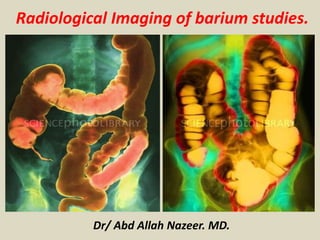
Presentation1, radiological imaging of barium studies.
•Als PPTX, PDF herunterladen•
161 gefällt mir•18,681 views

health&medicne.

Empfohlen
Empfohlen
Weitere ähnliche Inhalte
Was ist angesagt?
Was ist angesagt? (20)
Andere mochten auch
Andere mochten auch (20)
Ähnlich wie Presentation1, radiological imaging of barium studies.
Ähnlich wie Presentation1, radiological imaging of barium studies. (20)
Mehr von Abdellah Nazeer
Mehr von Abdellah Nazeer (20)
Kürzlich hochgeladen
Kürzlich hochgeladen (20)
Presentation1, radiological imaging of barium studies.
- 1. Dr/ Abd Allah Nazeer. MD. Radiological Imaging of barium studies.
- 2. Radiographic procedure. Barium Swallow Conventional Barium Esophagography (Barium Swallow) Modified Barium Swallow (Oral and Pharyngeal Function Study) Diagnosis of Food Impactions & Foreign Bodies in the Esophagus Upper G.I. Series; Biphasic-Contrast Examination Using "Bubbly Barium" Single-contrast examination Small Bowel Follow-Through Enteroclysis Peroral Pneumocolon Barium Enema Policy on Doing Barium Enemas Following Endoscopy +/- Biopsy Types of Barium Enema Double-Contrast Examination of the Colon Single-Contrast Examination of the Colon Water Soluble Contrast Enema Colon Transit Time (Colonic Motility Test) Defecography (Evacuation Proctography).
- 3. Barium Swallow: The hypopharynx & cervical esophagus AP and lat, views of the barium- coated pharynx and hypopharynx obtained during phonation demonstrates normal anatomy with aspiration of the contrast to larynx and trachea. p p vv p v
- 6. This oblique view of a normal barium swallow shows the normal impressions made by the (A) aortic arch, (B) left main stem bronchus, and (LA) left atrium on the esophagus. The esophagus is a muscular tube that is normally 25- 30 cm long and 2-3 cm wide. The esophagus is found anterior to the vertebral column and extends from approximately C6 down, following the curvature of the vertebral column. It passes through the esophageal hiatus of the diaphragm at approximately T10. The esophagus is divided into three segments: the cervical, thoracic, and abdominal segments. The cervical portion is separated from the cervical vertebrae by only a few mm of prevertebral soft tissue. There are several structures that are in close proximity to the esophagus and which make normal impressions on it. These structures are the aortic arch at T3-T4, the left main stem bronchus, the left inferior pulmonary veins and in some normal healthy people the left atrium, although a large impression is usually a sign of left atrial disease.
- 7. The following are additional diseases and conditions that affect the esophagus: Achalasia Acute esophageal necrosis Barrett's esophagus Boerhaave syndrome Caustic injury to the esophagus Chagas disease Diffuse esophageal spasm Esophageal atresia and Tracheoesophageal fistula Esophageal cancer Esophageal dysphagia Esophageal varices Esophageal web Esophagitis GERD Hiatus hernia Jackhammer esophagus (hypercontractile peristalsis) Killian–Jamieson diverticulum Mallory-Weiss syndrome Neurogenic dysphagia Nutcracker esophagus Schatzki's ring Zenker's Diverticulum
- 8. Esophageal duplication cyst Esophageal duplication cysts are a type of congenital foregut duplication cyst. Clinical presentation Patients are generally asymptomatic but may complain of dysphagia due to esophageal compression. They typically present in childhood. Location It mainly occurs within the thoracic esophagus. Radiographic features Plain radiograph Well defined soft tissue density in close association with the esophagus. Fluoroscopy On barium swallow the cyst may cause extrinsic compression of the esophagus. CT Well defined thick walled structure (fluid density) noted along the esophagus. MRI T1: low to intermediate signal intensity T2: high signal intensity
- 10. Plain X-ray showing upper mediastinal widening and vertebral defects. CT scan thorax shows enhancing rim of an esophageal duplication cyst. This was excised by right thoracotomy.
- 12. Pediatric intramural esophageal cyst.
- 15. Congenital tracheoesophageal (TE) fistulas result from failure of the esophageal lumen to develop completely separate from the trachea. Embryonically, the trachea and upper GI tract have a common origin at the caudal end of the embryonic pharynx. In normal development during the second month of gestation, the esophagus assumes a dorsal position, while the trachea lies ventrally. Failure of this complete separation leads to the development of TE fistulas. Below is a diagram depicting the different type of congenital TE fistulas and their frequency of occurrence. Esophageal atresia (EA) is a common cause of polyhydramnios in utero. At birth, the infant may have difficulty handling secretions and may have respiratory distress at first feeding. Attempts to pass a nasogastric tube are usually unsuccessful. Infants with TE fistulas tend to have rounded abdomens and bowel sounds, while those with EA without a fistula tend to have scaphoid abdomens and absent bowel sounds.
- 16. In image "A" we can see atresia of the upper esophagus as evidenced by failure to pass a feeding tube down the esophagus, but we still observe gas in the abdomen. These findings are likely due to a esophageal atresia with a distal tracheoesophageal fistula. Images "B, C, D" show contrast filling a blind pouch.
- 17. Esophageal atresia with TE fistula. Contrast was administered through a G tube into the stomach. The contrast refluxed into the distal esophagus across the tracheoesophageal fistula into the trachea and from the trachea into the esophageal pouch.
- 18. H-type fistula. A, Tracheoesophageal fistula (arrow) in a 7-day-old boy with imperforate anus. B, Demonstration of another H- type fistula (arrow) from the upper cervical esophagus to the trachea, using the technique of contrast injection through a feeding tube with very careful volume control. C, Large H- type fistula from the upper cervical esophagus to the trachea (T).
- 19. Connections between esophagus and airway. A, Congenital esophagobronchial fistula. Oblique view shows esophagus (arrows with 1) and bronchus to right upper lobe (arrow with 2). B, Esophageal bronchus. Frontal view from an esophagram demonstrates the origin of the right main bronchus from the distal esophagus.
- 20. A Schatzki ring, also called Schatzki-Gary ring, is symptomatically narrow esophageal B-ring occurring in the distal esophagus and usually associated with a hiatus hernia. Relatively common, lower esophageal rings are found in ~10% of esophagrams. Location: Schatzki rings are located at the gastro-esophageal junction. They should not be confused with A-rings, which are found a few centimeters proximal to the B- ring esophageal webs, which are lined on both sides by esophageal mucosa Associations More than half of patients will have an associated esophageal condition such as: hiatus hernia, reflux oesophagitis, esophageal web, esophageal diverticulum Radiographic features Fluoroscopy: barium swallow Single-contrast solid barium swallows (especially in the RAO prone position) are more sensitive than endoscopy in detecting Schatzki rings. On barium swallow the following features may be seen: full-column barium swallow will reveal a circumferential narrowing at the gastro- esophageal junction, often a few centimeters above the diaphragmatic hiatus thin smooth ring, 1-3 mm double contrast studies are less sensitive performing a Valsalva manoeuvre may improve sensitivity barium-tablet or barium-coated marshmallow may also improve sensitivity
- 21. The above esophagrams show a Schatzki ring (red arrow) at the distal esophagus.
- 24. Esophageal webs refer to an esophageal constriction caused by a thin mucosal membrane projecting into the lumen. Epidemiology Esophageal webs tend to affect middle-aged females. Clinical presentation Patients are usually asymptomatic and the finding may be incidental and unimportant. However, if the stenosis is severe symptoms include dysphagia and regurgitation of food. Pathology Location More commonly occur in the cervical esophagus near cricopharyngeus muscle than in the thoracic esophagus. They typically arise from the anterior wall and never from the posterior wall; they can also be circumferential. Occasionally, multiple webs are visualized during maximal distension. Associations Plummer-Vinson syndrome GORD/GERD (especially a distal esophagus web) external beam radiation Radiographic appearance Fluoroscopy: barium swallow may be demonstrated on high-volume barium esophagrams when the esophagus is fully distended a "jet effect" of contrast passing distal to the web may be seen.
- 25. Barium swallow shows circumferential radiolucent ring in upper esophagus. Proximal dilatation and jet phenomenon (Barium spurting through the ring on fluoroscopy) indicate partial obstruction.
- 26. Congenital esophageal web with tight upper esophageal stenosis with proximal dilatation, hold up of contrast and distal narrowing.
- 27. Anterior web (large arrow) and posterior impression (small arrow) due to cricopharyngeus spasm.Circumferential web (arrow).Anterior esophageal web (arrow).
- 28. Esophageal diverticula are sac or pouch projections arising from the esophagus. They can occur in all ages but more frequent in adults and elderly people. Pathology: Esophageal diverticula are either: true diverticula: include all esophageal layers false diverticula: contain only mucosa and submucosa herniating through the muscular layer (e.g. Zenker diverticulum) Esophageal diverticula are classified according to the mechanism of formation into: traction diverticula: occurs secondary to pulling forces on the outer aspect of the esophagus pulsion diverticula: occurs secondary to increased intraluminal pressure (e.g. Zenker diverticulum) Classification They can be classified according to their location: Upper esophageal diverticula Zenker diverticulum: actually pharyngeal but it is common practice to include it with esophageal diverticula Killian-Jamieson diverticulum Middle esophageal diverticula Traction diverticula: are (true diverticula) which occur secondary to scarring, fibrosis and inflammatory processes (tuberculous adenitis) in the mediastinum pulling on the esophageal wall pulsion diverticula: are usually false diverticula and occur secondary to abnormal increased intraluminal pressure against a weak esophageal segment Lower esophageal diverticula Epiphrenic diverticula
- 30. Image "A" depicts the frontal view of a large barium-filled sac (Z) below the level of the hypopharynx. Image "B" is a lateral view depicting a large Zenker's diverticula (Z) in the posterior cervical esophagus.
- 31. Zenker's diverticulum on chest film, barium study and CT.
- 34. Image "A" depicts multiple varices on esophagram. Image "B" is an angiographic demonstration of cavernous transformation of the portal vein (PV) with reversal of blood flow through the coronary veins (CV) and splenic vein (SV) producing esophageal varices (Var.)
- 35. Esophagram depict contrast extravasation from the distal esophagus in a patient with spontaneous perforation of the esophagus.
- 36. CT shows dilated esophagus (arrow) that led to esophagram. RIGHT: Esophagram shows narrowing (arrow) at level of hiatus. Achalasia.
- 37. Image "A" depicts a lateral view of the esophagus showing a massively dilated esophagus with retention of contrast in the distal portions of the esophagus. Image "B" shows the "bird's beak" appearance of the dysfunctional lower esophageal sphincter.
- 40. Scleroderma - Barium swallow of patient with scleroderma. Note the dilated esophagus (arrows).
- 41. Esophagrams showing the typical "corkscrew" or "beaded" appearance of diffuse esophageal spasms(DES).
- 42. Stricture - Patient with esophageal stricture, with green arrows showing area of stricture. Note the barium tablet indicated by the red arrows.
- 43. Barrett's - Upper GI swallow of patient with Barrett's esophagus. Arrow points to new transition point of squamo-columnar junction. Note the irregularities of the mucosa inferior to transition point. Barrett’s esophagus. Pathology: Columnar metaplasia of the esophageal stratified epithelium and there is a strong association with adenocarcinoma. Findings: 1- mild esophageal stricture. 2-reticular mucosal pattern.
- 45. Canida - Above is a characteristic "shaggy esophagus" associated with Canida infection. Image "A" depicts the longitudinally oriented plaque-like lesions visible in Candida esophagitis. Image "B" depicts the granular appearance of the esophageal mucosa secondary to edema and inflammation.
- 46. Image "A" and "B" both depict ulcerations of the distal esophageal mucosa secondary to lye ingestion. Image "C" depicts irregular narrowing of the esophagus with ulcerations.
- 48. Congenital esophageal stenosis (fibromuscular form) in a 25-year-old man with a 4-year history of dysphagia.
- 50. Gastro-esophageal reflux disease (GERD) is a spectrum of disease that occurs when gastric acid refluxes from the stomach into the lower end of the esophagus across the lower esophageal sphincter(LOS). Minor reflux disease In most patients with reflux disease, reflux is initiated by transient collapses of LOS pressure. This results in the lower end of the esophagus being bathed in gastric acid for longer than normal. Patients may be symptomatic without developing endoscopic appearances of oesophagitis (40% of cases). These patients will also have no detectable abnormality on a barium swallow. Advanced reflux disease In patients with a permanently low LOS pressure, symptoms are generally more severe and there is evidence of disease in endoscopic or barium studies. Abnormalities that are radiologically detectable include: free reflux impaired primary peristalsis and poor clearance abnormal esophageal contractions oesophagitis with scarring strictures, Barrett esophagus and aspiration sacculations and intramural pseudodiverticula
- 51. GERD
- 52. (Reflux) and a hiatal hernia (hh).
- 54. Ovoid filling defects caused by the leiomyoma. The smooth surface and obtuse angles formed are characteristic of submucosal masses.
- 55. A calcified esophageal mass is almost always a leiomyoma.
- 56. Esophageal carcinoma is relatively uncommon. It tends to present with increasing dysphagia, initially to solids and progressing to liquids as the tumour increases in size, obstructing the lumen of the esophagus. Chest radiograph Many indirect signs can be sought on a chest radiograph and these include: widened azygo-esophageal recess with convexity toward right lung (in 30% of distal and mid-esophageal cancers) thickening of posterior tracheal stripe and right paratracheal stripe >4 mm (if tumour located in the upper third of esophagus) tracheal deviation or posterior tracheal indentation/mass retrocardiac or posterior mediastinal mass esophageal air-fluid level lobulated mass extending into gastric air bubble (Kirklin sign) Fluoroscopy/Barium Swallow irregular stricture pre-stricture dilatation with 'hold up' shouldering of the stricture CT eccentric or circumferential wall thickening >5 mm peri-esophageal soft tissue and fat stranding dilated fluid- and debris-filled esophageal lumen is proximal to an obstructing lesion tracheobronchial invasion appears as a displacement of the airway (usually the trachea or left mainstem bronchus) as a result of mass effect by the esophageal tumour aortic invasion.
- 57. "A" we can see a Schatzki ring (red arrows) and filling defects (yellow arrows) proximal to the ring which was found to be squamous cell cancer. Images "B" and "C" show the same findings in a close-up view.
- 58. Irregular stricture in the esophagus with ulceration of the esophageal mucosa. Also noticed the shouldered margins of the lesions. CT images, one can see circumferential thickening of the esophageal wall (annular lesion).
- 59. "A" the red arrows show mucosal invasion with ulceration, whereas the yellow arrow points out a stricture at the GE junction. In image "B", we can further see an irregular filling defect in the distal esophagus associated with adenocarcinoma.
- 60. LEFT: Small polypoid carcinoma. RIGHT: Large polypoid lesion.
- 61. Esophageal carcinoma with ulcerations (arrows) and sharp right angle junction with esophageal wall (arrowheads).
- 62. Distal narrowing simulates achalasia, but narrowing is eccentric, shoulders are asymmetric (arrows), and the mucosa is irregular at the tip of narrowing. CT shows gastric fundus thickening (arrows) due to adenocarcinoma.
- 64. Esophageal lymphoma. (A) Right posterior oblique barium esophagogram demonstrating smooth stricture above mid esophagus, indicating circumferential tumor narrowing the lumen. (B), Axial enhanced CT Scan demonstrate markedly thickened esophagus and narrow slit-like lumen.
- 65. Diffuse large B-cell lymphoma of the esophagus. Axial (a) and coronal (b) fused PET/CT images show a large focus of FDG accumulation in an intraluminal esophageal mass (arrow) and a smaller focus in the left hilar nodes (arrowhead in b). Primary esophageal lymphoma is rare and is commonly mistaken for esophageal carcinoma. Biopsy provides the final diagnosis.
- 66. Stomach. Imaging Modalities and Procedures Fluoroscopy (Upper GI Series) Computed Tomography Normal Anatomy Iatrogenic Conditions Afferent Limb Syndrome Inflammatory Diseases Gastritis Ulcers Benign (Peptic Ulcer Disease) Malignancy Associated Zollinger-Ellison Syndrome Pseudotumors Gastric Varices Gastric Diverticulum Double Pylorus
- 67. Neoplastic Diseases Benign Gastric Polyps Malignant Linitis Plastica Gastric Carcinoma Gastric Lymphoma Metastases GI Stromal Tumors (GISTs) Others Hypertrophic Pyloric Stenosis Gastric Dilatation Bezoars Gastric Volvulus Menetrier's Disease
- 68. Anatomy of the stomach.
- 69. Hypertrophic pyloric stenosis (HPS) refers to the idiopathic thickening of gastric pyloric musculature which then results in progressive gastric outlet obstruction. Plain radiograph: Abdominal x-ray findings are non-specific but may show a distended stomach with minimal distal intestinal bowel gas. Fluoroscopy: An upper gastrointestinal series (barium meal) excludes other, more serious causes of pathology, but the findings of a UGI series infer rather than directly visualise the hypertrophied muscle. On upper gastrointestinal fluoroscopy: delayed gastric emptying peristaltic waves (caterpillar sign) elongated pylorus with a narrow lumen (string sign) which may appear duplicated due to puckering of the mucosa (double-track sign) the pylorus indents the contrast-filled antrum (shoulder sign) or base of the duodenal bulb (mushroom sign) the entrance to the pylorus may be beak-shaped (beak sign) Ultrasound: Ultrasound is the modality of choice in the right clinical setting because of its advantages over a barium meal are that it directly visualizes the pyloric muscle and does not use ionizing radiation. The hypertrophied muscle is hypoechoic, and the central mucosa is hyperechoic. Diagnostic measurements include (mnemonic "number pi"): pyloric muscle thickness, i.e. diameter of a single muscular wall on a transverse image: >3 mm (most accurate 3) length, i.e. longitudinal measurement: >15-17 mm pyloric volume: >1.5 cc pyloric transverse diameter: >13 mm With the patient right side down the pylorus should be watched and should not be seen to open. Described sonographic signs include: antral nipple sign, cervix sign, target sign.
- 70. UGIS Findings: Elongation and narrowing of the pyloric canal (2-4 cm in length) String Sign: Passage of small contrast through the narrowed pyloric channel Crowding of mucosal folds in pyloric channel producing a double or triple track sign Hypertrophic pyloric stenosis.
- 71. Ultrasound views of the stomach and pylorus in a 5-week-old boy with gastric distention as evidenced by hyperechoic gas in a fluid-filled stomach (A; arrow) and pyloric lengthening (B; arrow) and thickening (C; arrow).
- 75. Abdominal plain radiograph (A) and UGI series (B) in a 4-week-old boy demonstrating gross gastric dilatation (arrows) and a bird-beak appearance to the gastric outlet.
- 76. INFANTILE HYPERTROPHIC PYLORIC STENOSIS.
- 78. Duodenal atresia results from a congenital malformation of the duodenum and requires prompt correction in the neonatal period. It is considered to be one of the commonest causes of a fetal bowel obstruction. Epidemiology The prevalence of duodenal atresia is ~1 in 5,000-10,000 newborns, and there is no sex-associated difference in prevalence. Radiographic features Plain radiograph Abdominal radiographs may classically show a double bubble sign with gas filled distended stomach and duodenum with an absence of distal gas. A similar appearance (either filled with fluid or gas) can be seen in other modalities. Distal bowel gas although more classically associated with duodenal stenosis, however, it can be seen in duodenal atresia via anomalous bile duct anatomy. Ultrasound May also show a dilated stomach and duodenum giving a double bubble type appearance. This, however, may not be sonographically detectable until the mid to late second trimester. May also show evidence of polyhydramnios as an ancillary sonographic feature.
- 80. Abdominal examination showed a uterus size that is more than the gestational age calculated from the LMP. Ultrasound showed "double bubble sign" with polyhydramnios.
- 81. “Double bubble" sign (dilated stomach and duodenal bulb) -- dilated stomach and no gas distal to the proximal duodenum. Stated another way, there is no gas in the rest of the small or large bowel.
- 82. Duodenal atresia and web. A) Typical double-bubble appearance of gastric and duodenal airs (arrow) is well depicted on erect plain film in a case with duodenal atresia. B) Second portion of duodenum is partially obstructed by web (arrow) on barium study in another case.
- 84. Duodenal diverticula are outpouchings from the duodenal wall. They may result from mucosal prolapse or the prolapse of the entire duodenal wall and can be found at any point in the duodenum although are by far most commonly located along the medial wall of the second, or superior wall of the third part of the duodenum. Diverticula located at the ampulla of Vater may cause difficulty for endoscopists as they attempt to cannulate the biliary system. Clinical presentation Duodenal diverticula are very common, found in up to 23% of asymptomatic patients, and in the vast majority remain asymptomatic throughout life. In 10% of patients, some symptoms are attributable to them, with only a minority requiring surgical intervention. Pathology: There are a two of types of duodenal diverticula: primary and secondary diverticulum A primary duodenal diverticulum occurs where there is prolapse of mucosa through the muscularis propria. They usually occur within the 2nd part (62%) and less commonly in the 3rd (30%) and 4th (8%) parts. Unlike secondary diverticula they are rarely seen in the 1st part. When they occur in the 2nd part, most (88%) are seen on the medial wall around the ampulla, 8% are seen posteriorly and 4% on the lateral wall. A secondary duodenal diverticulum results from prolapse of the entire duodenal wall and almost invariably occurs in the 1st part of the duodenum. These are true diverticula and are usually secondary to duodenal or periduodenal inflammation, such as from previous ulcer disease. Location specific sub types periampullary diverticulum Radiographic features CT Diverticulae are seen as saccular outpouchings from the duodenum that may contain gas, fluid, contrast or food debris or any combination of these. They often contain a air-fluid or air-contrast level.
- 85. Intraluminal duodenal diverticulum with acute pancreatitis. (a) Upper gastrointestinal series image shows a contrast-filled intraluminal duodenal diverticulum with the “windsock” appearance (arrowhead). Oblique coronal reformatted image shows a debris-filled “windsock” intraluminal diverticulum (arrowheads) distorting the pancreas and the second and third portions of the duodenum.
- 86. Intraluminal duodenal diverticulum with situs anomaly with hematochezia. (a) Coronal reformatted contrast-enhanced CT image shows two intramural diverticula with “windsock” appearance (arrows) in the 3rd and 4th portion of the duodenum.
- 87. Duodenal diverticulum in 45-year-old woman. Axial CT scan obtained with IV and oral contrast materials at level of pancreatic head shows 10-mm cystic process with curvilinear area of increased attenuation (long arrow) that was initially thought to represent intraductal papillary mucinous tumor. Short arrow identifies duodenum. Spot radiograph from upper gastrointestinal barium series shows characteristic appearance of duodenal diverticulum (arrow).
- 88. Duodenal diverticulum. Coronal T2-weighted MR image obtained with HASTE sequence shows 15-mm cystic process (long arrow) in region of pancreatic head. Main pancreatic duct (short arrow) and common bile duct (arrowhead) are shown entering cystic process. This finding was initially thought to represent nonspecific cystic pancreatic neoplasm.
- 89. Extraluminal diverticulum (arrow) is typically shows a fluid-air level in medial wall of descending duodenum (D) beneath the pancreatic head on CT images. Double- contrasted barium study of the same case confirms the diverticula (arrow).
- 90. Transient duodenal hernia. CT shows A) Herniated small bowel loops (arrows) in the left upper quadrant, B) The engorged mesenteric vessels (arrow) towards the entrance of the hernia sac. C) Regression of the hernia sac on follow-up CT and D) regression of the engorged mesenteric vessels on follow-up CT.
- 91. Left paraduodenal hernia with abdominal pain. Contrast-enhanced CT shows a sac-like bowel loop (arrows) in the left paraduodenal fossa. Note anterolaterally displaced inferior mesenteric vein (arrowhead).
- 92. Annular pancreas. Fluoroscopy demonstrates concentric narrowing of the second portion of the duodenum. Annular pancreas is a rare congenital abnormality in which a ring of pancreatic tissue encircles the duodenum at or above the major papilla. Embryologically it is a sequelae of a persistent left ventral bud, which usually atrophies during embryological development. Plain films may demonstrate proximal small bowel obstruction. Fluoroscopy more clearly delineates the abnormality. It will show dilatation of the proximal duodenum, with eccentric or concentric narrowing of descending duodenum. In the most severe cases, mucosal effacement will be seen. Note that there is NO ulceration or mucosal destruction, differentiating it from neoplastic or inflammatory etiologies. CT is beneficial for diagnosis confirmation, as it will demonstrate the ring of pancreatic tissue surrounding and compressing the duodenum.
- 94. Annular pancreas with repeated episodes of vomiting. (a) Coronal thick-slab single-shot MRCP shows aberrant pancreatic duct (arrow) encircling the descending portion of the duodenum with dilatation of the proximal duodenum (*). Note mild dilatation of main pancreatic duct (arrowhead).
- 95. SMA syndrome: Note the dilated duodenum (D) with abrupt caliber change at D2/3 portion caused by compressive effects of SMA SMA syndrome: CT scan of the same patient showing duodenal obstruction (long arrow) at the level of SMA (short arrows).
- 96. Gastrointestinal stromal tumor (GIST). A solid and heterogeneously enhanced tumor (arrows) with smooth contours located on duodenum partially obliterates the lumen on CT scan. Concentrically narrowed duodenal lumen (arrows) with “apple core” appearance is seen on barium study.
- 97. Duodenal adenocarcinoma. A) US demonstrates pseudo kidney appearance of duodenal mural thickening. B) CT scan confirms concentric duodenal mural thickening and C) barium study also reveals irregular mucosal filling defects and mild dilation of duodenal genu.
- 98. Duodenal lymphoma. Non-contrast (A) and contrasted (B) CT scans show narrowed lumen and concentrically thickened horizontal portion of duodenum (arrows). C) US image demonstrates pseudo kidney appearance (arrow) of duodenal wall thickening. D) Barium study also reveals narrowed and irregular mucosa pattern (arrows) of horizontal duodenum.
- 99. Gastritis. Note the pronounced thickening of rugal folds throughout the stomach. Stomach.
- 100. Acute gastritis with Thickened gastric rugae (> 5mm) secondary to edema Mucosal nodularity Antral narrowing (indicative of h. pylori) Erosions: manifest by small mucosal defects that collect contrast
- 101. Axial (A) and coronal (B) contrast-enhanced CT in a 56-year-old woman with diffuse gastric mucosal thickening (arrows) caused by Antral gastritis. The fundus is relatively normal.
- 102. Coronal CT with soft tissue windows (A) and lung window settings (B) in a 67-year-old diabetic woman with mural gastric gas (arrow) due to emphysematous gastritis.
- 103. Ménétrier Disease (Hypertrophic Gastritis).
- 104. A featureless stomach due to atrophic gastritis. There is also a small antral polyp (arrow).
- 105. Barium study from a patient with known Crohn's shows a serrated appearance of the antrum (arrows) due to inflammatory involvement from Crohn's.
- 106. Gastric (peptic) ulcers can be detected on multiple imaging modalities, but are best evaluated on a double contrast barium upper GI study. Radiographic features Appearance The classic appearance for a benign gastric ulcer on a double contrast study is >2 mm oval mucosal defect (a "crater") Thin gastric folds radiating toward the crater There are however, multiple different appearances that an ulcer may take, including a linear shape or a serpentine shape. Mucosal defects <2 mm are termed "erosions". Ulcers are often associated with a ring of edema around the ulcer crater, which can give rise to a thin radiolucent "waist" to the ulcer crater. This has been termed a Hampton line, ulcer collar, or ulcer mound, as increasing amounts of edema are present. Ulcer location The vast majority (90-95%) of gastric ulcers are located on the lesser curvature and posterior stomach wall in the gastric body and antrum. They are uncommonly on the greater curvature (~5%).
- 108. UGI series in a 44-year-old man with thickened antral folds and a punctate collection of barium at the center (arrow) due to an antral ulcer.
- 109. Peptic ulcers (blue arrow). Benign peptic ulcer.
- 110. UGI series in a 76-year-old woman with a larger benign lesser curve ulcer (arrow). Large benign lesser curve ulcer (large arrow) with uniform fold convergence on the ulcer (small arrow).
- 111. Benign gastric ulcer. prominent radiating folds extend directly to the ulcer.
- 113. Barium meal demonstrates a giant gastric ulcer in profile. It is arising from the greater curvature of the pyloric antrum, has a deep but smooth ulcer crater, protrudes beyond the expected gastric contour, and has a prominent ulcer mound.
- 115. Upper GI series showing the differences between a malignant and benign gastric ulcer. Left panel: Malignant gastric ulcer of the distal lesser curvature. There is the biconvex meniscus sign with a nodular ulcer mound (arrow). Right panel: Benign gastric ulcer of the lesser curvature. The ulcer crater has smooth margins and projects beyond the gastric wall (arrow).
- 118. Duodenal Ulcer • 2-3 times more frequent than gastric ulcers • 3:1 male: female ratio Pathophysiology • Excessive acidity in duodenum from, Abnormally high gastric secretion, Inadequate neutralization Predisposing factors • Steroids, Severe head injury, Post-surgical, COPD. Location • Bulbar (95%) • Anterior wall– 50%, • Posterior wall– 23% • Inferior fornix– 22% • Superior fornix– 5% Post bulbar (3-5%) • Majority on medial wall just proximal to ampulla, Tendency for hemorrhage in 66% • Male: female ration 7:1 X-ray • Small round, ovoid or linear crater • Kissing ulcers–ulcers opposite from each other on the anterior and posterior walls • Giant duodenal ulcer–>3cm (rare) with higher morbidity and mortality • May be mistaken for the duodenal bulb itself and missed • Clover-leaf deformity–healed central ulcer of the bulb with four-leaf clover-like deformity Complications • Hemorrhage 15% melena>hematemesis • Perforation <10% anterior>posterior /may fistulize to GB • Obstruction 5% • Penetration <5% walled-off perforation
- 119. Double-contrast upper gastrointestinal series. Posterior wall duodenal ulcer.
- 120. Duodenal Ulcer. There is a collection of barium on the dependent surface of the duodenal bulb (white arrows) on this double contrast (air-contrast) upper GI examination. This represent barium in an ulcer crater.
- 123. Chronic duodenal ulcer disease. Typical cloverleaf deformity is visible (arrows).
- 124. UGI series in a 72-year-old man with a paraesophageal hernia (arrow). The GE junction lies below the diaphragm. Hiatus hernias (HH) occur when there is herniation of abdominal contents through the esophageal hiatus of the diaphragm into the thoracic cavity. The most common content of a hiatus hernia is the stomach. There are two main types of hiatus hernia (although they may co-exist): sliding hiatus hernia (>90%) rolling (para-esophageal) hiatus hernia (<10%)
- 125. Coronal (A) and axial (B) CT in a 47-year-old woman with a sliding hiatal hernia.
- 126. GI series and axial contrast-enhanced CT in a 44-year-old woman with both a paraesophageal (large arrows) and sliding hiatal hernia (small arrows). The GE junction lies below the diaphragm.
- 127. Gastric Diverticula
- 128. Gastric Polyps - Upper GI fluoroscopy showing gastric polyps (arrows).
- 129. UGI in a 54-year-old man with a lesser curve smooth mucosal filling defect (arrow) due to a gastric adenomatous polyp.
- 130. Gardner syndrome and multiple gastric adenomatous polyps.
- 131. A. UGIS double contrast study. The arrows outline the area of irregular mucosa which was caused by an invasive gastric carcinoma. B. Single contrast study from the same patient showing the apple core appearance of the stomach due to the invasive gastric adenocarcinoma.
- 132. Linitis Plastica(scirrhous adenocarcinoma). UGIS demonstrates luminal narrowing, wall thickening, and rigidity. Rugal fold effacement. Mucosal nodularity or ulceration.
- 133. Mets - Patient with metastatic breast cancer with stomach lining infiltration. Note the enlarged diameter of the wall (arrows).
- 134. Leiomyoma of the stomach(GIST). Leiomyosarcoma of the stomach(GIST).
- 135. A smooth, rounded submucosal mass (arrow) that proved to be a benign GIST. Axial (A) and coronal (B) CT in a 44-year-old man with a smooth intraluminal submucosal filling defect at the gastric fundus (arrows) due to a GIST.
- 136. A CT image of a well-defined GIST confirmed by pathology. There is no apparent central necrosis and the tumor is not enhancing because only oral contrast was given. Axial (A) and coronal (B) CT in a 55-year-old woman with a transmural gastric mass with ulceration (arrows) due to a benign GIST.
- 137. Gastric dilatation without evidence of obstruction Gastric emphysema: linear streaks of gas within the stomach wall.
- 138. Gastric Bezoars.
- 139. Gastric volvulus.(b) Mesenteroaxial gastric volvulus. Gastric volvulus.(a)Organoaxial gastric volvulus.
- 140. UGI series (A) and coronal CT (B) in a 59-year-old woman with an organoaxial volvulus. The greater curvature (large arrow) is superior (cephalad) and the lesser curvature inferior (small arrow). The GE junction is indicated by the arrowhead.
- 142. UGI swallow in a 68-year-old woman with mesenteroaxial volvulus. The GE junction is inferior (large arrow) and the pylorus, superior (small arrow).
- 144. Perforated gastric volvulus (mesenteroaxial type) with emphysematous gastritis with abdominal pain.
- 145. Zollinger-Ellison syndrome (ZES) is a clinical syndrome that occurs secondary to a gastrinoma. (Hypervascular pancreatic mass with multiple peptic ulcer and thickened wall). Clinical presentation Diagnosis of ZES is often delayed by 5-7 years after the onset of symptoms. Pathology Gastrinomas are usually multiple and typically located in the duodenum (more common) or pancreas (less common). These tumours secrete gastrin that results in hypersecretion of gastric acid, which in turn results in diarrhea, gastritis, severe gastro-esophageal reflux disease and peptic ulcer disease. Associations multiple endocrine neoplasia (MEN) type 1: ZES occurs when gastrinoma is functional Radiographic features Fluoroscopy On double-contrast upper gastrointestinal studies the following features may be seen: Thickened rugal folds Multinodular gastric contour Erosions and ulcers, especially in atypical locations Barium may be diluted by the high volume of fluid in the stomach CT: negative contrast may be used to distend the stomach thickened rugal folds multiple gastric nodules/masses
- 147. Marked hypervascularity and thickening of the gastric wall image. Multiple liver metastases are present image. The serum gastrin levels were strikingly elevated, confirming ZES, though the gastrinoma was not identified on CT.
- 148. A, Axial contrast-enhanced CT in a 70-year-old man with diffuse gastric mucosal thickening due to Zollinger-Ellison syndrome (arrow). B, A 4-cm pancreatic tail gastrinoma is present (arrow).
- 149. Small Bowel. Congenital Anomalies Meckel's Diverticulum Atresia (Duodenum, Jejunum, Ileum) Annular Pancreas Ischemic Diseases Mesenteric Ischemia Shock Bowel Inflammatory Diseases Crohn's Disease Ulcers Zollinger-Ellinson Syndrome
- 150. Infectious Diseases Structural Abnormalities Intussusceptions Small Bowel Obstruction Ileus Diverticula Functional Abnormalities Scleroderma Sprue Neoplastic Diseases Carcinoid Lymphoma Carcinoma Leiomyosarcoma Metastases
- 152. Jejunal-ileal atresia is a segmental atresia of the jejunum or the ileum. It is associated with malrotation and volvulus (25%) and cystic fibrosis (10%). Patients present within the first days of life with vomiting or a distended abdomen. Multiple distended loops of bowel. Barium enema demonstrates unused microcolon in a patient with distal ileal atresia.
- 154. Midgut volvulus in a 68-year-old man with acute abdominal pain. (a) Contrast- enhanced CT shows superior mesenteric vein (arrow) lying to the left to the superior mesenteric artery (arrowhead) (reversal of the normal relationship between superior mesenteric artery and superior mesenteric vein).
- 155. Whirl sign associated with postoperative adhesion in a 55-year-old man who underwent small bowel resection due to traumatic injury 22 years earlier. Axial contrast-enhanced CT shows the “whirl appearance” (arrows) around the superior mesenteric artery. Note normal position of the ascending and descending colon.
- 156. Small Bowel Obstruction Radiographic - Plain Film Supine Distended loops of bowel Valvulae conniventes are present "Stepladder" pattern Variable amount of gas in colon depending on severity and duration of obstruction Gasless abdomen will be seen if distended loops are fluid- filled Erect / Lateral Decubitus Plain Film "String of pearls" sign from small collections of air Air-fluid levels Differential levels - air-fluid levels are at different heights Closed-loop obstruction - entrapment of a loop of bowel by obstruction (can occur with adhesions, hernias, volvulus)
- 157. Small bowel obstruction secondary to adhesions. Note the linear impression at the site of obstruction.
- 158. Adynamic ileus in scleroderma, manifest as diffuse dilatation. Note the pseudo-diverticula and featureless pattern of the loops of small bowel. Diffuse dilatation of loops in chronic idiopathic intestinal pseudo-obstruction.
- 159. Upright abdominal radiograph demonstrates air-fluid levels and small bowel dilatation. Supine abdominal plain film demonstrates dilated loops of small bowel.
- 160. Focal ileus - Abdominal x-ray of patient with focal ileus associated with pancreatitis.
- 161. Intussusception occurs when one segment of bowel is pulled into itself or a neighboring loop of bowel by peristalsis. It is also known as bowel telescoping into itself. It is an important cause of an acute abdomen in children and merits timely ultrasound examination and reduction to preclude significant sequelae including bowel necrosis. Radiographic features Intussusception can occur essentially anywhere. In adults, no such distribution is present as in the vast majority of cases a lead point lesion is present, and thus the location will depend on the location of that lesion. In children there is a strong predilection for the ileocolic region: ileocolic: most common (75-95%), presumably due to the abundance of lymphoid tissue related to the terminal ileum and the anatomy of the ileocecal region ileoileocolic: second most common ileoileal and colocolic: uncommon gastric intussusception: rare, but documented. Plain radiograph Abdominal x-rays may demonstrate an elongated soft tissue mass (typically in the upper right quadrant in children) with a bowel obstruction (and therefore air-fluid levels and bowel dilation) proximal to it. There may be an absence of gas in the distal collapsed bowel.
- 162. Ultrasonography has a false-negative rate approaching zero and is a reliable screening tool for children at low risk for intussusception. Children with classic findings of intussusception, however, need to be investigated with contrast enema, which is both diagnostic (the gold standard in the diagnosis of intussusception) and therapeutic. Ultrasound signs include: target sign (also known as the doughnut sign) pseudokidney sign crescent in a doughnut sign Fluoroscopy A contrast enema remains the gold standard, demonstrating the intussusception as an occluding mass prolapsing into the lumen, giving the "coiled spring” appearance (barium in the lumen of the intussusceptum and in the intraluminal space). The main contraindication for an enema is a perforation. CT: Has become the modality of choice for assessment of acute abdomen in adults, and thus most frequently images intussusception. Also, short length transient intussusception is a frequent incidental finding. The appearance of intussusception on CT is characteristic and depends on the imaging plane and where along the bowel, the images are obtained. Best known is the so-called bowel-within-bowel configuration, in which the layers of the bowel are duplicated forming concentric rings (CT equivalent of the ultrasonographic target sign) when imaged at right angles to the lumen, and a soft tissue sausage when imaged longitudinally At the proximal end of the intussusception, there will be two concentric enhancing/hyperdense rings, formed by the inner bowel and the folded edge of the outer bowel. As one images further along the intussusception the mesentery (fat and vessels) will form a crescent of tissue around the compressed innermost lumen, surrounded by the two layers of the outer enveloping bowel. Even further distally the lead point (if present) will be visualized.
- 163. A: Barium enema reveals the intussusceptum in the transverse colon (arrow). B: With further pressure the intussusceptum is reduced into the ascending colon.
- 166. Intussusception. A, A 3-month-old boy with intussusception. A transverse ultrasound image through the intussusceptum complex shows the donut or target sign, with intussusceptum composed of small bowel, nodes, and mesentery surrounded by the intussuscipiens. B, A longitudinal section of intussusception in the same patient as depicted in part A. The image shows the terminal end of the intussusception, with the inner and outer sleeves of the intussusceptum (white arrows) containing the intussuscepted mesentery (M). Black arrows outline the outer edge of the intussuscipiens. Note that no lead point is present.
- 168. Intussusception. CT demonstrates edematous bowel wall with a target appearance. The intussusceptum forms the inner part of the bull’s eye, while the intussuscipiens forms the outer layer.
- 169. Intussusception.
- 170. Coeliac disease, also known as non-tropical sprue, is a T-cell mediated autoimmune chronic gluten intolerance condition characterized by loss of villi in the proximal small bowel and gastrointestinal malabsorption (sprue). It should always be considered as a possible underlying etiology in cases of iron deficiency anemia of uncertain cause. Clinical presentation Many patients have a paucity of symptoms with no GI upset. However, abdominal pain is considered the most common symptom. Other manifestations include: iron deficiency anemia and guaiac-positive stools Diarrhea, constipation, malabsorption, including fat-soluble vitamins and weight loss. Fluoroscopy Features of small bowel barium studies are not sensitive enough for confident diagnosis, but the following changes may be seen: small intestinal dilatation due to excess fluid dilution of contrast multiple non-obstructing intussusceptions jejunoileal fold pattern reversal mosaic pattern flocculation segmentation CT enteroclysis Features present on CT enteroclysis may include: jejunoileal fold pattern reversal: thought to have the highest specificity is considered the most discriminating independent variable for the diagnosis of uncomplicated coeliac disease ileal fold thickening vascular engorgement prominent mesenteric lymph nodes may cavitate with a fluid fat level submucosal fat deposition in long standing cases.
- 171. Small bowel follow-through in a patient with celiac sprue. Initial imaging (left) demonstrates mild dilatation and “jejunalization” of the ileum. Subsequent imaging at 30 minutes (right) demonstrates a rapid transit time, with barium dilution and flocculations.
- 172. Findings of malabsorption at barium examination. (a) Image shows duodenitis with nodularity in a fold-free duodenum (arrow). (b) Image shows flocculation (within oval at upper right), dilution (single arrow), and dilatation (double arrow). (c) Image shows moulage (within oval), which is a featureless bald appearance of the jejunum caused by atrophy of folds and wall edema. (d) Image shows reversal of the fold pattern (within oval), with more prominent folds in the ileum than in the jejunum.
- 173. Meckel's Diverticulum Clinical Meckel's diverticulum is the failed obliteration of intestinal end of omphalomesenteric duct, a finding reportedly present in 2-3% of autopsies. This true diverticulum (containing all three bowel wall layers) is found 40-150 cm proximal to ileocecal valve, within the ileum. Clinical presentation is variable, and symptomatology can arise in children or adults. 50% contain heterotopic mucosa (usually gastric), and the most common adult manifestation is ulceration and bleeding. In children, SBO (usually from intussusception), pseudoappendicitis (diverticulitis), or rarely, perforation, can also occur. Radiological Plain Films Plain films are nonspecific and include distal SBO, sentinel loop in the right lower quadrant, or occasionally, enteroliths within the diverticulum. Fluoroscopy Fluoroscopy is a more sensitive evaluation for Meckel's diverticulum. It usually presents as a contrast-filled outpouching containing a triangular fold pattern or rugae. With intussusception, a polypoid filling defect can be observed projecting into the bowel lumen. When a Meckel's diverticulum presents as bleeding, an ulcer can occasionally be found. Nuclear Medicine Technetium-99m pertechnetate selectively localizes to gastric mucosa. Because a large percentage of Meckel's diverticula contain gastric mucosa, Tc-99m pertechnetate can be an effective means of evaluation. The study is dubbed a "Meckel's scan". A positive test entails a "hot spot" of increased activity, usually in the right lower quadrant. Note, however, that because only 50% of Meckel's diverticula contain gastric mucosa, a negative Meckel's scan does not exclude the diagnosis.
- 174. Prone and supine radiographs of the right side of the abdomen obtained during an upper GI barium series in a 13-year-old boy shows the terminal small bowel and a Meckel diverticulum (arrow).
- 175. Meckel's Diverticulum. Reflux into the small bowel has occurred during a single-contrast barium enema examination. Black arrow points to Meckel's diverticulum arising from small bowel near terminal ileum.
- 176. A 36-year-old female with chronic diarrhea and abdominal pain. Axial CT examination shows a banana-shaped, low- attenuation, well- circumscribed lesion (arrow) within the pelvis continuous with the bowel. Post-operatively, this was found to be a mucocele of the Meckel's diverticulum. The patient also had Crohn's disease with bowel wall thickening involving the large bowel (*).
- 177. A 45-year-old asymptomatic male with staging CT for lymphoma. Coronal contrast- enhanced CT image showing the Meckel's diverticulum as a tubular structure arising from the antimesenteric border of the ileum pointing in the pelvis (white arrow).
- 178. A 19-year-old male with painless rectal bleeding. 99mTc-pertechnetate scintigraphy showing uptake (arrows) of ectopic gastric mucosa in a Meckel's diverticulum.
- 179. Peutz-Jeghers syndrome with multiple small bowel polyps, mainly located in jejunum.
- 180. Patient with Peutz-Jeghers syndrome with ileal polyp as lead point for intussusception.
- 181. Ascariasis. SBFT (left ) demonstrates a linear filling defect (arrows). Enteroclysis (right) shows multiple long, tubular filling defects (arrow).
- 182. Acute radiation enteritis with regular fold thickening and effacement.
- 183. Mesenteric ischemia. Note the separation of small bowel loops and fold thickening from edema.
- 184. Mesenteric neoplasm causing small bowel ischemia. Separation of loops caused by wall edema. Note also diffuse fold thickening.
- 185. Shock bowel represents an ischemic insult to the intestines resulting from decompensated hypovolemic shock. As a result, the bowel becomes dilated and fluid-filled. The bowel walls become edematous and thickened, with marked enhancement on CT. Shock bowel is usually associated with significant ischemic injuries to other vital organs, and it carries a high mortality.
- 186. Crohn's disease is an idiopathic inflammatory bowel disease (IBD) characterized by widespread gastrointestinal tract involvement typically with skip lesions. Fluoroscopy Features on barium small bowel follow-through include: mucosal ulcers, aphthous ulcers initially deep ulcers (more than 3mm depth) longitudinal fissures, transverse stripes when severe leads to cobblestone appearance may lead to sinus tracts and fistulae pseudodiverticula formation: due to contraction at the site of ulcer with ballooning of the opposite site string sign: tubular narrowing due to spasm or stricture depending on chronicity partial obstruction CT fat halo sign, comb sign bowel wall enhancement bowel wall thickening (1-2 cm) which is most frequently seen in the terminal ileum strictures and fistulae mesenteric/intra-abdominal abscess or phlegmon formation abscesses are eventually seen in 15-20% of patients
- 194. PA proven hemangioma: coronal T1 FS post contrast and coronal T2 show enhancing well defined intraluminal jejunal mass.
- 195. Lipoma of the small bowel.
- 196. Spontaneous mesenteric hematoma causing separation of loops. Arrows (above) and “H” (below) delineate the extrinsic mass.
- 197. Desmoid of the small bowel
- 198. Burkett’s lymphoma. Enteroclysis (left) demonstrates separation of bowel loops with irregular fold thickening and luminal narrowing. CT (right) of the same patient confirms the presence of a large cavitary mass in the left abdomen.
- 199. Lymphoma in the terminal ileum.
- 200. Lymphoma in the proximal jejunum.
- 201. Ileal-ileal intussusception (yellow arrow), in a patient with multifocal small bowel lymphoma (not all lesions shown here). Mesenteric lymphadenopathy (red arrows).
- 202. Irregular, nodular thickened folds in lymphocytic lymphoma.
- 203. Lymphoid hyperplasia. Note the innumerous tiny filling defects.
- 204. Primary adenocarcinoma. Fluoroscopy (left) demonstrates annular luminal narrowing with shouldered margins (arrow). CT (right) demonstrates marked, irregular bowel wall thickening causing the “apple-core” appearance seen on fluoroscopy.
- 205. Adenocarcinoma in the jejunum.
- 207. Small intraluminal mass in the ileum (yellow arrow). Associated spiculated mesenteric mass with adjacent desmoplastic reaction in small bowel carcinoid.
- 208. Metastatic melanoma. Note the multiple large filling defects of varying size and shape.
- 209. Ulceration. This image demonstrates barium pooling in the base of an ileal ulceration. The atypical location of this ulcer should raise the suspicion for something other than an uncomplicated ulcer. Other complicating features include luminal narrowing and fold thickening.
- 210. Appendicitis Approximately 20% of patient visits to the emergency department for non-traumatic acute abdominal symptoms are related to the appendix. In fact, appendicitis is the most common reason for emergency abdominal surgery in the young adults and especially in the pediatric population. Therefore it is important to be able to quickly and correctly identify pathology of the appendix and treat it. Pathophysiology Appendiceal obstruction leads to venous and lymphatic obstruction producing an edematous, inflamed appendix. The resulting ischemia and mucosal breakdown allows bacteria to invade the appendix wall. Gangrene with rupture and peritonitis may ensue. Clinical Presentation Migration of pain from periumbilical region to RLQ. Right lower quadrant pain or pain at McBurneyâs point. Rebound tenderness at McBurneyâs point. Anorexia. Abdominal rigidity. Fever. Laboratory Data Leukocytosis with leftward shift. Can also have some hematuria secondary to ureteral inflammation. Diagnosis Most often diagnosed clinically, imaging can help in atypical of equivocal cases.
- 211. Abdominal ultrasound showing an elongated, blind ended tube. Highly suspicious for appendicitis. Abdominal CT demonstrating a fluid filled appendix, surrounded by an appendiceal abscess(fluid around the appendix surrounded by an enhancing rim).
- 212. Abdominal CT showing a cystic lesion in the expected region of the appendix. A second image from the same patient shows mural calcifications within the lesion. This is highly suggestive of a mucocele from a mucinous adenocarcinoma(although pathology on this patient revealed this lesion to actually be a mucinous cystadenoma).
- 213. Barium meal x ray shows elongated opacified appendix with multiple Distal filling defects, related to chronic appendicitis.
- 214. Colon. Congenital Anomalies Hirschsprung's disease Malrotation Duplication Vascular Complication Ischemic Colitis Diverticular Bleed Inflammatory Diseases Crohn's Disease Ulcerative Colitis
- 215. Infection diseases. Amebic Colitis Pseudomembranous colitis Diverticulitis Structural Abnormalities Intussusception Large Bowel Obstruction Megacolon Diverticulosis Volvulus Neoplastic Diseases Colon Polyps Adenoma Carcinoma Metastases
- 217. Microcolon: Barium enema examination demonstrating typical microcolon. This can be secondary to meconium ileus, ileal/ jejunal atresia or Hirschsprung's disease.
- 218. Meconium ileus: Supine abdominal radiograph showing Multiple dilated loops of small bowel. Soap bubble appearance of meconium mixed with gas (arrow) noted in Right side of abdomen. Note the absence of air fluid level despite distal intestinal obstruction. Meconium ileus is caused by thick, tenacious meconium that adheres to the wall of the small bowel and causes obstruction most often at the level of the ileocecal valve in a neonate. Almost all patients with meconium ileus have cystic fibrosis; 10-15% of CF patients present with meconium ileus. Complications include ileal atresia and/or stenosis, volvulus, perforation, and meconium peritonitis (due to obstruction and ischemia from tenacious meconium). It can be treated nonsurgically with water-soluble enemas to relieve the obstruction or be treated surgically.
- 220. Meconium Ileus: Water soluble contrast enema showing filling defects (arrow) within the distal ileum representing meconium and functional micro colon (unused).
- 222. Hirschsprung disease is the most common cause of neonatal colonic obstruction (15-20%). It is commonly characterized by a short segment of colonic aganglionosis affecting term neonates, especially boys. Clinical presentation The condition typically presents in term neonates with failure to pass meconium in the first 1-2 days after birth, although later presentation is also common. Overall ~75% of cases present within six weeks of birth 4, and over 90% of cases present within the first five years of life. A definitive diagnosis requires a full thickness rectal biopsy. Pathology Hirschsprung disease is characterized by aganglionosis (absence of ganglion cells) in the distal colon and rectum. It can be anatomically divided into four types according to the length of the aganglionic segment: short segment disease: ~75% * rectal and distal sigmoid colonic involvement only long segment: ~15% typically extends to splenic flexure / transverse colon total colonic aganglionosis: ~7.5% (range 2-13%) occasional extension of aganglionosis into the small bowel ultrashort segment disease 3-4 cm of internal anal sphincter only.
- 223. Radiographic features Radiograph Findings are primarily those of a bowel obstruction. The affected bowel is of smaller calibre and thus depending on the length of segment affected variable amounts of colonic distension are present. In protracted cases marked dilatation can develop, which may progress to enterocolitis and perforation. Fluoroscopy A carefully performed contrast enema is indispensable in both the diagnosis of Hirschsprung disease but also in assessing the length of involvement. It should be noted however that the depicted transition zone on the contrast enema is not accurate at determining the transition between absent and present ganglion cells. The affected segment is of small calibre with proximal dilatation. Fasciculation/saw-tooth irregularity of the aganglionic segment is frequently seen. Views of particular importance include: early filling views that include rectum and sigmoid colon allowing for rectosigmoid ratio to be determined. transition zone Antenatal ultrasound in particular cases there may be evidence of fetal colonic dilatation.
- 224. Short narrowed segment indicated between the yellow dotted lines; TZ = transition zone. Yellow arrows indicate the small bowel (jejunal) pattern to the descending colon.
- 227. Total colonic aganglionosis showing dilatation of the small bowel (arrow) proximal to transition zone. The large bowel is shortened with peculiar contours. There is marked regurgitation of barium into the dilated small bowel.
- 228. Sigmoid volvulus is a cause of large bowel obstruction and occurs when the sigmoid colon twists on the sigmoid mesocolon. Sigmoid Volvulus. Dilated loop of sigmoid colon has a "coffee-bean" shape and the wall between the two volvulated loops of sigmoid (black arrow) "points" towards the right upper quadrant. There is a considerable amount of stool in the colon from chronic constipation.
- 229. Sigmoid Volvulus, Bird peak Sign.
- 230. Sigmoid volvulus with abdominal pain. (a) Plain abdominal radiography shows an air-filled, dilated sigmoid colon (*) arising from the pelvis. Note a percutaneous endoscopic gastrostomy tube. (b) Coronal reformatted contrast-enhanced CT image shows dilated sigmoid colon (*) with the beak sign (arrow).
- 232. Classic "bird of prey" appearance of Sigmoid Volvulus on Barium study (arrow)Sigmoid Volvulus on plain film
- 233. Cecal volvulus with abdominal distention. (a) Abdominal radiograph shows air-distended cecum in the coffee-bean shape (*) in the left abdomen. (b) Axial contrast-enhanced CT shows dilated cecum (*) in the left abdomen and the whirl sign (arrows). c) Coronal reformatted contrast- enhanced CT shows dilated cecum (*) with beak-like tapering (arrow) in the left abdomen.
- 234. Cecal Volvulus
- 239. Abdominal X-ray of Crohn's disease patient showing transmural colonic inflammation (arrows) and ileal abnormalities
- 240. Ulcerative colitis is an inflammatory bowel disease that not only predominantly affects the colon, but also has extraintestinal manifestations. Fluoroscopy Double contrast barium enema allows for exquisite detail of the colonic mucosa and also allows the bowel proximal to strictures to be assessed. Mucosal inflammation leads a granular appearance to the surface of the bowel. As inflammation increases, the bowel wall and haustra thicken. Mucosal ulcers are undermined (button-shaped ulcers). When most of the mucosa has been lost, islands of mucosa remain giving it a pseudopolyp appearance. In chronic cases, the bowel becomes featureless with the loss of normal haustral markings, luminal narrowing and bowel shortening (lead pipe sign). Small islands of residual mucosa can grow into thin worm-like structures (so-called filiform polyps) Colorectal carcinoma in the setting of ulcerative colitis is more frequently sessile and may appear to be a simple stricture.
- 241. Ulcerative colitis with lead pipe colon.
- 242. Ulcerative colitis with loss of haustral pattern and lead pipe appearance
- 243. Ulcerative colitis with pseudo-polyp.
- 244. CT showing inflammation and bowel wall thickening in ulcerative colitis
- 245. CT showing diffuse inflammation in Amebic Colitis.
- 246. Pseudomembranous colitis. (Left) Axial CT scan of the mid abdomen utilizing oral but not intravenous contrast demonstrates marked thickening of the colonic wall (white arrows) producing the so-called "accordion sign." There is a small amount of pericolonic stranding (red arrow) and ascites (green arrow). (Right) Axial CT scan through the pelvis shows marked thickening of the wall of the rectum (yellow arrows) indicating this is a pan-colitis
- 249. Typhlitis in a patient with neutropenia.
- 250. CT showing inflamed diverticula Barium study of a perforated diverticula showing extravasation of blood into the abdominal cavity in a Diverticulitis patient
- 251. Complicated sigmoid diverticulitis with two paracolic abscesses(white arrows).
- 252. Adenomatous polyp on plain film (center)
- 254. Colorectal carcinoma (CRC) is the most common cancer of the gastrointestinal tract and the second most frequently diagnosed malignancy in adults. CT and MRI are the modalities most frequently used for staging. Barium enema sensitivities for polyps >1 cm single contrast: 77-94% double contrast: 82-98% polyps <1 cm: < 50% detection Appearances will reflect macroscopic appearance, with lesions seen as filling defects. These need to be differentiated from residual fecal matter. Typically they appear as exophytic or sessile masses, or may be circumferential (apple core sign). Fistulas to bladder, vagina or bowel may also be demonstrated. Rarely the stenotic segment will be long particularly with scirrhous adenocarcinomas. CT: CT is the modality most used for staging colorectal carcinoma, with an accuracy of only between 45-77%, able to asses nodes and metastases. It is often able to diagnose tumours although it is insensitive to small masses. CT colonography is increasing in popularity as an alternative to colonoscopy. Most colorectal carcinomas are of soft tissue density that narrow the bowel lumen. Ulceration in larger mass is also seen. Occasionally low-density masses with low-density lymph nodes are seen in mucinous adenocarcinoma, due to the majority of the tumour composed of extracellular mucin. Psammomatous calcifications in mucinous adenocarcinoma can also be present. Complications may also be evident, e.g. fistulae, obstruction, intussusception, perforation. MRI: Has a staging accuracy of 73% with a 40% sensitivity for lymph node metastases. MR is having an increasing role to play in the staging of rectal cancer.
- 255. Apple core lesion in ascending colon (arrow) Mucinous Colon Cancer on CT (arrow)
- 260. Thank You.