

Empfohlen
Weitere ähnliche Inhalte
Was ist angesagt?
Was ist angesagt? (20)
Ähnlich wie Leukoplakia
Ähnlich wie Leukoplakia (20)
Kürzlich hochgeladen
Kürzlich hochgeladen (20)
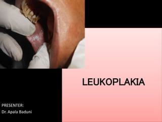
Leukoplakia
- 2. CONTENTS: • INTRODUCTION • CLASSIFICATION • CLINCAL FEATURES • TREATMENT MODALITIES
- 3. Introduction • The oral mucosa is normally semi-translucent, allowing the color of underlying tissues to show through to a variable degree. Lesions most commonly appear white secondary to • Increased thickness of the epithelium caused by Epithelial hypertrophy or hyperplasia, Edema
- 4. Increased production of surface keratin(hyperkeratosis). Thickening specifically in the spinous layer Collapsed bullae or ulcerative lesions covered with a surface layer of fibrin may also appear white. Keratin production as a protective response • Decreased submucosal vascularity
- 5. • (Wood and Goaz)
- 6. • Keratotic Leukoedema Linea alba Leukoplakia Nicotina stomatitis Migratory glossitis Lichen Planus Lichenoid Reaction Papilloma Verrucous Vulgaris Smokers tobacco lesion Carcinoma Cigarette smokers lip lesion
- 7. • Non keratotic (Wood and Goaz) Pyogenic Granuloma, Traumatic ulcer, Skin Grafts, Chemical burns, ANUG, Candidiasis, Hairy Leukoplakia, White Sponge Nevus, Diffuse gangrenous stomatitis, White Exophytic Squamous Cell Carcinoma, Verrucous Carcinoma, Necrotic Ulcers of Systemic Diseases, Rarities
- 8. • According to Burket’s
- 9. • Hereditary • Reactive/ Inflammatory Burket’s 1.Leukoedema 2.White sponge nevus 3.Heriditary benign intraepithelial dyskeratosis • Linea alba [white line] • Frictional [traumatic] keratosis • Cheek chewing • Chemical injuries of the oral mucosa • Actinic keratosis [cheilitis] • Smokeless tobacco –induced keratosis • Nicotine stomatitis • Sanguinaria –induced leukoplakia
- 10. • Infectious • Idiopathic • Miscellaneous Burket’s 1.Oral hairy leukoplakia 2.Candidiasis 3.Mucous patches 4.Parulis Leukoplakia • Geographic Tongue • Hairy Tongue (Black) • Oral Submucous Fibrosis
- 11. Leukoplakia literally means white patch Sir James Paget had recognized the lesion's cancer-transforming potential and its relationship to pipe smoking, reporting on "leukokeratosis" and "smoker's patch" as early as 1851. Schwimmer of Budapest coined the term "leukoplakia“.- As a white lesion of the tongue which probably represented a syphylitic glossitis(1877) More than any other oral disease, leukoplakia has suffered from an excess of diagnostic terms and definitions; at least 75 have been used thus far. HISTORY
- 12. Definition 1stdefinition (1961 Shafer) Leukoplakia should be used as a clinical term. It has no specific histopathological connotation & it should never be used as a microscopic diagnosis WHO definition (1978) a white patch or plaque that cannot be characterized clinically or pathologically as any other disease Modified Definition (Axell et al 1984): a white patch/plaque that cannot be characterized clinically or histopathologically as any other disease & is not associated with any physical/ chemical causative agent except the use of tobacco
- 13. Modified Definition (Pindborg 1997):a predominantly white lesion of the oral mucosa that cannot be characterized as any other definable lesion Revised WHO Definition (2007):the term leukoplakia should be used to recognize white patches of questionable risk having excluded other known diseases/ disorders that carry no increased risk of cancer
- 14. Epidemiology More prevalent in Southeast Asia Prevalence in India=0.2% to 4.9% Tobacco abuse is the most common etiological factor MT –<smokeless leukoplakia
- 15. Epidemiology Malignant transformation rate=3% to 6% Age: b/w40-70 years Sex: males>>> females Site: buccal/vestibular mucosa (habit related)>>Floor of mouth (least)
- 16. Etiology & Pathogenesis Local factors: Tobacco abuse •Commonest •~ 80% tobacco users develop leukoplakia •Prevalence: 1.8 % who used; 0.03% who did not use (Mehta, Bhonsle) •Both smoke & smokeless tobacco •Dose & duration dependant •Cause & effect relation confirmed by studies
- 17. Etiology &pathogenesis •Tobacco Chewing •Prevalence: 1.8% - betel tobacco chewers •Risk for leukoplakia - 60 times >>in chewers than in non chewers •The risk ↑s with- •↑ in frequency of chewing • Earlier initiation of habit • Duration of exposure to quid. Various chemicals leach out like- nitrosonornicotine, nicotine, pyridine, picoline and collidin- chemical damage which causes epithelial hyperplasia
- 18. Etiology &pathogenesis Smoking • Development is dose and duration dependent • Old age group - higher prevalence • Site correlation exists •Heavy cigarette smokers – more palatal lesions • Majority of lesions - cheeks. (due to heat & combustion products) (Jean M. Baric et al OOO 1982 -54; 424-429) Polycyclic hydrocarbons, betanaphthylamine, nitrosamines, carbon monooxide, nicotine- irritation along with heat.
- 19. Alcohol: • Not associated with ↑d risk of leukoplakia development alone. Acts synergistically with tobacco. • It facilitates the entry of carcinogen into exposed cells and thus alters the epithelium and its metabolism.
- 20. Sanguinaria- maxillary vestibule or on the alveolar mucosa. The affected epithelium may demonstrate dyslpasia identical to that seen in other leukoplakia. Etiology &pathogenesis
- 21. Candida Infection Etiology &pathogenesis •Found frequently in leukoplakic lesions (candidal leukoplakia) • 60% nodular type, 3% homogenous type •Can colonize the superficial epithelial layers of the mucosa. • Longstanding discussion - candida infection is a cause or a superimposed infection in a pre-existing lesion
- 22. Viruses Etiology &pathogenesis HSV & HPV – HPV 16 & 18 strains • Role remains questionable BUT evidence exists of association of HPV 16 strain with ↑d risk of MT likelihood of detecting HPV • 2-3 times >r in pre-cancerous lesion • 4-5 times >r in OSCC than in normal oral epithelium. Induce dysplasia like changes.
- 23. Syphilis – a higher incidence of leukoplakia seen in patients with syphilic glossitis- tongue becomes stiff and frequently has extensive dorsal leukoplakia. Etiology &pathogenesis
- 24. Sunlight: UV radiation from sun rays – leukoplakia development at vermillion border of lower lips and associated with actinic chelitis. Etiology &pathogenesis
- 25. Stages of leukoplakia (Sharp et al) Earliest lesion: non palpable, faintly translucent, white discoloration Later: localized/difuse, slightly elevated plaques with irregular outline, opaque white with fine granular texture Progressive lesion: thick white lesion showing induration, fissuring & ulcer formation
- 26. Leukoplakia 14. Clinical Classification Scuibba, 1995 • Homogenous • Focal/ Smooth/ Pumice like • low risk malignant transformation • Non Homogenous/ Heterogenous • Interspersed with red/ atrophic areas • Speckled, erosive, ulcerative, verrucous • High risk of dysplasia/ malignant transformation
- 27. Leukoplakia 14. Clinical Classification Scuibba, 1995 WHO (Pindborg, 1997) • Homogenous * Flat, Corrugated, Wrinkled, Pumice like * Low risk MT • Non Homogenous * Verrucous * Nodular * Ulcerated * Erythroplakic
- 28. 14. Clinical Classification Scuibba, 1995 WHO (Pindborg, 1997) Amagasa et al, 2006 Type 1 - flat, white patch/ plaque without red component Type 2 - flat, white patch/ plaque with red component Type 3 - slightly raised/ elevated white patch/ plaque Type 4 - markedly raised/ elevated white patch/ plaque
- 29. 14. Clinical Classification Scuibba, 1995 WHO (Pindborg, 1997) Amagasa et al, 2006 Wood & Goaz * Homogenous * Reversible * Speckled * Irreversible * Erythroplakic * Verrucous
- 30. Leukoplakia 14. Clinical Classification Scuibba, 1995 WHO (Pindborg, 1997) Amagasa et al, 2006 Wood & Goaz WHO 2007 • Homogenous • Non Homogenous Speckled Nodular Verrucous Proliferative Verrucous Leukoplakia (PVL)
- 31. Homogenous Leukoplakia Clinical features Homogenous white patch/ plaque No red component Fine, white, grainy texture. Low risk malignant transformation rate Localized or extensive “Beach at ebbing tide appearance, cracked mud appearance” Histopathological features •Hyperkeratosis •Acanthosis •Lymphocytes •Dysplasia
- 34. Nodular Leukoplakia Clinical features White lesion with raised, pebbly surface with polypoid outgrowths (nodules) on an erythematous base Very fine, pin head sized or larger, rounded, red or white Associated with high malignant transformation rate showing ED/ carcinoma. Appearance often similar to Candidiasis Mild complaints of localized pain/ discomfort Histopathological features Irregular hyperkeratosis Bulbous rete pegs Lymphocytes Moderate/Severe dysplasia
- 35. Verrucous Leukoplakia Clinical features White lesion with raised corrugated surface White component is thicker than red & protrudes over surface mucosa The surface exhibits multiple papillary projections which are heavily keratinized 60 – 80 years Histopathological features Irregular hyperkeratosis- verruciform hyperkeratosis Bulbous rete pegs Lymphocytes Moderate/Severe dysplasia Congested vessels
- 36. Non Homogenous/ Heterogenous Leukoplakia/ Speckled leukoplakia Clinical features Red and white mixed lesion White lesions interspersed with red or atrophic areas Higher risk of malignant transformation Histopathological features Irregular hyperkeratosis Bulbous and crowded rete pegs Epithelial atrophy Lymphocytes Severe dysplasia
- 37. Proliferative Verrucous Leukoplakia Clinical features 1st recognized in 1985 by Hansen Persistent , extensive verrucoid/ exophytic/ wart like appearance Begins as simple hyperkeratosis, slow- growing, persistent & irreversible, but tends to spread, becomes multifocal with an exophytic surface. Most commonly resistant to all forms of therapy as recurrence is the rule. Strong female predilection > 60 years Highest risk of malignant transformation. Site- often bilateral & affects mandibular alveolar &buccal mucosa Histopathological features Irregular hyperkeratosis- verruciform hyperkeratosis Bulbous rete pegs Lymphocytes Moderate/Severe dysplasia Congested vessels
- 38. Oral Hairy Leukoplakia Histopathological features HPK, acanthosis, koliocytic cells (viral infected Balloon Cells) in spinous layer Homogenous viral nuclear inclusions with residual rim of normal chromatin Clinical features Corrugated white lesion on lateral/ ventral tongue surface in immunodeficient patients Association with HIV +v adults, AIDS, organ transplant, prolonged steroid therapy 25% - HIV +v adults; not common in HIV children 80% - AIDS Lateral/ ventral/ dorsal border of tongue Corrugated, shaggy/ frayed appearance or plaque like. Bilateral
- 39. Candidal Leukoplakia Clinical features Lesion: Firm, white leathery plaques Site: Cheeks, lips, palate, tongue Diagnosis: PAS (Periodic Acid Schiff) +. Histopathological features Epithelial proliferation (inflammatory/ reactive changes in epithelium) Epithelial Dysplasia hyphae in leukoplakic lesions
- 40. Histopathologically, a distinction can be made between dysplastic and non-dysplastic leukoplakia
- 41. Criteria used for diagnosing dysplasia • Architecture • Irregular epithelial stratification • Loss of polarity of basal cells • Drop-shaped rete ridges • Increased number of mitotic figures • Abnormal superficial mitoses • Premature keratinization in single cells (dyskeratosis) • Keratin pearls within rete pegs van der Waal I, Potentially malignant disorders of the oral and oropharyngeal mucosa; OralOncol(2008),doi:10.1016/j.oraloncology.2008.05.016
- 42. • Cytology • Abnormal variation in nuclear size (anisonucleosis) • Abnormal variation in nuclear shape (nuclear pleomorphism) • Abnormal variation in cell size (anisocytosis) • Abnormal variation in cell shape (cellular pleomorphism) • Increased nuclear-cytoplasmic ratio • Increased nuclear size • Atypical mitotic figures • Increased number and size of nucleoli • Hyperchromasia
- 44. Histopathological stages in epithelial precursor lesions
- 46. 46
- 47. IncisionalBiopsy Brush biopsy Vital staining Superficial smear T pallidum serology Chemiluminescent Spectroscopy Molecular Markers Investigations
- 48. 48 Investigations DNA content (ploidy) The DNA content (DNA ploidy) of a cell gives a rough measurement of genetic instability & DNA aberration In cancers, genetically stable diploid cells are replaced by genetically unstable aneuploid cells In leukoplakias, aneuploid populations have also been reported with/out correlation to the grade of dysplasia DNA aneuploidy is a powerful predictor of malignant development in oral leukoplakias&erythroplakias FISH, Karyotyping, PCR
- 49. 49 Investigations Loss of Heterozygosity (LOH) Loss of genomic material in 1 pair of chromosomes is = loss of heterozygosity (LOH) LOH at chromosomal regions containing tumor suppressor genes might be related to the process of malignant development LOH, particularly at chromosome arms 3 & 9, has been associated with a possibility of malignant development of premalignant lesions Microsatellite analysis, RFLP
- 50. 50 Investigations p 53 Prevents accumulation of genetic damage in cells by allowing repair of damage or by causing cell death Half-life & quantity in normal cells - extremely small P53 over expression in oral precancers – 47% - very high Clear expression of p53 above the basal cell layer - indicator of a developing carcinoma, even in the absence of obvious dysplasia
- 51. 51 Investigations Keratins Proteins that constitute intermediate filament cytoskeleton of epithelial cells K5/K14 is present in basal cells; K4/K13, K1/K10 in spinouscells in non-cornified&cornified epithelium respectively In dysplastic epithelia - K5/K14 keratins are also expressed in parabasal&spinous cell layers In Severe dysplasia - K4/K13 & K1/K10 - completely lost
- 52. • Prevention • Treatment – Surgical – Medical • Studies/ Trials Management
- 53. The term leukoplakia can be used at different levels of certainty (C-factor) as a clinical term only (C1 or C2) or as a clinicopathological term (C3 or C4). van der Waal I, Potentially malignant disorders of the oral and oropharyngeal mucosa; ..., Oral Oncol(2008), doi:10.1016/j.oraloncology.2008.05.016
- 55. objective of treatment ↓ detect and prevent malignant change. • Choice of treatment – based on – known natural history – careful evaluation of possible a/e of & expected levels of compliance with, any treatment modality 55 Treatment
- 56. Treatment • Spontaneous disappearance after removal of irritant – NO FURTHER TREATMENT required • Anti-inflammatory & Anti-mycotic agents • Elimination of habits • Lesion persists – Biopsy – Adjunctive methods (toluidine blue staining, cyto brush technique) Management
- 57. Systemic Approaches • Antioxidants, Vitamins • Antioxidants local application & photodynamic therapy • Indication- – recurrence after surgical excision – Pts with widespread leukoplakia involving larger area – Pts with extensive surgical risks 57
- 58. Mechanism of action of free radical Free radicals are formed by dissociation of neutral molecule generating two free radicals or loss or addition of single electron to neutral molecule
- 59. • Classification of free radicals First classification • Examples of free radicals include sigma, pi-delocalized, carbon- centered, oxygen-centered, sulfur-centered, nitrogen-centered, reducing radicals, oxidizing radicals. Second classification Hydroperoxyl (per hydroxyl) radical • Superoxide radical • Hydrogen peroxide • Singlet oxygen and triplet oxygen.
- 60. • Antioxidants Antioxidants may be regarded as those substances which will significantly delay or inhibit the oxidation of a substance and protect the body against oxidative damage. • Mode of action The antioxidants act by breakage of chain reaction, reducing concentration of reactive oxygen species, scavenging initiating radicals or chelation of transition metal catalyst
- 61. • Classification of antioxidants Enzymatic antioxidants • include superoxide dismutase, malondialdehyde, glutathione peroxides, catalase, glutathione reductase, glutathione transferase. Non-enzymatic antioxidants • Nutrient-alpha tocopherol, beta carotene, vitamin A, vitamin E, acorbate, glutathione, selenium Non-nutrient • ceruloplasmin, transferrin, uric acid, peptides camosine.
- 62. Antioxidants (AO) • Choice is usually complex as there is no combination proved superior • Combinations: – Single & combination dosages of vitamins A, C & E – Beta carotene (BC) – Analogues of vitamin A – Diets high in AO & cell growth suppressor proteins (fruits, vegetables) • Disadvantage: – Use not been reproducibly effective in management – Patterns of response & relapse - quite similar (showing partial & complete remission in 40/60% cases) 62
- 63. CAROTENOIDS • The carotenoids are a group of extremely hydrophobic molecules with little or no solubility in water. Beta carotene Betacarotene is a vitamin A precursor The potential benefits and protective effects against cancer are possibly related to its antioxidizing action. This function is accomplished through a ligation between beta-carotene and oxygen, which is an unstable reactive molecule, thus diminishing the damaging effects of free radicals . Liede et al 1998, Sankaranarayanan et al. 1997, Garewal et al. 1990, K. Malaker 1991 Lycopene Lycopene is a carotenoid without provitamin A action. In addition to its antioxidizing property, lycopene also has the capacity to modify intercellular exchange junctions, and this is considered to be an anticancer mechanism Lycopene has the uncommon feature of becoming bound to chemical species that react to oxygen, thus being the most efficient biological antioxidizing agent. Singh et al 2004., Hoppe et al. 2003, Nagao et al.2000,
- 65. Vitamins L-Ascorbic Acid (Vitamin C). L-AA has antioxidizing properties and reacts with superoxide produced as a result of the cells’ normal metabolic processes; This inactivation of superoxide inhibits the formation of nitrosamines during protein digestion and helps avoid damage to DNA and cellular proteins. T. J. Barth- 1997 . α-Tocoferol (Vitamin E). Benner et al 1993. Erhardt et al 2002. G. E. Kaugars 1994, Α- Tocoferol is an effective antioxidant at high levels of oxygen, protecting cellular membranes from lipidic peroxidation.
- 67. Retinoic Acid (Vitamin A). The current definition of retinoid includes all the natural and synthetic compounds with an activity similar to that of Vitamin A. 13-cRA was used for the first time against acne, in 1969. . S. M. Lippman, 2006 At the cellular-level, retinoids interact with surface receptors and penetrate the cell. They are subsequently metabolized and transported to the nucleus by various proteins. Retinoids affect diverse processes, such as keratin production immunologiaccal and inflammatory response. Kaugars et al., Toma et al., Gorsky and Epstein, Scardina et al., Fenretinide. Fenretinide (4-HPR) or N-(4- hydroxyphenyl) vitamin A analogue . A characteristic feature of 4-HPR is its ability to inhibit cell growth through the induction of apoptosis with mechanisms that may be both receptor-dependent and receptor-independent.
- 68. Topical applications- Bleomycin for dysplastic oral leukoplakia • Bleomycin, a cytotoxic antibiotic, was first used for the treatment of neoplasms of the penis and scrotum, but has also been employed for squamous cell carcinoma of the head and neck region, oesophagus, and skin • Epstein in 1998 used topical 1% bleomycin in dimethylsulfoxide for treating dysplastic oral lesions – once/day X 14 days in 19 patients • 75% - resolution of dysplasia at follow-up biopsy • 94% - partial responses • 2 patients – MT – after 1.75 years of treatment. • After a follow-up period of 3.4 years: – 31.6% - no clinically visible lesions – 47.4% - clinically benign lesions
- 71. Topical applications - Calcipotriol • Femiano et al in 2001 compared clinical efficacy of 50mg/g topical calcipotriol & topical tretinoin in oral leukoplakia for 5 weeks • Calcipotriol – Vit D3 analogue - forms a molecule receptor complex with keratinocyte& T-lymphocyte specific cytoplasmic receptor - inhibits cellular proliferation by an apoptotic mechanism • Results: 2 weeks - lesions regressed, with leveling of hyperkeratotic surfaces, softening & an attenuation of whiteness. • Conclusion: topical Calcipotriol is effective for treating oral leukplakia topically 71
- 72. Green tea • Green tea contains four major polyphenols: Epicatechin (EC) Epigallocatechin (EGC) Epicatechin-3-gallate (ECG) Epigallocatechin-3-gallate (EGCG) • Inhibit a variety of processes associated with cancer cell growth, survival as well as metastasis.
- 73. Molecular pathways altered by green tea extract. • Epigallocatechin-3-gallate (EGCG) modulates the mitogen- activated protein kinase (MAPK) pathway bringing about growth inhibition. • Cell cycle arrest =EGCG. • Promotion of apoptosis by inhibition of bcl2 and bcl-xl Ramshankar V, Krishnamurthy A. Chemopreventionof oral cancer: Green tea experience. J Nat Sc Biol Med 2014;5:3-7 • GT suppressing oral premalignant lesions by blocking angiogenesis stimuli. Leong H, Mathur PS, Greene GL. Green tea catechins inhibits angiogenesis through suppression of STAT3 activation. Breast Cancer Res Treat 2009;117:505-15.
- 74. • The first clinical trial using green tea for oral premalignant lesions was a double-blind, placebo-controlled randomized trial in patients with oral leukoplakia • 760 mg of mixed tea capsules along with mixed tea ointment topically vs placebo with topical glycerin. • The treatment groups showed 37.9% response rate after 6 months vs the control arm.
- 75. • The dose limiting toxicities reported were tremors, cough, constipation, and headache; attributed to the caffeine components of GTE. • Oral GTE dose of 1 gm/m2 thrice daily for at least 6 months was recommended. • This study showed that applying tea extracts directly to the lesions may help in improving the local concentrations of the active constituents. Tsao AS, Liu D, Martin J, Tang XM, Lee JJ, El-Naggar AK, et al.Phase II randomized, placebo-controlled trial of green tea extractin patients with high-risk oral premalignant lesions. Cancer PrevRes (Phila) 2009;2:931-41.
- 76. Possible action mechanism for curcumin in pre-cancerous lesions based on serum and salivary markers of oxidative stress Journal of Oral Science Vol. 52 (2010) No. 2 June P 251-256 • Patients with oral leukoplakia, oral submucous fibrosis or lichen planus, and healthy individuals (n = 25 for each group) • 17-50 years were selected. • Salivary and serum oxidative markers such as malonaldehyde (MDA), 8-hydroxydeoxyguanosine (8-OHdG), vitamins C and E were measured just prior to the intake of curcumin • After one week of curcumin intake and following clinical cure of precancerous lesions.
- 77. • Serum and salivary vitamins C and E showed increase • MDA and 8-OHdG levels showed decreases in patients with oral leukoplakia, submucous fibrosis and lichen planus after intake of curcumin for all categories of precancerous lesions • The changes in these values were observed to be statistically significant after clinical cure of the disease (P < 0.05). • The five-point rating scale for pain, as well as lesion size in oral leukoplakia, submucous fibrosis and lichen planus, improved significantly (P < 0.05).
- 78. • Values for serum and salivary vitamins C and E showed a significant decrease in oral leukoplakia, submucous fibrosis and lichen planus, in contrast to healthy individuals, but increased significantly in all groups subsequent to curcumin administration after clinical cure of lesions • Conclusion: curcumin mediates its anti-pre-cancer activities by increasing levels of vitamins C and E, and preventing lipid peroxidation and DNA damage.
- 79. Curcuma species contains turmerin, turmerons, atlantones zingiberene and curcumin regarded as most active constituent Curcumin also affect carcinogenesis process associated with the growth and dissemination of established malignancy. Angionesis is a now regarded as critical to the transition of premalignant lesion in a hyperproliferative state to malignant phenotype this facilitating tumour growth and metastasis Singh V, Pal M, Gupta S, Tiwari SK, Malkunje L,Das S. Turmeric - A new treatment option for lichen planus: A pilot study. Natl J Maxillofac Surg 2013;4:198-201.
- 80. • Treatment – Homogenous: 1. Patient education and motivation to discontinue tobacco habit. 2. Identification and removal of any predisposing factors. 3. Anti-fungal agents for 2 weeks may reduce lesion size. 4. Periodic observation. 5. Increase in lesion size or change in appearance indicates need for histological examination. 6. Dysplastic changes indicate surgical removal&long term follow-up. Management
- 81. • Treatment – Non Homogenous 1. Patient education and motivation to discontinue tobacco habit 2. Identification and removal of any predisposing factors 3. Histological examination 4. Dysplastic changes indicate surgical removal&long term follow-up 5. Non-dysplastic lesions may be treated similar to homogenous variety Management
- 82. • Surgical removal of the lesion, including surgical excision, laser surgery, cryotherapy. • Topical medical treatment, including anti-inflammatory agents, antimycotic agents, carotenoids and retinoids, cytotoxic agents, etc. • Systemic medical treatment. • Removal of predisposing habits (e.g. tobacco, alcohol, etc.). • Other treatment (e.g. photodynamic therapy). • Combined treatment.
- 83. In the absence of histologically demonstrable dysplastic changes ↓ careful routine follow up with elimination of risk associated habits (alcohol, tobacco, irritation, trauma is a mandatory 1st step) 83
- 84. Surgery • Recommended treatment (traditionally), with/out epithelial dysplasia • Surgical modalities - – Scalpel – Cryosurgery – Laser surgeries • Recurrence: – Rate - 20 to 35%. – Site - adjacent to previous excised lesion 84
- 85. Surgery • Advantages: – Most effective – Safe means of removing pathological tissue • Disadvantage: – Difficulty in determining proper margin of lesion – dysplastic epithelium extending into salivary ducts (possible explanations for the comparatively high recurrence rate ) 85
- 86. Surgery • M. Pandey et al in 2001 - cold knife surgical excision of 59 cases of non-homogeneous leukoplakia of the oral cavity in Kerala. • After a 12 month follow-up - 44 (74.8%) became disease free with no evidence of recurrent/new lesions • During follow-up - 3 (5%) developed new & 6 (10.1%) - recurrent lesions • No event of malignant change during follow-up • Conclusion:long-term follow-up of large number of surgically treated cases may provide valuable leads to management of oral leukoplakia 86
- 87. Surgery • Saito et al in 2001 studied 142 patients with oral leukoplakia – – 75 - surgical excision – 12 - cryosurgery – 4 - cryosurgery, followed by surgical excision – 51 – no surgical treatments • MTR – Surgical group < non surgical group (4 – 16 yr follow up) • Results: surgical excision may ↓ risk of carcinoma development 87
- 88. Surgery • P. Holmstrup in 2006 studied 269 lesions in 236 patients to learn the long term outcome of leukoplakia&erythroplakia, with/out surgical intervention & to relate the outcome to factors supposed to be significant for malignant development (clinical type, demarcation, size, site, presence of ED, smoking & surgery) • Without surgical intervention 16% lesions disappeared, 4% developed Carcinoma (mean follow up - 6.6 yrs) • No other examined variables including presence of any degree of epithelial dysplasia, site, demarcation, smoking and surgical intervention were statistically significant factors for malignant development 88
- 89. CO2- laser surgery • Causes excision of lesion & part of underlying tissue or evaporation of surface epithelium • Entire lesion can be sent for h/p examination • Advantages: – magnification & precise beam control by microscope – healing by secondary intention & epithelial regeneration – ↓s wound contraction & impairment of functions due to scar formation – ↓d morbidity 89
- 90. Photodynamic Therapy • Photochemical reactions are mediated via interaction of photosensitizing agents, light & oxygen • Indication: treatment of malignant & benign diseases • Advantages: – potentially curative procedures – minimal host toxicity. • Procedure: – Photosensitizer administration by 1 of several routes (topical, oral, iv) - taken up by target cells – photosensitizer activation in presence of O2 with a specific wavelength of light - direct damage to cytoplasmic structures 90
- 91. • The principle of PDT is a nonthermal photochemical reaction, • Which requires the simultaneous presence of a photosensitising drug (photosensitiser), oxygen, and visible light. • After a period to allow the photosensitiser to collect in the target tissue • The photosensitiser is activated by exposure to low-power visible light of a drug-specificwavelength. • The light source consists of a portable diode laser and the light is transmitted via laser fibres to or into the tumour
- 92. • Illumination of the tumour by light at the activating wavelength results in the destruction of cells by a nonfree radical oxidative process. • These reactive oxygen species may damage crucial cell components, such as structural proteins, enzymes, DNA, and phospholipids. • PDT is a cold photochemical reaction, and the photosensitising agents are of inherently low systemic toxicity. PDT damage heals mainly by regeneration rather than scarring.
- 93. Topical application of 5-aminolevulinic acid& Photodynamic Therapy • 5 ALA - a precursor in the biosynthesis of haeme - induces the production of the endogenous photosensitizer protoporphyrin IX which can be used for PDT • ALA - used as a topical photosensitizer (20% ALA cream applied for 2 hours - light activated) • complete response – 45% • partial response – 33% • Unsuccessful – 22% 93
- 94. Cryosurgery • Deliberate destruction of tissue by application of extreme cold • Advantages: – as an easily applicable outpatient technique – very low incidence of infection – can be repeated without permanent side effects • Disadvantages – – Lack of visual control over extent in depth of treatment – Unavailability of intact specimen for h/p examination – pain & edematous swelling – 1st 2 postoperative weeks
- 95. • Monitoring Follow up • Regular follow ups • Careful examination every 4 6 months • Re-biopsy: any evidence of cliniical change • Screening: brush/ conventional biopsy at selected sites & visits
- 96. • Monitoring • Patient instructions Follow up • Self examination of area in question • Elimination of risk behaviour
- 97. • Monitoring • Patient instructions • Complications Follow up • Squamous Cell Carcinoma • Verrucous Carcinoma (malignant transformation into carcinoma depends upon clinical form, duration & site)
- 98. • SPECKLED LEUKOPLAKIA carries the highest average transformation potential followed by verrucous leukoplakia • HOMOGENOUS LEUKOPLAKIA carries the lowest risk. • The greater degree of dysplasia, the greater the degree of malignant change. Prognosis
- 99. DIFFERENTIAL DIAGNOSIS OF HOMOGENOUS LEUKOPLAKIA • Lichen Planus(Wickham’s striae, skin lesions, feather margins, > women) • Leukoedema (milky opalescense, extent, elimination on stretching) • Cheek-biting lesion (history & clinical examination, jagged tooth) • Smokeless tobacco lesion (h/o smokeless tobacco use, lesion in vestibule) • Hyperplastic/ Hypertrophic Candidiasis(clinical & h/p examination)
- 100. • Hairy leukoplakia (tongue, immunosuppresed patient, HIV tests) • Electrogalvanic/ mercury contact allergy (metal filling) • White Sponge Nevus (least common, < 15 years, familial pattern, clinical appearance,bilateral mostly)
- 101. DIFFERENTIAL DIAGNOSIS OF SPECKELED LEUKOPLAKIA •MIGRATORY GLOSSITIS (identified by its classicfeatures: location on the tongue, red patches with raised whitish rims, the changing patterns CHRONIC MECHANICAL TRAUMA (close inspection reveals rough tissue tags that surround red areas. These tissue tags are not characteristic of speckled leukoplakia)
- 102. NICOTINE STOMATITIS Characteristically occurs on the hard palate of smokers, usually pipe smokers. The white keratotic component may have a cobblestone appearance speckled with red dots that may be the size of pinpoints. Occasionally the soft palate also has an appearance similar to the reticular pattern of lichen planus with reddened mucosa between the striae. A cobblestone appearance with dots in any area of the lesion, most commonly near the junction of the hard palate, permits the diagnosis. A definitive diagnosis be made if the lesion disappears on discontinuation of smoking.
- 103. EROSIVE LICHEN PLANUS Erosive lichen planus bilateral or generalized distribution in the mouth are usually readily identifiable SQUAMOUS CELL CARCINOMA The classic ulcerative squamous cell carcinoma is described as a crater like lesion having a velvety red base and a rolled, indurated border •HYPERTROPHIC AND HYPERPLASTIC CANDIDIASIS Hypertrophic and hyperplastic candidiasis are conditions in which leukoplakial lesions are associated with or caused by low-grade candidiasis. A red component is frequently part of the clinical picture. Hyperplastic candidiasis is most commonly seen at the oral commissures as angular cheilosis or mucocutaneous candidiasis
- 104. LUPUS ERYTHEMATOSUS Lupus erythematosus produces variable red and white lesions, which may be similar to those of lichen planus but usually adopt a broader, less linear pattern with feathered borders . the lesions in lichen planus. these lesions have a wide distribution in the oral cavity and lips and may involve the skin. Systemic workup and biopsy are usually necessary to establish the diagnosis
- 105. Differential diagnosis of verrucous leukoplakia • Verrucavulgaris(uncommon orally, small, raised) • VERRUCOUS CARCINOMA vs Verrucous hyperplasia • In 1980, Shear and Pindborg introduced the concept of verrucous hyperplasia, describing how this differed from verrucous carcinoma. They described hyperplasia as a proliferative epithelial lesion with the epithelial hyperplastic folds extending above the margins of the surrounding mucosa, whereas in carcinoma the folds invade down into the connective tissue and below the surrounding normal mucosal margins.
- 106. CONCLUSION • Several clinical trials have investigated the treatment of OL with use of supplements. • At this time, randomized controlled trials for nonsurgical treatment of OL demonstrate no evidence of effective treatment in preventing malignant transformation and recurrence. • It reinforces that after clinical resolution, OL should be regularly followed.
- 107. Reference Material 1. Burket’s Oral Medicine by Malcolm Lynch, 11th Edition 2. Oral Diseases in the Tropics by Prabhu& Wilson 3. Differential Diagnosis of Oral Lesions by Goaz& Wood, 5th Edition 4. Dental clinics of north America - Oral soft tissue lesions. Jan 2005, vol 49, no.1 5. Cawson’s essential’s of Oral pathology & Oral medicine, 7th edition. Cawson, Odell 6. Mehta et al; report on investigations of oral cancer & precancerous conditions in indian rural populations 1966- 1969 munksgardcopenhagen. 7. Nielsen et al., 1996; Eur J Cancer B Oral Oncol32(B):264-270. 8. Praetorius, 1997; HPV-associated diseases of oral mucosa. ClinDermatol15:399-413. 9. Jepsen and Winther, 1965; ActaOdontol Scand 23:239-256. 10. Renstrup, 1970; ActaPatholMicrobiol Scand [B] MicrobiolImmunol78:421-424. 11. Bánóczy and Sugar, 1972 J Oral Pathol1:265-272. 12. The results of CO2 laser surgery in patients with oral leukoplakia: a 25 year follow up . P.S. van der Hemet al Oral Oncology (2005) 41, 31–37
- 108. 13. Evaluation of surgical excision of non-homogeneous oral leukoplakia in a screening intervention trial, Kerala, India M. Pandey et al Oral Oncology 37 (2001) 103- 109 14. long-term treatment outcome of oral premalignant lesions P. Holmstrup et al Oral Oncology (2006) 42, 461–474 15. Oral proliferative verrucous leukoplakia (PVL); open trial of surgery compared with combined therapy using surgery and methisoprinol in papillomavirus-related PVL F. Femiano, et al Int. J. Oral Maxillofac. Surg. 2001; 30: 318–322 16. Binu J Jacob et al oral oncology 2004; 40 ;697-704 17. Lumerman Oral Oral O 1995, 79, 321-329. 18. M. Pandey et al Oral Oncology 37 (2001) 103-109 19. TohruSaito,et al Int. J. Oral Maxillofac. Surg. 2001; 30: 49–53 20. P. Holmstrup Oral Oncology (2006) 42, 461–474 21. P.S. van der Hem et al Oral Oncology (2005) 41, 31–37 22. Alexander Kübler et al I JO M S, 27, 6, 1998, 466-469 23. Femianoetal- Int J . Oral MaxfacSurg; 2001;30;402-406 24. Joel B. Epstein, et al Cancer 1998;83:629-634
Hinweis der Redaktion
- B carotene antioxidizing action [32–34]. This function is accomplished through a ligation between beta-carotene and oxygen, which is an unstable reactive molecule, thus diminishing the damaging effects of free radicals Lycopene In vitro experiments have shown the inhibition of the process of human neoplastic cellular growth by lycopene, since this protein interferes in growth factor receptor signaling and, thus, in cellular cycle progression, as previously demonstrated for neoplastic cells from the prostate glandLycopene is better absorbed in oil resin capsules and in tomato juice than in the form of raw tomatoes
- Vit c The current US recommended daily allowance for ascorbic acid ranges between 100–120 mg/per day for adults suggested that a daily intake of at least 140 mg/day is required for smokers because they usually present a reduction of the L-AA concentration in serum leukocytes Vit e recommended daily limit rates are 10 mg/day for adult men and 8 mg/day for adult womenBenner et al. [66] evaluated the toxicity and efficacy of AT in 43 patients with OL in use of 400 IU twice daily for 24 weeks. Follow-up was performed at 6, 12, and 24 weeks after the beginning of treatment to assess toxicity,clinical response, and serum AT levels. It was observed that 10 patients (23%) had complete clinical remission of lesion and 10 (23%) had a partial clinical response. Nine (21%) had histologic responses (complete reversal of dysplasia to normal epithelium).
- Various studies have evaluated the therapeutic effectiveness of vitamin A derivatives in the treatment of OL, although not all studies have shown concordant results (Table 1). In one study, of the 45 patients registered, 7 (15.5%) had OL. Patients received a fixed dose of 13-cRA (10 mg/day) plus an escalating dose (beginning at 800 IU/day, until 2000 IU/day) for 4 months. Seventyone percent of OL patients had complete clinical responses [71]. A study conducted with retinoic supplementation (300.000 IU retinol acetate) for OL treatment demonstrated complete resolution in 52% of patients. Side effects observed included six patients with headaches, five patients reported muscle pain, and two patients reported dry mouthThe use of systemic retinoids is not indicated in cases of (1) pregnancy or probability of pregnancy; (2) noncompliance with the use of contraceptives; (3) breast feeding;(4) hypersensitivity to parabeno (in isotretinoin capsules).It is, relatively, not indicated in cases of (1) leukopenia; (2) hypothyroidism (patients using bexarotene); (3) high levels of cholesterol and triglyceride; (4) hepatic malfunction; (5)renal malfunction [67, 69]. Absorption of systemic retinoids is boosted by up to 60% when they are taken together with the meals.