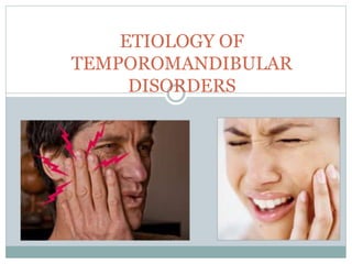
8. etiology of tmd
•Als PPTX, PDF herunterladen•
9 gefällt mir•2,669 views

The document discusses various theories regarding the etiology of temporomandibular disorders (TMDs). It covers early theories that emphasized occlusal factors or trauma, as well as more modern theories that consider TMDs to have a multifactorial etiology influenced by predisposing, initiating, and perpetuating factors. These factors include occlusal condition, trauma, emotional stress, deep pain input, and parafunctional habits. The document also discusses how functional and parafunctional muscle activities can be affected by the occlusal condition.

Empfohlen
Weitere ähnliche Inhalte
Was ist angesagt?
Was ist angesagt? (20)
Andere mochten auch
Andere mochten auch (20)
Ähnlich wie 8. etiology of tmd
Ähnlich wie 8. etiology of tmd (20)
Mehr von dr zarir ruttonji
Mehr von dr zarir ruttonji (10)
Kürzlich hochgeladen
Kürzlich hochgeladen (20)
8. etiology of tmd
- 2. CONTENTS 1. Introduction 2. Terminology 3. History of TMDs 4. Epidemiology of TMDs 5. Theories of Etiology 6. Development of functional disturbances in the masticatory system
- 3. 7. Etiologic Considerations Of Temporomandibular Disorders Occlusal Factors Trauma Emotional stress Deep Pain input Parafunctional habits 8. Food for thought 9. Conclusion 10. References
- 4. Introduction
- 5. TERMINOLOGY 1934 James Costen....Costen Syndrome 1959 Shore coined temporomandibular joint disturbances temporomandibular joint dysfunction syndrome Ramfjord and Ash- functional temporomandibular joint disturbances Bell suggested the term temporomadibular disorders (TMDs) Jeffrey P Okeson. Management of temporomandibular disorder and occlusion. 4th editionc
- 6. HISTORY OF TMDS In 1934 Dr James Costen, an otolaryngologist In 1930s and 1940s ...raise the bite as suggested by Costen. Late 1940s and 1950s...role of occlusal interferences. Importance was given to masticatoy muscle disorders In 1960s and into 1970s occlusion and later emotional stress In 1970s disorders arising from intracapsular sources Jeffrey P Okeson. Management of temporomandibular disorder and occlusion. 4th edition
- 7. EPIDEMIOLOGY OF TMDS1 1. 41% at least one symptom 2. 56% at least one clinical sign 3. Only 10% symptoms that were severe enough to make the patient seek treatment. 4. The greatest factor for seeking treatment is the degree of pain experienced. 5. Most symptoms are seen in 20-40 year age group.
- 8. THEORIES OF ETIOLOGY OF TMDS Biomedical model comprising • The mechanical displacement theory2 • The trauma theory3 • The biomedical theory4 • The osteoarthritic theory5 • The muscle theory6 The Neuromuscular Theory7 The Psychophysiological Theory8-10 The Multifactorial Theory1,11,12 The biopsychosocial Model13
- 9. THE MECHANICAL DISPLACEMENT THEORY Etiology oftemporomandibular disorders:the journey so far . INTERNATIONAL DENTISTRY SA VOL. 12, NO. 4
- 10. THE TRAUMA THEORY Zarb and Speck Micro-/Macrotrauma as the principal factor. no critical appraisal for the multitude of factors involved Etiology oftemporomandibular disorders:the journey so far . INTERNATIONAL DENTISTRY SA VOL. 12, NO. 4
- 11. THE BIOMEDICAL THEORY Reade Supported the role of trauma in the initiation of the disorder. The condition will either resolve or in presence of certain factors like disrupted occlusion, parafunctional habits and occupational activities, will progress further. Psychological elements were recognized as important maintaining influences. Etiology oftemporomandibular disorders:the journey so far . INTERNATIONAL DENTISTRY SA VOL. 12, NO. 4
- 12. THE OSTEOARTHRITIC THEORY Stegenga Osteoarthrosis as the causative factor for TMD. Muscular symptoms and internal derangement were secondary to joint pathology. Can explain some subcategories of TMD but fails to explain the rest. Eitiology of Temporomandibular disorders: the journey so far . INTERNATIONAL DENTISTRY SA VOL. 12, NO. 4
- 13. THE MUSCLE THEORY Travell and Rinzler Primary etiologic factor was in the masticatory muscles themselves. The myalgia in the facial region is caused by chronic myospasm which is secondary to parafunctional habits. Denied any influence of the occlusion. Etiology oftemporomandibular disorders:the journey so far . INTERNATIONAL DENTISTRY SA VOL. 12, NO. 4
- 14. THE NEUROMUSCULAR THEORY Ramjford. Occlusal interferences were the causative factor for the disorder. Occlusal interferences caused an altered proprioceptive feedback, leading to incoordination and spasm of some of the masticatory muscles. Etiology oftemporomandibular disorders:the journey so far . INTERNATIONAL DENTISTRY SA VOL. 12, NO. 4
- 15. THE PSYCHOPHYSIOLOGICAL THEORY Schwartz and Laskin. Suggested that the psychological factors are more important than the occlusal disturbances in initiating the disorder. It is the interaction between physiological predisposition, and psychological stress which causes TMD. The effect on the individual depended on their ability to cope with stress Etiology oftemporomandibular disorders:the journey so far . INTERNATIONAL DENTISTRY SA VOL. 12, NO. 4
- 16. THE PSYCHOLOGICAL THEORY Emotional disturbances initiating centrally, induced muscular hyperactivity which led to parafunctional habits and so indirectly to occlusal abnormalities. In TMD patient the behavioural aspect of the patient needs to be studied. It is still not clear whether they are the cause or the consequence of pain. Etiology of temporomandibular disorders: the journey so far . INTERNATIONAL DENTISTRY SA VOL. 12, NO. 4
- 17. THE MULTIFACTORIAL THEORY The etiology is multifactorial for TMD even though finding the primary etiologic factor can be difficult for the individual patient. The etiology will be different in young and in older patients. Factors influencing the disorder have been categorized by Bell into the predisposing, initiating and perpetuating factors. Etiology oftemporomandibular disorders:the journey so far . INTERNATIONAL DENTISTRY SA VOL. 12, NO. 4
- 18. THE BIOPSYCHOSOCIAL MODEL - 1992, DWORKIN integrates both the physical disorder factors, i.e., biological factors and the illness impact factors, i.e., psychological and social factors.
- 19. RDC/TMD
- 20. disturbances in the masticatory system1 • Normal + An event Physiologic TMD function tolerance symptoms
- 21. Normal Function complex neuromuscular control system. The brainstem (specifically the central pattern generator) regulates muscle action by the way of muscle engrams When sudden, unexpected sensory input is received, protective reflex mechanism is activated, creating a decrease in muscle activity in the area of the input.
- 22. Events During normal function of the masticatory system events can occur that may influence function. The events can be of local or systemic origin. Local Events 1. Placement of an improperly occluding crown. 2. Secondary to trauma involving local tissues, such as the postinjection response following local anesthesia. 3. Trauma can also arise from opening the mouth too wide 4. Unaccustomed use ---- bruxism 5. Constant deep pain input. 6. unknown cause (idiopathic pain)
- 23. Systemic Events The entire body and/or the Central Nervous System may be involved as an event. One of the most common types of systemic alterations is the increased level of emotional stress.
- 24. Physiologic Tolerance All individuals do not respond in the same manner to the same event. After how long or after what severity is the event perceived as unpleasant describes the physiologic tolerance. Physiologic tolerance can be influenced by both local and systemic factors.
- 25. Local Factors How the masticatory system responds to local factors is influenced by its orthopaedic stability. Orthopaedic instability related to the occlusion, the joints or both. 1. genetic, developemental or iatrogenic causes. 2. alterations in the normal anatomic form, such as a disc displacement or an arthritic condition. 3. disharmony between the ICP and the MS position of the joints.
- 26. Systemic Factors Influenced by diet, gender and genetics. Presence of acute or chronic diseases. Effectiveness of the pain modulation system
- 27. Temporomandibular symptoms •When functional change exceeds a critical level, alterations in the tissues begin, this level is known as the structural tolerance. •If the structural tolerance of any component is exceeded breakdown will occur. initial breakdown --- the lowest structural tolerance
- 29. Etiologic Considerations Of Temporomandibular Disorders1,14 • Predisposing factor – factors that increase the risk of TMD or orofacial pain. • Initiating factors – factors that cause the onset of disorder. • Perpetuating factors – factors that interfere with healing and complicate management. • Contributing factors –factors that initiate, perpetuate or result in a disorder.
- 30. Predisposing factors Systemic factor – Medical conditions such as rheumatic infections, nutritional and metabolic disorders Psychologic factors – Personality, behaviour Structural factors – All types of occlusal discrepancies, improper dental treatment, postural abnormalities, skeletal deformation, past-injuries etc. Genetic factors
- 31. Initiating factors:- Trauma – Micro or macro trauma Overloading of joint structures – Parafunctional habits etc. Perpetuating factors:- Mechanical and muscular stress Metabolic problems
- 32. Occlusal Condition Trauma Emotional Stress Deep Pain Input Parafunctional Habits
- 33. Occlusal Condition Earlier : Main causative factor Now: Little or no role Divided the fraternity into Occlusionists and non- occlusionists.
- 34. Reasons for the controversy The evaluation of the relationship of occlusion and TMDs should be done in both static and dynamic conditions. Faulty study design? Occlusion and Temporomandibular Disorders (TMD): Still Unsolved Question? - Alanen15
- 35. Pullinger et al16 used a blinded multifactorial analysis of each factor in combination with the other factors. They concluded that no single occlusal factor was able to differentiate patients from healthy subjects. 4 occlusal features however occurred mainly in TMD patients 1. presence of skeletal anterior open bite 2. retruded contact position and intercuspal position slides of greater than 2 mm. 3. overjets of greater than 4mm 4. five or more missing and unreplaced posterior teeth.
- 36. They concluded that although the relative odds for disease were elevated with several occlusal variables, clear definition of disease groups was evident only in selective extreme ranges, thus occlusion cannot be considered as the most important factor in the definition of TMDs.
- 37. Dynamic functional relationship between occlusion and TMD When considering the dynamic relation, the occlusal condition can affect some TMDs in 2 ways 1. effect of occlusal factors on orthopaedic stability 2. effect of acute changes in the occlusal condition and TMD
- 38. 1. Effect of occlusal factors on orthopaedic stability
- 39. 2 factors determine whether an intracapsular disorder will develop 1. the degree of orthopaedic instability: a discrepancy of 1-2mm.... shifts greater than 3mm present as significant risk factors. 2. the amount of loading: bruxism patients with orthopaedic instability. Patients with unilateral chewing patterns.
- 40. 2. Effect of acute changes in the occlusal condition and TMD activities of the masticatory system can be divided into 2 basic types: 1) Functional which includes chewing, speaking and swallowing; and 2) Parafunctional which includes clenching or grinding of the teeth.
- 41. Functional Muscle Activities : Controlled activities Protective reflexes are constantly present, guarding against potential damaging contacts. Interfering tooth contacts during function have inhibitory effects on functional muscle activity. Functional activities are directly influenced by the occlusal condition.
- 42. Parafunctional activities Totally different controlling mechanism. Instead of being inhibited by tooth contacts, earlier concepts suggested that parafunctional activities are actually provoked by certain tooth contacts. However these concepts for the most parts have been disapproved.
- 43. How do occlusal interferences affect muscle symptoms? An acute change will precipitate protective co- contraction. and at the same time has an inhibitory effect on the parafunctional activity. As the occlusal interferences become chronic the muscle response is altered in 2 ways: 1. alter the muscle engram so as to avoid the potentially damaging contact. 2. tooth movement to accommodate for the heavy loading
- 44. Trauma • Ample evidence supports the fact that trauma can lead to TMDs. • Greater impact on intracapsular disorders than muscular disorders. Trauma can be divided into 2 basic types Macrotrauma Microtrauma
- 45. Macrotrauma17 most common structural alterations affecting the TMJ are elongation of the discal ligaments. Can be further divided into Direct INDIRECT
- 46. Direct trauma - a blow to the chin, can instantly create an intracapsular disorder. The open mouth trauma can lead to discal displacement and/or dislocation. Closed mouth trauma is less injurious to the condyle disc complex as the intercuspation of the teeth maintains the jaw position. Iatrogenic trauma: intubation procedures during general anesthesia, third molar extraction procedures.
- 47. Indirect trauma injury that may occur to the TMJ secondary to a sudden force that does not directly impact or contact the mandible. The most common type is associated with cervical flexion-extension (whiplash) injuries seen in road traffic high speed accidents.
- 48. Microtrauma Any small force that is repeatedly applied to the joint structures over a long period of time. Mechanism of Microtrauma: loading exceeds the functional limit of the tissues, collagen fibrils become fragmented decrease in the stiffness of the collagen network. proteoglycan water gel swells and flow out into the joint space,
- 49. Softening of the articular surface called chondromalacia. Early stage of chondromalacia is reversible if the excessive loading is reduced. Loading continues to exceed the capacity of the articular tissues, irreversible changes can occur.
- 50. Clenching or bruxism leads to posterior border thinning of the disc, which causes elongation of the inferior retrodiscal ligaments which eventually leads to disc displacement. Disharmony between ICP and MS positions.
- 51. Emotional Stress The hypothalamus, the reticular system, and particularly the limbic system are primarily responsible for the emotional state of the individual. Mechanism of Action Stress activates the hypothalamus prepares the body to respond by the autonomic system increases the activity of the gamma efferents
- 52. intrafusal fibers of the muscle spindle contract. sensitizes the spindle so that any slight stretching of the muscle will cause a reflex contracture. The overall effect is an increase in tonicity of the muscle.
- 53. Stress is released by two mechanisms: 1. External: released by shouting, cursing, hitting. A more positive way to release stress is by engaging in physical exercises. 2. Internal: stress manifests as irritable bowel syndrome, Hypertension, cardiac arythmias, increased activity of head and neck muscles. Emotional stress also leads to decreased physiologic tolerance due to increased sympathetic tone.
- 54. Deep Pain Input Deep pain input can centrally excite the brainstem, producing protective co-contracture Patient suffering from toothache will have a limited mouth opening. sinus pain , ear pain , pain sources remote to the face like cervical pain input.
- 55. Parafunctional Activities This includes bruxism, clenching and certain oral habits. Parafunctional activities can b divided into 2 basic categories 1. Diurnal: occuring through the day. 2. Noctural: occurring through the night.
- 56. Diurnal activity: parafunctional activity during the day clenching and grinding tongue or cheek biting finger and thumb sucking unusual postural habits occupation realted activities sub-conscious level therefore merely questioning the patient is not a reliable way to assess the presence or absence of these activities.
- 57. Nocturnal activity single episodes (clenching) rhythmic contractions (Bruxism).
- 58. Bruxism: the subconscious, non-functional grinding of teeth Associated with a change from deeper to lighter sleep. Bruxism may be closely associated with the arousal phases of sleep.
- 59. Sleep REM sleep : 80% Non-REM sleep: 20% REM sleep: restoring function of the cortex and brainstem activity. Psychic Rest Non-REM sleep: restoring function of the system. Increased synthesis of protein and RNA. Physical Rest
- 60. Deprived of REM sleep: greater anxiety and irritability Non-REM sleep: musculoskeletal tenderness, aching and tenderness. Duration of Bruxing Event: Average bruxing event lasts for 9 secs. Total bruxing time -40 secs per hour. Bruxism occurs only 5 times during entire sleep with an average of 8 secs per event.
- 61. Intensity of Bruxing Events: average bruxing event involves 60% of the maximum clenching power before the patient went to sleep. Sleep Position and Bruxing: Earlier :persons sleeping on the sides experience more bruxing activity Now: research has clarified that bruxing events are moe when the person is sleeping on his/her back.
- 62. Bruxing Events and Masticatory Symptoms: Ware and Rugh studied a group of bruxism patients without pain and a group with pain. Concluded that the latter group had a significantly higher number of bruxing events during REM sleep than did the former. Both groups however bruxed more than a control group
- 63. Food for thought… Symptom based treatment???? Greene18 concluded from his review that not only are the old mechanistic etiologic concepts incorrect, but also that 2 of the most popular current concepts (biopsychosocial and multifactorial) are seriously flawed. Even in the absence of a perfect understanding of etiology, we still can provide good conservative care, and we should avoid aggressive and irreversible treatments,
- 64. Conclusion To manage TMD effectively the clinician must be able to determine its etiology. TMDs are dependant on a variety of factors which have a complex interplay between them. Occlusion is not the sole factor responsible for TMDs and the clinician should recognize that as the Clinician who only evaluates the occlusion is likely missing as much as the clinician who never evaluates the occlusion.
- 65. References 1. Jeffrey P Okeson. Management of temporomandibular disorder and occlusion. 4th edition: Mosby year book inc. Missouri 2. Molin C. From Bite to mind: A personal and literature review. Int J Prosthodont. 1999: 12 :279-288 3. Zarb GA, Speck JE. The treatment of mandibular dysfunction. In: Zarb GA, Carlsson GE, editors. Temporomandipular joint function and dysfunction. Copenhagen: Munksgaard; 1979. 4. Reade PC. An approach to the management of temporomandibular joint pain-dysfunction syndrome. J Prosthet Dent 1984;51:91–6. 5. Stegenga B, de Bont LGM, Boering G. Osteoarthrosis as the cause of craniomandibular pain and dysfunction. A unifying concept. J Oral Maxillofac Surg 1989;47:249–56.
- 66. 6. Laskin. Etiology of the pain dysfunction syndrome. J Am Dent Assoc.1979:79:147 7. Ash and Ramjford. Occlusion:: 4th edition: 1995: W B Saunders company: Philadelphia 8. Greene CS. Etiology of temporomandibular disorders. Sem Orthod 1995;1:222–8. 9. McNeill C. Management of temporomandibular disorders: concepts and controversies. J Prosthet Dent 1997;77:510–22. 10. Turner JA, Dworkin SF. Recent developments in psychological diagnostic procedures: screening for psychological risk factors forpoor outcomes. JADA 2004;135:1119–25. 11. Pertes and Gross clinical management of temporomandibular disorders and orofacial pain: 1995: quintessence publishing co inc: Illionis.92 INTERNATIONAL DENTISTRY SA VOL. 12, NO. 4
- 67. 12. Goldstein. TMD : A review of current understanding. J Prosthet Dent 1999;88:56-67 13. Dworkin. Perspectives on the interaction of biological, psychological and social factors in TMD. J Am Dent Assoc. 1994:125:856-863 14. Bell WE. Temporomandibular disorders. Classification, diagnosis, management. 3rd ed.. Chicago: Year Book; 1990. 15. Pentti Alanen .Occlusion and Temporomandibular Disorders (TMD): Still Unsolved Question? J Dent Res 81(8):518-519, 2002 16. Pullinger AG, Seligman DA: A multiple logistic regression analysis of the risk and relative odds of TMDs as a function of common occlusal features, J Dent Res72:968-79,1993 17. Sonia Bhat. Etiology of temporomandibular disorders: the journey so far. international dentistry sa vol. 12, no. 4.88-98 18. Charles S. Greene,.The Etiology of Temporomandibular Disorders: Implications for Treatment. J OROFAC PAIN 2001;15:93–105.
- 68. We must do the right as we see the right THANK YOU
Hinweis der Redaktion
- The masticatory system is extremely complex More complex a system, the greater the likelihood of breakdown.
- Based on 11 case studies
- According to these studies, the percentage of population with some type of TMD is between 40-60%, a figure so high that it might lead one to doubt the validity of these studies. Only 5% comprised a group that would be typically described as TMD patients seen in dental offices
- Thus brought tmd into the broader awspect of muscle disorders
- He noted that regional pain associated with bruxism and myalgia was completely eliminated in subjects after occlusal equilibration.
- Spasm of the masticatory muscles caused by overextension, overcontraction or muscle fatigue due to parafunctions was used by patients as a means to relieve stress.
- Several authors have confirmed the role of psychological factors in TMD.
- Gradually, concepts based on a single factor lost their scientific and clinical credibility. As it became more and more apparent that the etiology was multifactorial and that none of these theories in isolation could explain the etiologic mechanisms in TMD patients. The theories advanced from a pure mechanistic view, and expanded to a wider arena inclusive of psychological and behavioral With increasing age, there is an increased risk of age-related joint changes and systemic conditions affecting the TMJ--reparative capacity of the articular cartilage is significantly reduced
- this model showed the dynamic nature of intrinsic intrapersonal factors (such as nociception, pain perception, pain appraisal) and extrinsic interpersonal factors (behaviour responses to pain, social roles for the person in pain within the context of the family, the health care delivery system, the workplace, and the social welfare system) in chronic pain, including TMD
- most widely studied instruments in this orientation is the RDC/TMD, which conceptualizes TMD according to a two-axis system, one for the physical disorder factors (Axis I) (Figure 7) and the other for the psychosocial illness impact factors (Axis II)
- that are appropriately selected according to the sensory input received from the peripheral structures.
- may represent any change in sensoy or proprioceptive input, which is the subconscious, non-functional grinding of teeth. Pain felt in the masticatory or associated structures often alters normal muscle function by the way of central excitatory effects
- The most stable orthopaedic relationship between the mandible and the maxilla is achieved when the mandible closes with the condyles in their most superoanterior position, resting against the posterior slopes of the articular eminence with the discs properly interposed, there is even and simultaneous contact of all the posterior teeth directing forces towards the long axes of those teeth. From that position, when the mandible moves eccentrically, the anterior teeth contact and disocclude the posterior teeth.
- When an event exceeds the physiologic tolerance of an individual, the system begins to reveal certain changes.
- Therefore breakdown sites can be: the muscles, the TMJs, the supportive structures of the teeth, and the teeth themselves. If the weakest structure is the muscle then the symptoms felt are mucle tenderness, pain and restricted jaw movements. If TMj is the weakest link then joint tenderness, pain and clicks will be reported. If the weakest links are the teeth then periodontal destruction resulting in mobility and wear of the tooth surfaces is reported.
- All the above factors can be broadly grouped into 3 major factors:- Anatomic Psychologic Neuromuscular
- This issue is extremely critical because if occlusion plays an important role in TMDs then the dentist has the sole responsibility of treating such findings but if occlusion does not play any role then the dentist should refrain from treating TMDs by making changes in the occlusion. This debate does not in any way relate to the importance of occlusion in dentisty, as occlusion is the basis of dentistry.
- is not likely to cause an intracapsular disorder,
- Occlusal contact pattern of teeth influences the activity of masticatory muscles for example, introducing a slightly high contact can induce masticatory muscle activity which eventually results in pain.
- that allow the masticatory system to perform necessary function with minimum damage to any structure.
- 1. the most common way is to alter the muscle engram . In most cases the patients can adapt to change. However if altered muscle engrams cannot adapt, a continued muscle co-contraction can produce a muscle pain disorder.
- The dense fibrous connective tissues that cover the articular surfaces of the joints can well tolerate the loading forces but only within certain limits. The delivery of vital nutrients and the elimination of metabolic products by the synovial fluid may be impaired if articular tissues are subjected to excessive stress.
- The emotional centers of the brain influence muscle function. These centers influence the muscle activity by the gamma efferent pathways
- Once the toothache is resolved normal mouth opening will resume but if the dentist fails to comprehend the reason for limited mouth opening as the tooth ache he might try to alter the existing condition and cause more harm than good.
- However there is a great variation observed between patients and uncertainty exists as to the number and duration of bruxing events that creates muscle symptoms.
- . Other studies by the same authors established that more sustained contraction occurs in bruxism was much higher during REM than non-REM phases of sleep.
- Therefore, what we really have at the individual TMD patient evel is nearly always an idiopathic situation—we simply do not know enough, or cannot measure enough, or cannot precisely determine why each patient has a TMD.