ANATOMY OF THE LYMPHATIC SYSTEM
•Als PPTX, PDF herunterladen•
38 gefällt mir•8,374 views

Anatomy of the Lymphatic System, a prerequisite for the Bsc. degree in Biology

Empfohlen
Weitere ähnliche Inhalte
Was ist angesagt?
Was ist angesagt? (20)
Ähnlich wie ANATOMY OF THE LYMPHATIC SYSTEM
Ähnlich wie ANATOMY OF THE LYMPHATIC SYSTEM (20)
Mehr von Fasama H. Kollie
Mehr von Fasama H. Kollie (20)
Kürzlich hochgeladen
Kürzlich hochgeladen (20)
ANATOMY OF THE LYMPHATIC SYSTEM
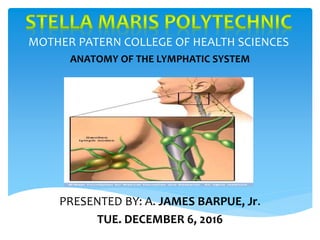
- 1. MOTHER PATERN COLLEGE OF HEALTH SCIENCES ANATOMY OF THE LYMPHATIC SYSTEM PRESENTED BY: A. JAMES BARPUE, Jr. TUE. DECEMBER 6, 2016
- 2. The lymphatic system consists of the following components: Lymph lymphatic vessels lymphatic tissue lymphatic organs Components of the lymphatic system
- 3. Lymph is usually a clear, colorless fluid, similar to blood. plasma but low in protein It originates as tissue fluid that has been taken up by the lymphatic vessels. Lymph leaving the lymph nodes contains a large number of lymphocytes Lymph can also contain macrophages, hormones, bacteria, viruses, cellular debris, and even traveling cancer cells. Lymph
- 4. Lymph flows through a system of lymphatic vessels (lymphatics) similar to blood vessels These begin with microscopic lymphatic capillaries The lymphatic capillary penetrates nearly every tissue of the body They are closely associated with blood capillaries A lymphatic capillary consists of a sac of thin endothelial cells Lymphatic Vessels
- 5. lymphatic endothelial cells are not joined by tight junctions the gaps between them are so large that bacteria, lymphocytes, and other cells and particles can enter along with the tissue fluid The overlapping edges of the endothelial cells act as valvular flaps that can open and close The overlapping edges of the endothelial cells act as valvular flaps that can open and close Lymphatic Capillaries cont’d
- 7. The larger lymphatic vessels are similar to veins in their histology. They have a tunica interna tunica media tunica externa. As the lymphatic vessels converge along their path, they become larger and larger vessels with changing names. Lymphatic Vessels cont’d
- 8. The route from the tissue fluid back to the bloodstream is: lymphatic capillaries —> collecting vessels —> six lymphatic trunks— two collecting ducts —> subclavian veins The lymphatic capillaries converge to form collecting vessels At irregular intervals, they empty into lymph nodes The lymph trickles slowly through the node Lymphatic Vessels cont’d
- 10. It leaves the other side of the node through another collecting vessel traveling on and often encountering additional lymph nodes before it finally returns to the bloodstream Eventually, the collecting vessels converge to form larger lymphatic trunks each of which drains a major portion of the body Lymphatic Vessels cont’d
- 11. There are six lymphatic trunks: Jugular Subclavian Branchomediastinal Intercostal Intestinal lumbar Lymphatic Vessels cont’d
- 13. The lymphatic trunks converge to form two collecting ducts: 1. The right lymphatic duct 2. The thoracic duct The thoracic duct, on the left, is larger and longer. It begins just below the diaphragm, anterior to the vertebral column at the level of the second lumbar vertebra Lymphatic Vessels cont’d
- 14. Here, the two lumbar trunks and the intestinal trunk join and form the cisterna chyli cisterna chyli , named for the large amount of chyle that it collects after a meal. Lymphatic Vessels cont’d
- 17. Lymph flows under forces similar to those that govern venous return except that the lymphatic system has no pump like the heart. It flows at even lower pressure and speed than venous blood. The valves of lymphatic vessels prevent the fluid from flowing backward The Flow Of Lymph
- 18. The Flow Of Lymph cont’d
- 19. Aside from cells playing purely structural roles, the lymphatic system has six principal categories of defensive cells: I. Natural killer (NK) cells II. T lymphocytes (T cells) III. B lymphocytes (B cells) IV. Macrophages V. Dendritic cells VI. Reticular cells Macrophages, B lymphocytes, and reticular cells are collectively called APCs. Lymphatic Cells
- 20. They are aggregations of lymphocytes in the connective tissues of mucous membranes and various organs The simplest form is diffuse lymphatic tissue It is prevalent in the respiratory, digestive, urinary, and reproductive tracts In these areas, it is called mucosa-associated lymphatic tissue (MALT) It is sometimes called BALT and GALT based on the location. Lymphatic Tissues
- 21. Lymphatic Nodule in the Mucous Membrane of the small intestine
- 22. In some places, lymphocytes and macrophages congregate in dense masses called lymphatic nodules (follicles) lymphatic nodules (follicles), come and go as pathogens invade the tissues and the immune system answers the challenge Abundant lymphatic nodules are a constant feature of the lymph nodes, tonsils, and appendix In the ileum, they form clusters called Peyer patches. Lymphatic Tissues cont’d
- 23. lymphatic (lymphoid) organs have well-defined anatomical sites These organs include the red bone marrow, thymus, lymph nodes, tonsils, and spleen The red bone marrow and thymus are regarded as primary lymphatic organs The lymph nodes, tonsils, and spleen are called secondary lymphatic organs Lymphatic Organs
- 25. Red bone marrow is involved in hemopoiesis and immunity In children, it occupies the medullary spaces of nearly the entire skeleton In adults, it is limited to parts of the axial skeleton and the proximal heads of the humerus and femur is an important supplier of lymphocytes to the immune system Red bone marrow is a soft, loosely organized, highly vascular material, separated from osseous tissue by the endosteum of the bone Red Bone Marrrow
- 26. it produces all classes of formed elements of the blood its red color comes from the abundance of erythrocytes Numerous small arteries enter nutrient foramina on the bone surface It penetrates the bone, and empty into large sinusoids in the marrow The sinusoids drain into a central longitudinal vein that exits the bone via the same route that the arteries entered Red Bone Marrow cont’d
- 27. The sinusoids, 45 to 80 µm wide They are lined by endothelial cells are surrounded by reticular cells and reticular fibers. The spaces between the sinusoids are occupied by islands(cords) of hemopoietic cells, composed of macrophages and blood cell stages of development As blood cells mature, they push their way through the reticular and endothelial cells to enter the sinus and flow away in the bloodstream. Red Bone Marrrow cont’d
- 30. It is a bilobed organ located between the sternum and aortic arch in the upper mediastinum The two lobes are connected by a median bridge of tissue The fibrous capsule of the thymus gives off trabeculae (septa) that penetrate into the gland It houses developing lymphocytes Thymus
- 31. Trabeculae divide it into several angular lobules. Each lobule has a dense, dark-staining cortex and a lighter medulla inhabited by T lymphocytes Reticular epithelial cells seal off the cortex from the medulla It surround the blood vessels and lymphocyte clusters in the cortex They thereby form a blood—thymus barrier that isolates developing lymphocytes from blood-borne antigens. Thymus cont’d
- 32. In the medulla, the reticular epithelial cells form whorls called thymic (Hassall) corpuscles reticular epithelial cells secrete several signaling molecules that promote the development and action of T cells Thymus cont’d
- 34. A lymph node is-an elongated or bean-shaped structure It is usually less than 3 cm long with an indentation called the hilum on one side Lymph nodes are the most numerous lymphatic organs Lymph Nodes
- 35. It is enclosed in a fibrous capsule with trabeculae The subcapsular sinus contains reticular fibers, macrophages, and dendritic cells. The parenchyma is divided into : Cortex Medulla Lymph Nodes
- 38. Lymph nodes are widespread but especially concentrated in the following locations: Cervical lymph nodes Axillary lymph nodes Thoracic lymph nodes Abdominal lymph nodes Intestinal and mesenteric lymph Inguinal lymph nodes Popliteal lymph nodes Lymph Nodes
- 39. The tonsils are patches of lymphatic tissue They are located at the entrance to the pharynx Each is covered by an epithelium It has deep pits called tonsillar crypts lined by lymphatic nodules. There are three main sets of tonsils: 1. A single median pharyngeal tonsil Tonsils
- 40. 2. A pair of palatine tonsils 3. numerous lingual tonsils Tonsils
- 42. The Spleen
- 43. The spleen is about 12 cm long (5 in.) it weighs up to 160 g (5.6 oz). The spleen is located in the left hypochondriac region, just inferior to the diaphragm . It is posterolateral to the stomach It is protected by ribs 10 through 12 It is attached to the lateral border of the stomach by a broad mesenteric band The Spleen
- 44. It lies wedged between the stomach, the left kidney, and the muscular diaphragm Two types of tissues found in the spleen includes: red pulp white pulp The Spleen Cont’d
- 46. Are located in the mucosa and submucosa throughout the small intestine They contain mostly B cells. Peyer’s Patches
- 47. The appendix is a pouch of lymphatic tissue that is attached to the large intestine It is located in the lower-right area of the abdomen The appendix
- 48. The lymphatic system consists of the following components: (1) lymph (2). lymphatic vessels and (3). lymphatic tissue Lymphatic organs include: the red bone marrow, thymus, lymph nodes, tonsils, and spleen. The red bone marrow and thymus are regarded as primary lymphatic organs. The lymph nodes, tonsils, and spleen are called secondary lymphatic organs. Summary