
Empfohlen
Weitere ähnliche Inhalte
Ähnlich wie case report Mr J english day-2.pptx
Ähnlich wie case report Mr J english day-2.pptx (20)
Kürzlich hochgeladen
Kürzlich hochgeladen (20)
case report Mr J english day-2.pptx
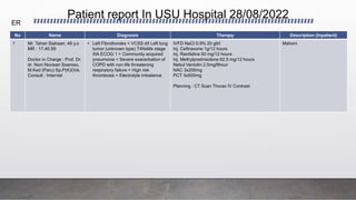
- 1. Patient report In USU Hospital 28/08/2022 No Name Diagnosis Therapy Description (Inpatient) 1 Mr. Tahan Siahaan, 48 y.o MR : 17.40.59 Doctor in Charge : Prof. Dr. dr. Noni Novisari Soeroso, M.Ked (Paru) Sp.P(K)Onk. Consult : Internist • Left Fibrothoraks + VCSS d/t Left lung tumor (unknown type) T4NxMx stage IIIA ECOG 1 + Community acquired pneumonia + Severe exacerbation of COPD with non life threatening respiratory failure + High risk thrombosis + Electrolyte imbalance IVFD NaCl 0.9% 20 gtt/i inj. Ceftriaxone 1g/12 hours inj. Ranitidine 50 mg/12 hours inj. Methylprednisolone 62.5 mg/12 hours Nebul Ventolin 2.5mg/8hour NAC 3x200mg PCT 3x500mg Planning : CT Scan Thorax IV Contrast Mahoni ER
- 2. Patient report In USU Hospital 28/08/2022 No Name Diagnosis Therapy Description (Inpatient) 1 Mr. Bruly Mael Sirait, 43 y.o MR : 17.40.54 Doctor in Charge : Internist Consult : Dr. dr. Pandiaman Pandia, Sp.P(K) • Loss to follow up case of Pulmonary TB + Functional Dyspepsia dd Organic + DM type II + HHD NAC 3x200mg Curcuma 1x1 tab R/H/Z/E : 600/300/1500/1250 Vit. B6 2x10mg Planning : Molecular test of sputum Meranti 3 WARD
- 3. No Name Diagnosis Therapy Description (Inpatient) 1. Mrs. Kaliyem 63 years old MR : 174143 Doctor in Charge : Prof. Dr. dr. Noni N. Soeroso, Sp.P(K).Onk (with admission letter)) Consultant : Cardiologist Mild Left Pleural Effusion + Left Lung Tumor (type?) T4N3M1a (pleura, pericardium, contralateral) Stage IV A ECOG II + Severe COPD Exacerbation without respiratory failure in COPD Stable Group D + High Risk Thrombosis + Hypocalemia IVFD NaCl 0.9% 20 gtt/i inj. Ceftriaxone 1g/12 hours Inj. Gentamicin 240mg/24 hours Inj. Metronidazole 500mg/8 hours inj. Ranitidine 50 mg/12 hours Inj. Ketorolac 30 mg/12 hours inj. Methylprednisolone 62.5 mg/12 hours Nebul Ventolin 2.5mg/8hour Nebul Pulmicort 0,5mg/12 hour Retaphyl SR 2x150mg NAC 3x200mg Plan: Lymph Glands FNAB on Supraclavicle CT Scan Thorax IV Contrast Stable Mahoni ER Patient report in USU Hospital 29/08/2022
- 4. No Name Diagnosis Therapy Description (Inpatient) Ward Patient report in USU Hospital 29/08/2022
- 5. Patient report in Adam Malik Hospital 29/08/2022 No Name Diagnosis Therapy Description (Inpatient) 1. Mr. Jiban Capah 64 years old MR : 871760 Doctor in Charge : Neurosurgeon Consultant : Dr. dr. Pandiaman Pandia, Sp.P(K), anesthetist, internist Sepsis d/t Community Acquired Pneumonia + Mild ARDS + loss of consciousness + d/t Multiple SOL Intracranial dd Brain Metastasis dd Tuberculoma + Hepatic Cirrhosis Decompensated Stadium + Upper GI Tract Bleeding d/t variceal dd non variceal bleeding - Acc to treat together - Inj. Ceftriaxone 1g/12 hour - Neb. Salbutamol 2,5mg/8 hour - Neb. Budesonide 1mg/12 houras - N-Acetylsistein 200mg/8 hour Unstable ICU 2. Mr. Pawer Sitompul 66 years old MR : 871773 Doctor in Charge : Neurologist Consultant : dr. Ade Rahmaini, Sp.P(K) Left lung tumor dd Lung tumor metastasis + Secondary Headache + Left hemiparesis d/t Susp. Brain metastasis - Acc to treat together - Kodein 20mg/12 hour Plan: CT Scan Thorax IV Contrast Stable RA 4 ER
- 6. No Name Diagnosis Therapy Description (Inpatient) 3. Mr. Rian Rinaldi 22 years old MR : 871778 Doctor in Charge : dr. Parluhutan Siagian, Sp.P(K) Consultant : Susp. Moderate Covid 19 + Atelectasis d/t New case of Pulmonary TB on ATT intensive phase IVFD NaCl 20gtt/I Inj. Omeprazole 40mg/24 hour Inj. Vit C 1000mg/24 hour N-Acetylsistein 200mg/8 hour Paracetamol 500mg/8 hour Vit D 5000 IU/24 hour Plan: Swab RT PCR Covid 19 2x Stable Still in ER 4. Mrs. Surya Asih 64 years old MR : 639393 Doctor in Charge : Surgeon Consultant : dr. Parluhutan Siagian, Sp.P(K) Susp. Mild Covid 19 + Susp. (L) Ca Breast + Susp. Brain Metastasis - Acc to treat together - N-Acetylsistein 200mg/8 hour - Paracetamol 500mg/8 hour - Vit D 5000 IU/24 hour Plan: Swab RT PCR Covid 19 Stable Still in ER ER Patient report in Adam Malik Hospital 29/08/2022
- 7. No Name Diagnosis Therapy Description (Inpatient) 1. Mr. Tunggul Hutahaean 55 years old MR : 871757 Doctor in Charge : Cardiologist Consultant : Dr. dr. Amira Permatasari Tarigan, Sp.P(K) Community Acquired Pneumonia dd Viral Pneumonia dd New Case of Pulmonary Tuberculosis + Severe COPD Exacerbation without respiratory failure on COPD Stable Group B + Pericardial Effusion Post Pericardiocentesis - Acc to treat together - Inj. Levofloxacin 750mg/24 hour - Inj. Methylprednisolone 32mg/24 hour - Neb. Salbutamol 2,5mg/8 hour - Neb. Budesonide 0,5mg/12 hour - N-Acetylsistein 200mg/8 hour - Vit B Complex/8 hour Plan: Chest X-ray RT PCR Covid-19 2x Check D-dimer Rapid Molecular Testing and Sputum Culture Spirometry and DLCO if Stable Stable RIC Level 4 Ward Patient report in Adam Malik Hospital 29/08/2022
- 8. DAILY MORNING REPORT Tuesday, 30th August 2022 • Residents on duty : dr. Redha, dr. Aulia, dr. Minarni, dr. Mona, dr. Selfi, dr. Faiz, dr. Elfia, dr. Novi • Doctor in charge : dr. Setia Putra Tarigan, Sp.P(K).Onk • Consultant : - • Working diagnosis : 1. Superior Vena Cava Syndrome 2. Malignant Pleural Effusion due to Right Lung Cancer (type?) T4N3M1a (pleura) Stage IV A ECOG I 3. Severe COPD Exacerbation without Respiratory Failure on Stable COPD group B 4. Community Acquired Pneumonia 5. High Risk Thrombosis 6. Hypoalbuminemia
- 9. Patient Record Name : Mr. JPH Age : 62 y.o Sex : Male Occupation : Construction worker Ethnic : Bataknese Religion : Islam Address : Sembada Ujung Chief Complaint: Shortness of breath Registration date : 27/08/2022
- 10. Course of Disease • Patient came to Medan Lung Hospital with main complaint shortness of breath (SOB). SOB was experienced since two months ago. SOB was associated with activities such as walking 100m (mMRC = 3). SOB is not related to position or weather. There were no history of wheezing. • Coughing was found in the last two weeks with productive sputum. The color of sputum was white and volume half tablespoon per cough. There was no Coughing up blood or history of coughing up blood. • Chest pain was experienced since a month ago. Pain was felt like prickling around the right lower chest and did not radiate anywhere. Pain worsened during coughing or inspiration. (VAS = 2-3) • Chest X-ray and rapid molecular test for tuberculosis from sputum were performed. The result was negative tuberculosis from the sputum. Patient was given Anti Tuberculosis Treatment (ATT). Patient was referred to Bina Kasih Hospital to get further examination July 2022 August 2022 • Patient was admitted to Bina Kasih Hospital with main complaint shortness of breath (SOB). SOB was experienced since three months ago. SOB was associated with activities such as walking 100m (mMRC = 3). SOB is not related to position or weather. There were no history of wheezing. • Coughing was found in the last month with productive sputum. The color of sputum was white and volume half tablespoon per cough. There was no Coughing up blood or history of coughing up blood. • Chest pain was experienced since two months ago. Pain was felt like prickling around the right lower chest and did not radiate anywhere. Pain worsened during coughing or inspiration. (VAS = 5-6) • The ATT was stopped by the pulmonologist. • Chest X-ray and Thorax CT-Scan with contrast was performed. A central mass was found on the CT-Scan. Patient was referred to Adam Malik Hospital to get further examination.
- 11. Course of Disease • Patient was referred to Adam Malik Hospital with main complaint shortness of breath (SOB). SOB was experienced since three months ago. SOB was associated with activities such as walking 100m (mMRC = 3). SOB is not related to position or weather. There were no history of wheezing. • Coughing was found in the last month with productive sputum. The color of sputum was white and volume half tablespoon per cough. There was no Coughing up blood or history of coughing up blood. • Chest pain was experienced since two month ago. Pain was felt like prickling around the right lower chest and did not radiate anywhere. Pain worsened during coughing or inspiration. (VAS = 7-8) • Patient has gotten emergency radiotherapy 5 times. Patient was planned for bronchoscopy but rejected by anesthetic department as the patient cannot lie down for extended time. Then the patient was discharged as his condition gotten better and was planned bronchoscopy from polyclinic 6th August 2022 27th August 2022 • Patient came to Adam Malik Hospital with main complaint shortness of breath (SOB). SOB was experienced since four months ago. SOB was associated with activities such as walking 100m (mMRC = 3). SOB is not related to position or weather. There were no history of wheezing. • Coughing was found in 2 months with productive sputum. The color of sputum was yellowish and volume a tablespoon per cough. There was no Coughing up blood or history of coughing up blood. • Chest pain was experienced since three months ago. Pain was felt like prickling around the right lower chest and did not radiate anywhere. Pain worsened during coughing or inspiration. (VAS = 7-8)
- 12. Additional complaints • No loss of smell was experienced • No loss of taste was experienced • Hoarseness was experienced since two months ago • No sore throat was experienced • No difficult of swallowing was experienced • No fever was experienced • Losing appetite and weight loss around 12 kg within 3 months. • No nausea and vomiting were experienced. • No muscle pain and seizure were experienced. • No history of night sweating was found
- 13. The history of Previous Illness • Smoker with severe IB level (45 years x 24 cigarettes = 1080) • No history of drinking alcohol • No history of drug abuse was found. • No history of having pets with fur was found. • Exposure of biomass was found (asbestos since 25 years ago) • No history of Hypertension • No history of malignancy in the family • No history of contact to person with chronic cough • No history of Diabetes Mellitus • No history of asthma was experienced. • No history of COVID-19 was experienced with complete vaccination. • No history of allergy was experienced. • History of nebulization during hospitalization • History of the ATT consumption was found in July 2022 given by pulmonologist. ATT was stopped by pulmonologist in Bina Kasih Hospital after 2 weeks consumption
- 14. Risk Factor Tn.AS/73 thn 1. Smoking status: Heavy smoker (Brinkmann Index: 45 years x 24 cigarettes = 1080, severe IB) cigarette, deep inhalation. 2. Biomass exposure : Asbestos since 25 years ago 3. The history of malignancy in the family: - 4. The history of Pulmonary Tuberculosis: ATT consumption was found in July 2022 given by pulmonologist. ATT was stopped by pulmonologist in Bina Kasih Hospital after 2 weeks consumption 5. The history of COPD: - 6. The Home environment condition: good 7. The work environment: Bad 8. The use of oral contraception (female): - Mr. JPH/ 62 y.o
- 15. OCCUPATIONAL RISK FACTOR No 1. Determine Clinical Diagnostic Superior Vena Cava Syndrome + Malignant Pleural Effusion due to Right Lung Cancer (type?) T4N3M1a (pleura) Stage IV A ECOG I + Community Acquired Pneumonia + Severe COPD Exacerbation on COPD Stable group B + High Risk Thrombosis + Hypoalbuminemia 2. Exposure at work Asbestos since 25 years ago 3. Risk Factor Not using a facial mask 4. Amount of exposure 6 days a week 6 hours a day 5. Individual Factor at work Smoker with severe IB level 6. Another Factor - Mr. JPH/ 62 y.o
- 16. DIAGNOSIS OF OCCUPATIONAL LUNG DISEASE Clinical Diagnostic Superior Vena Cava Syndrome + Malignant Pleural Effusion due to Right Lung Cancer (type?) T4N3M1a (pleura) Stage IV A ECOG I + Community Acquired Pneumonia + Severe COPD Exacerbation on COPD Stable group B + High Risk Thrombosis + Hypoalbuminemia Exposure • Occupational : Construction worker • Type of exposure : asbestos • Smoking cessation : - Relationship with exposure Lacourt A, Pintos J, Lavoué J, Richardson L, Siemiatycki J. Lung cancer risk among workers in the construction industry: results from two case-control studies in Montreal. BMC Public Health. 2015 Sep 22;15:941. doi: 10.1186/s12889-015- 2237-9. PMID: 26395169; PMCID: PMC4580354. Amount of exposure 6 days a week 6 hours a day Individual Factor - Not using personal protective environment during work Another factor outside of work - Smoker with severe IB level Conclusion There is no associations between the occupational exposure with Secondary Spontaneous Pneumothorax Dekstra ec. Suspect relapse case of pulmonary TB Mr. JPH/ 62 y.o
- 17. Oncology Emergency (Respiratory System) No. Characteristics Presented Oncology Emergency 1. Pleural Effusions + - 2. Vena Cava Superior Syndrome + + 3. Brain Metastasis - - 4. Myasthenia gravis - - 5. Cardiac Tamponade - - 6. Large Airway Obstructions - - 7. Haemoptysis - - 8. Spinal cord Compression - - 9. Tumor Lysis Syndrome - - 10. Hypercalsemia - - 11. Neutropenia - - Mr. JPH/ 62 y.o
- 18. Problems / Clinical Details • Chronic shortness of breath • Chronic cough with sputum production • Severe Chest pain • Loss of appetite and weight about 12 kgs within 2 months • Heavy Smoker • History of the ATT consumption for two weeks in July 2022 • Exposure of biomass since 25 years ago Mr. JPH/ 62 y.o
- 19. Vital Signs Level of consciousness : Alert, GCS 15 E4V5M6 BP : 120/70 mmHg Pulse : 110 x/minutes, regular, equal for 4 extremities RR : 24 x/minutes, abdominalthoracal breathing type, without use of respiratory muscle Temp : 36.2 0C axilla SpO² : 94 % Room Air VAS : 7-8 Weight : 50 kg Height : 165 cm BMI : 18,4 kg/m2 (Normoweight) Mr. JPH/ 62 y.o
- 20. Physical examination General Inspection 1. Head • Deformity : - • Face : Moon face (-), anhidrosis (-) • Eyes : Anemia inferior conjunctivae palpebra (-/-), sclera icteric (-/-), myosis (-/-), ptosis (-/-), enophthalmos (-/-) • Nose : Septum deviation (-) , hypertrophy conchae (-), mucosal redness (-) • Mouth : Cyanosis (-), pursed lip breathing (-) • Tongue : Oral candidiasis (-), cyanosis (-) Mr. JPH/ 62 y.o
- 21. 2. Neck : JVP R-2 cmH2O, nuchal rigidity (-). Use of respiratory accessory muscle (-), Middle trachea, Thyroid enlargement (-) 3. Thorax : Cor : S1 (+), S2 (+), S3 (-), S4 (-), activity : adequate, regularity : regular Murmur : (-) Heart border : Upper : 2nd ICS Left Parasternal Line Right : Difficult to assess Left : 5th ICS Left Mid Axillary Line Lower : Diaphragm
- 22. Chest examination Anterior Posterior Inspection Static: Asymmetrical, deformity (-), collateral vein (+), vein enlargement (+), tattoo (-) Dynamic: Symmetrical Breathing pattern : prolonged expiration Static: Asymmetrical, deformity (-), collateral vein (+), vein enlargement (+), tattoo (-) Dynamic: Symmetrical Breathing pattern : prolonged expiration Palpation Trachea: medial Chest Expansion: Asymmetrical Tactile Fremitus : Upper Lung Right = Left Middle Lung Right < Left, weakened Lower lung Right < Left, weakened Subcutaneous Emphysema (-) crepitation (-) Lump (-), Tenderness (-) Chest Expansion: Asymmetrical Tactile Fremitus : Upper Lung Right = Left Middle Lung Right < Left, weakened Lower lung Right < Left, weakened Subcutaneous Emphysema (-) crepitation (-) Lump (-), Tenderness (-) Percussion Upper lung: sonor Middle Lung: dullness Lower Lung: dullness Liver border : Unable to assess Upper lung: sonor Middle Lung: dullness Lower Lung: dullness
- 23. Interpretation Right Left Auscultation • Upper hemithorax: Breath sound: vesicular/bronchovesicular/bronchial/Thraceal. Additional sound : fine / coarse, late / early , inspiratory / expiratory crakles, poliphonic / / monophonic high pitch/ low pitch, localized/generalized wheezing (-/-), pleural friction rub (-/-), Hippocrates succusion (-/-) Vocal resonance: egophony (-/-), bronchophony (-/-), whispered Pectoriloquy (-), hamman sign, d’espine sign (-/-) • Middle Hemithorax: Breath sound: Diminished/bronchovesicular/bronchial/Thraceal. Additional sound : fine / coarse, late / early , inspiratory / expiratory crakles, poliphonic / / monophonic high pitch/ low pitch, localized/generalized wheezing (-/-), pleural friction rub (-/-), Hippocrates succusion (-/-) Vocal resonance: egophony (-/-), bronchophony (-/-), whispered Pectoriloquy (-), hamman sign, d’espine sign (-/-) • Lower Hemithorax: Breath sound: Diminished/bronchovesicular/bronchial/Thraceal. Additional sound : fine / coarse, late / early , inspiratory / expiratory crakles, poliphonic / / monophonic high pitch/ low pitch, localized/generalized wheezing (-/-), pleural friction rub (-/-), Hippocrates succusion (-/-) Vocal resonance: egophony (-/-), bronchophony (-/-), whispered Pectoriloquy (-), hamman sign, d’espine sign (-/-) • Upper hemithorax: Breath sound: Vesicular/bronchovesicular/bronchial/Thraceal. Additional sound : fine / coarse, late / early , inspiratory / expiratory crakles, poliphonic / / monophonic high pitch/ low pitch, localized/generalized wheezing (-/-), pleural friction rub (-/-), Hippocrates succusion (-/-) Vocal resonance: egophony (-/-), bronchophony (-/-), whispered Pectoriloquy (-), hamman sign, d’espine sign (-/-) • Middle Hemithorax: Breath sound: Vesicularbronchovesicular/bronchial/Thraceal Additional sound : fine / coarse, late / early , inspiratory / expiratory crakles, poliphonic / / monophonic high pitch/ low pitch, localized/generalized wheezing (-/-), pleural friction rub (-/-), Hippocrates succusion (-/-) Vocal resonance: egophony (-/-), bronchophony (-/-), whispered Pectoriloquy (-), hamman sign, d’espine sign (-/-) • Lower Hemithorax: Breath sound: Vesicular/bronchovesicular/bronchial/Thraceal. Additional sound : fine / coarse, late / early , inspiratory / expiratory crakles, poliphonic / / monophonic high pitch/ low pitch, localized/generalized wheezing (-/-), pleural friction rub (-/-), Hippocrates succusion (-/-) Vocal resonance: egophony (-/-), bronchophony (-/-), whispered Pectoriloquy (-), hamman sign, d’espine sign (-/-) Chest Examination (Anterior)
- 24. Interpretation Right Left Auscultation • Upper hemithorax: Breath sound: vesicular/bronchovesicular/bronchial/Thraceal. Additional sound : fine / coarse, late / early , inspiratory / expiratory crakles, poliphonic / / monophonic high pitch/ low pitch, localized/generalized wheezing (-/-), pleural friction rub (-/-), Hippocrates succusion (-/-) Vocal resonance: egophony (-/-), bronchophony (-/-), whispered Pectoriloquy (-), hamman sign, d’espine sign (-/-) • Middle Hemithorax: Breath sound: Diminished/bronchovesicular/bronchial/Thraceal. Additional sound : fine / coarse, late / early , inspiratory / expiratory crakles, poliphonic / / monophonic high pitch/ low pitch, localized/generalized wheezing (-/-), pleural friction rub (-/-), Hippocrates succusion (-/-) Vocal resonance: egophony (-/-), bronchophony (-/-), whispered Pectoriloquy (-), hamman sign, d’espine sign (-/-) • Lower Hemithorax: Breath sound: Diminished/bronchovesicular/bronchial/Thraceal. Additional sound : fine / coarse, late / early , inspiratory / expiratory crakles, poliphonic / / monophonic high pitch/ low pitch, localized/generalized wheezing (-/-), pleural friction rub (-/-), Hippocrates succusion (-/-) Vocal resonance: egophony (-/-), bronchophony (-/-), whispered Pectoriloquy (-), hamman sign, d’espine sign (-/-) • Upper hemithorax: Breath sound: Vesicular/bronchovesicular/bronchial/Thraceal. Additional sound : fine / coarse, late / early , inspiratory / expiratory crakles, poliphonic / / monophonic high pitch/ low pitch, localized/generalized wheezing (-/-), pleural friction rub (-/-), Hippocrates succusion (-/-) Vocal resonance: egophony (-/-), bronchophony (-/-), whispered Pectoriloquy (-), hamman sign, d’espine sign (-/-) • Middle Hemithorax: Breath sound: Vesicularbronchovesicular/bronchial/Thraceal Additional sound : fine / coarse, late / early , inspiratory / expiratory crakles, poliphonic / / monophonic high pitch/ low pitch, localized/generalized wheezing (-/-), pleural friction rub (-/-), Hippocrates succusion (-/-) Vocal resonance: egophony (-/-), bronchophony (-/-), whispered Pectoriloquy (-), hamman sign, d’espine sign (-/-) • Lower Hemithorax: Breath sound: Vesicular/bronchovesicular/bronchial/Thraceal. Additional sound : fine / coarse, late / early , inspiratory / expiratory crakles, poliphonic / / monophonic high pitch/ low pitch, localized/generalized wheezing (-/-), pleural friction rub (-/-), Hippocrates succusion (-/-) Vocal resonance: egophony (-/-), bronchophony (-/-), whispered Pectoriloquy (-), hamman sign, d’espine sign (-/-) Chest Examination (Posterior)
- 25. Physical Examination 4. Abdomen : • Inspection : Symmetric • Palpation : fluctuation (-), Liver/spleen/kidney : not palpable • Percussion : timpani • Auscultation : normal peristaltic 5. Upper extremity : Clubbed fingers (-/-), palmar erythema (-), edema (-/-), resting tremor (-) weakness of the hand (-/-), cyanosis (-) 6. Lower extremity : Clubbed fingers (-/-), pretibial edema (-/-), weakness of the lower extremity (-/-)
- 26. Clinical Appearance Mr. JPH/ 62 y.o Vein Enlargement
- 27. Problem • Tachycardia • Tachypnea • Desaturation • Severe pain (VAS 7-8) • Thorax : o Inspection : Asymmetrical, delayed right chest movement, prolonged expiration, vein enlargement , collateral vein were found o Fremitus weakened in middle to lower right lung o Dullness percussion in middle to lower right lung o Breath sound bronchial on upper right lung, diminished on middle to lower right lung with no additional sound
- 28. STAGING BASED ON HISTORY TAKING AND PHYSICAL EXAMINATION • T4 : Hoarseness was found, Superior vena cava syndrome • Nx : Lymph node enlargement was not found • M1a : Pleural effusion Right Lung Tumor (type?) T4NxM1a (pleura) Stage IVA ECOG I
- 29. DIFFERENTIAL DIAGNOSIS OF HISTORY TAKING AND PHYSICAL EXAMINATION Superior Vena Cava Syndrome + Right Lung Tumor Right Pleural Effusion Pulmonary Tuberculosis
- 30. LABORATORY RESULTS IN H. ADAM MALIK GENERAL HOSPITAL 27/08/2022 Results NNormalrmal HGB 11.4 g% 13-18 g/dL WBC 12.000 /mm3 4,5-11,0 x 10³/mm³ RBC 3.96 x 10⁶/mm³ 4,50-6,50 x 10⁶/mm³ Hematokrit 34.7 % 39-54 % PLT 491 x 10³/mm³ 150-450 x 10³/mm³ Neutrofil absolut 10.51 x 10³ /µl 2,7-6,5 x 10³/µL Limfosit absolut 0,62 x 10³ /µL 1,5-3,7 x 10³/µL Monosit absolut 0,82 x 10³ /µL 0,2-0,4 x 10³/µL Eosinofil absolut 0,03 x 10³ /µL 0-0,10 x 10³/µL Basofil absolut 0,02 x 10³ /µL 0-0,10 x 10³/µL Ad random BGS 97 mg/dl < 200 mg/dL Ureum/creatinine 47/0.74 mg/dl 10-20/20-43/<1,1 mg/dL Na/K/Cl 134/3.3/94 135-155/3.6-5.5/96-106 Conclusions Anemia + Leukocytosis + Thrombocytosis + Neutrophilia + Lympophenia + Monocytosis + Hyponatremia + Hypokalemia + Hypochloride
- 31. Blood Gas Analysis in H. Adam Malik General Hospital 27/08/2022 O Room AirLPM RESULTS Normal pH 7,570 mmHg 7,35 – 7,45 pCO2 22,0 mmHg 38 – 42 pO2 191,0 mmol/L 85 – 100 Bicarbonat (HCO3) 20,2 mmol/L 22 – 26 Total CO2 20,9 mmol/L 19 – 25 BE -0,2mmol/L (-2) – (+2) O2 Saturation 100 % 95 – 100 PO2/FiO2 : 191/0,21 = 909,5 Conclusions Respiratoric alcalosis with partially compensated of metabolic
- 32. LABORATORY RESULTS IN H. ADAM MALIK GENERAL HOSPITAL 20/08/2022 Results Normal APTT Patient Control 38,5 36,5 27-39 INR 0,99 0,8-1,30 PT Patient Control 14,6 14,8 TT Patient Control 20,5 17,0 D-dimer 2430 < 500 Albumin 2,8 3,5-5,0 Conclusions Hypercoagulopathy + Hypoalbuminemia
- 33. PLEURAL FLUID CHEMICAL ANALYSIS RESULTS AT H. ADAM MALIK GENERAL HOSPITAL 29/08/2022 Results Reference Color Serrous Protein Total 3,6 g/dL Transudate (<3 g/dl) Exudate ( >3 g/dl) LDH 247 U/L Transudate (<200 U/L) Exudate ( >200 U/L) Glucose 123 55 - 140 pH 8 7 - 8 WBC sel 0,547 x 103 /uL3 < 0,5 RBC sel 0,003 x 106/uL3 MN sel 40,4 % PMN sel 59,6 % Conclusion Acute Exudate
- 34. Quick SOFA score • Criteria : Consciousness : Alert = 0 RR > 24 (20) = 1 SBP < 100 (130) = 0 Qsofa score : 1 point (low risk) Mr. JPH/ 62 y.o
- 35. EWS Score • Respiration Rate (24) : 2 • % SpO2 scale (94 %) : 2 • Supply Oxygen : 0 • Systolic BP (120) : 0 • Heart Rate (110) : 1 • Conciousness (Alert) : 0 • Temperature (36.2) : 0 Score : 5 point (Medium Alert) Mr. JPH/ 62 y.o
- 36. Pneumonia Severity Index Score • Age: 62 years old (score : 62 ) • Home care : - (score: 0) • Neoplastic disease: Yes (score : 30) • Liver disease: - (score : 0) • Cardiovascular disease: - (score : 0) • Cerebrovascular disease: - (score: 0) • Chronic renal disease: - (score : 0) • Acutely altered mental state: - (score : 0) • Respiratory rate : 24 (Score ): 0 • Heart rate : 110 (score : 0) • Systolic pressure : 120 (score : 0) • Temperature : 36,2 (score : 0) • PH : 7,550 (score : 0 ) • BUN : 22 (Score : 0 ) • Natrium : 134 (Score : 0 ) • GDS : 97 (Score : 10 ) • Hematocrit : 39,4 (Score : 0) • PaO2 : 191,0 (Score : 0) • Pleural effusion : Yes (Score : 10) Total : 102 = Class IV (inpatient, mortality rate 8.2-9.3%) Mr. JPH/ 62 y.o
- 37. CURB 65 • Confusion : CM : 0 • BUN > 20 : 22 : 1 • RR ≥ 30 : 24 : 0 • SBP < 90 DBP < 60 :120/70 : 0 • Age ≥ 65 : 62 : 0 • Total : 0 1 Low Risk Mr. JPH/ 62 y.o
- 38. WELL’S SCORE Characteristics Score Score in Patient Clinical signs and symptoms of DVT 0 0 PE is #1 diagnosis or equally likely 0 0 Heart rate > 100 beats/ minutes 1,5 1,5 Immobilization for 3 or more consecutive days or surgery in the previous 4 weeks 0 0 Previous objectively diagnosed PE or DVT 0 0 Hemoptysis 0 0 Malignancy (on treatment, treatment in last 6 months or palliative) 1 1 Total 2,5 Total Score : 0,0 Low Probability: 0-1 Intermediate probability : 2-6 High Probability : 7 or > Mr. JPH/ 62 y.o
- 39. Serial Chest X-Ray Medan Lung Hospital Infiltrate on right paracardial Conclusions: Pneumonia like appearance Mr. JPH/ 62 y.o Date : 13/07/2022 Medan Lung Hospital Infiltrate on right paracardial Conclusions: Pneumonia like appearance Date : 22/07/2022
- 40. Serial Chest X-Ray Bina Kasih Hospital Homogenous consolidation on right paracardial Conclusions: Suspected mass on right lung Mr. JPH/ 62 y.o Date : 01/08/2022 Adam Malik Hospital 1. Infiltrate on right parahilar 2. Homogenous consolidation on right paracardial Conclusions: Right pleural effusion + pneumonia Worsened infiltrate and consolidation on right lung Date : 27/08/2022 1 2 Adam Malik Hospital Homogenous consolidation on right lung Conclusions: Suspected mass on right lung Date : 05/08/2022 1
- 41. STAGING BASED ON CHEST XRAY • T2 : Atelectasis • Nx : Lymph node enlargement was not found • M1a : Pleural effusion Right Lung Tumor (type?) T2NxM1a Stage IV A ECOG I
- 42. CT Scan Thorax IV Contrast at Bina Kasih Hospital 03/08/2022
- 43. CT Scan Thorax IV Contrast at Bina Kasih Hospital 03/08/2022
- 44. CT Scan Thorax IV Contrast at Bina Kasih Hospital 03/08/2022 Conclusions: Solid mass with size 2x3x4 cm with partial atelectasis in right middle lung Enlargement of lymph node in superior, anterior and medial mediastinum that press superior vena cava, artery and vein pulmonary.
- 45. Transplenic view : Anechoic appearance was found Transversal & Longitudinal view of Right Mid Axillary Line ICS V : Anechoic appearance was found, with septa and fibrin. Estimated biggest fluid pocket was 42cc. Chest Ultrasound 29/08/2022 Mr. JPH/ 62 y.o
- 46. Chest Ultrasound 29/08/2022 Mr. JPH/ 62 y.o
- 47. Pleural Tapping Pleural tapping was performed on Right Mid Axillary Line ICS V Fluid (+) free flowing, positive pressure (+) Air (-) Color: Serous Hemorrhagic 470cc of pleural fluid was evacuated, stopped because the patient coughed. Samples are sent to clinical pathology, microbiology, and anatomical pathology laboratory
- 48. STAGING BASED ON THORAX CT SCAN • T2 : Tumor size 2x3x4cm • N2 : Lymph node enlargement on mediastinal superior, inferior, medial • Mx : Metastases was not determined Right Lung Tumor (type?) T2N2Mx Stage IIIA ECOG I
- 49. Electrocardiography 27/ 08/ 2022 Conclusion: Arrythmia Mr. JPH/ 62 y.o
- 50. PROBLEMS 1. Laboratory : Anemia + Leukocytosis + Thrombocytosis + Neutrophilia + Lympophenia + Monocytosis + Hyponatremia + Hypokalemia + Hypochloride 2. Blood Gas analysis: Respiratoric alcalosis with partially compensated of metabolic 3. Hypercoagulopathy, Hypoalbuminemia 4. Chest X-Ray : Right pleural effusion + pneumonia 5. Chest ultra sound: Anechoic appearance was found, with septa and fibrin. Estimated biggest fluid pocket was 42cc.
- 51. OVERALL STAGING • T4 : Superior vena cava syndrome, hoarseness (Physical Examination) • N2 : Lymph node enlargement on mediastinal superior, inferior, medial (CT Scan) • M1a : Pleural effusion (Chest X-ray and Ultrasound) Right Lung Tumor (type?) T4N2M1a Stage IV A ECOG I
- 52. Differential diagnosis Primary Diagnosis: Superior Vena Cava Syndrome + Malignant Pleural Effusion due to Right Lung Tumor (Type ?) T4N2M1a (Pleura) stage IVA ECOG I DD 1. Mesothelioma Tumor 2. Mediastinal Tumor Secondary Diagnosis: Severe COPD Exacerbation without respiratory failure on COPD Stable Group B Community Acquired Pneumonia Tertiary Diagnosis: 1. High Risk Thrombosis 2. Hypoalbuminemia Other Diagnosis: -
- 53. WORKING DIAGNOSIS Superior Vena Cava Syndrome + Malignant Pleural Effusion due to Right Lung Cancer (type?) T4N2M1a (pleura) Stage IV A ECOG I + Severe COPD Exacerbation without respiratory failure on COPD Stable Group B + Community Acquired Pneumonia + High Risk Thrombosis + Hypoalbuminemia
- 54. TREATMENT IN ER Non Pharmacology • Bedrest • Education Pharmacology • Nacl 0.9 % 20 drops/min • Inj. Levofloxacin 750mg • Inj. Ranitidine 50mg • Inj. Ketorolac 30mg • Inj. Furosemide 20mg • Inj. Dexamethasone 5mg • Neb. Salbutamol 2,5mg • Neb. Budesonide 1mg • N-Acetylsistein 200mg • Paracetamol 1000mg
- 55. TREATMENT IN WARD Non Pharmacology • Bedrest • Education Pharmacology • Nacl 0.9 % 20 drops/min • Inj. Levofloxacin 750mg/ 24 hour • Inj. Ranitidine 50mg/ 12 hour • Inj. Ketorolac 30mg/ 8 hour • Inj. Furosemide 20mg/24 hour • Inj. Dexamethasone 5mg/12 hour • Neb. Salbutamol 2,5mg/8 hour • Neb. Budesonide 1mg/12 hour • N-Acetylsistein 200mg/8 hour • Paracetamol 1000mg/8 hour
- 56. PLANNING • Cytology of Pleural Fluid • Microbiology of Pleural Fluid (Aerob, anaerob m.o., fungal, ST/RT) • Sputum Culture (sputum induction with NaCl 3% if needed) • Bronchoscopy • Radiotherapy • Spirometry and DLCO if patient is stable • Serial Chest X-Ray (Post Pleural Tapping) • Serial Chest Ultrasound • Consult to Pleura intervention division • Consult to Bronchoscopy intervention division • Consult to Interna-HOM • Consult to Nutritionist • Consult to Anesthetic for Pain Management
- 58. 27/08/22 PLAN D-2 • Consult to intervention division (Pleural intervention division) • Sputum Culture (sputum induction with NaCl 3% if needed) Therapy : • Nacl 0.9 % 20 drops/min • Inj. Levofloxacin 750mg/ 24 hour • Inj. Ranitidine 50mg/ 12 hour • Inj. Ketorolac 30mg/ 8 hour • Inj. Dexamethasone 5mg/12 hour • Inj. Furosemide 20mg/24 hour • Neb. Salbutamol 2,5mg/8 hour • Neb. Budesonide 1mg/12 hour • N-Asetylsistein 200mg/8 hour • Paracetamol 1000mg/8 hour v ASSESMENT : Superior Vena Cava Syndrome + Malignant Pleural Effusion due to Right Lung Cancer (type?) T4N2M1a (pleura) Stage IV A ECOG I + Severe COPD Exacerbation without respiratory failure on COPD Stable Group B + Community Acquired Pneumonia + High Risk Thrombosis + Hypoalbuminemia D-1 Shortness of breath (+) Chest pain (+) Physical Examination: diminished on middle and lower right hemithorax Spo2 : 96% room air D-2 Shortness of breath (+) Chest pain (+) Physical Examination: diminished on middle and lower right hemithorax Spo2 : 97% room air v 28/08/22 29/08/22 PLAN D-3 Consult to Internist-HOM Consult to Nutritionist Consult to Anesthetic for Pain Management D-3 Shortness of breath (+) Chest pain (+) Physical Examination: diminished on middle and lower right hemithorax Spo2 : 97% room air v
- 59. CONSULTATIONS Division Assessment Treatment Internist - HOM High Risk Trombosis • Inj. Lovenox Postponed • Planning : Check HST, Fibrinogen and D-Dimer 5 days • Clotting time • Albumin
- 60. Thank you