Application Note: Angiogenesis
•
0 gefällt mir•374 views

Radiotherapy and chemotherapy aim at killing tumor cells or at least stopping their multiplication. Those therapies have strong limitations: first, their inherent toxicity is not limited to tumoral cells, but also affects healthy tissue; second, only the strongest and most resistant tumoral cells are able to survive, leading to increasingly aggressive tumors.

Empfohlen
Weitere ähnliche Inhalte
Was ist angesagt?
Was ist angesagt? (19)
Andere mochten auch
Ähnlich wie Application Note: Angiogenesis
Ähnlich wie Application Note: Angiogenesis (20)
Mehr von FUJIFILM VisualSonics Inc.
Mehr von FUJIFILM VisualSonics Inc. (16)
Kürzlich hochgeladen
Kürzlich hochgeladen (20)
Application Note: Angiogenesis
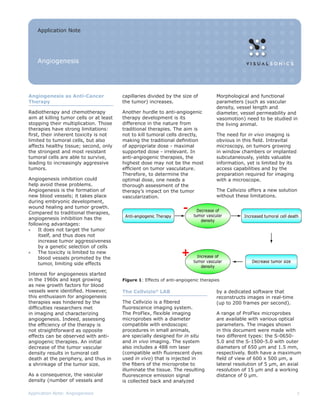
- 1. Application Note Angiogenesis Angiogenesis as Anti-Cancer capillaries divided by the size of Morphological and functional Therapy the tumor) increases. parameters (such as vascular density, vessel length and Radiotherapy and chemotherapy Another hurdle to anti-angiogenic diameter, vessel permeability and aim at killing tumor cells or at least therapy development is its vasomotion) need to be studied in stopping their multiplication. Those difference in the nature from the living animal. therapies have strong limitations: traditional therapies. The aim is first, their inherent toxicity is not not to kill tumoral cells directly, The need for in vivo imaging is limited to tumoral cells, but also making the traditional definition obvious in this field. Intravital affects healthy tissue; second, only of appropriate dose - maximal microscopy, on tumors growing the strongest and most resistant supported dose - irrelevant. In in window chambers or implanted tumoral cells are able to survive, anti-angiogenic therapies, the subcutaneously, yields valuable leading to increasingly aggressive highest dose may not be the most information, yet is limited by its tumors. efficient on tumor vasculature. access capabilities and by the Therefore, to determine the preparation required for imaging Angiogenesis inhibition could optimal dose, one needs a with a microscope. help avoid these problems. thorough assessment of the Angiogenesis is the formation of therapy’s impact on the tumor The Cellvizio offers a new solution new blood vessels; it takes place vascularization. without these limitations. during embryonic development, wound healing and tumor growth. Compared to traditional therapies, angiogenesis inhibition has the following advantages: » It does not target the tumor itself, and thus does not increase tumor aggressiveness by a genetic selection of cells » The toxicity is limited to new blood vessels promoted by the tumor, limiting side effects Interest for angiogenesis started in the 1960s and kept growing Figure 1: Effects of anti-angiogenic therapies as new growth factors for blood vessels were identified. However, The Cellvizio® LAB by a dedicated software that this enthusiasm for angiogenesis reconstructs images in real-time therapies was hindered by the The Cellvizio is a fibered (up to 200 frames per second). difficulties researchers met fluorescence imaging system. in imaging and characterizing The ProFlex, flexible imaging A range of ProFlex microprobes angiogenesis. Indeed, assessing microprobes with a diameter are available with various optical the efficiency of the therapy is compatible with endoscopic parameters. The images shown not straightforward as opposite procedures in small animals, in this document were made with effects can be observed with anti- are specially designed for in situ two different types: the S-0650- angiogenic therapies. An initial and in vivo imaging. The system 5.0 and the S-1500-5.0 with outer decrease of the tumor vascular also includes a 488 nm laser diameters of 650 μm and 1.5 mm, density results in tumoral cell (compatible with fluorescent dyes respectively. Both have a maximum death at the periphery, and thus in used in vivo) that is injected in field of view of 600 x 500 μm, a a shrinkage of the tumor size. the fibers of the microprobe to lateral resolution of 5 μm, an axial illuminate the tissue. The resulting resolution of 15 μm and a working As a consequence, the vascular fluorescence emission signal distance of 0 μm. density (number of vessels and is collected back and analyzed Application Note: Angiogenesis 1
- 2. The system is controlled by an Apple computer running OS X. ImageCell, the sophisticated image processing software developed by Mauna Kea Technologies, controls image acquisition, processing and ProFlex microprobes with display. Quantification and file different orientations export tools are included in the package. Suitability of the Cellvizio for Angiogenesis Studies The Cellvizio offers a new way to image and characterize tumoral Figure 2: Illustration of the facilitated access with ProFlex microprobes. A angiogenesis with numerous conventional lens has a diameter of a few millimeters and can only move in the X, Y advantages: and Z directions. ProFlex microprobes, with an outer diameter inferior to 1 mm, can also be oriented with theta and phi movements. Easy Imaging Longitudinal Studies Made Possible Intravital microscopy imaging most often requires a long and tedious The new type of access offered by the Cellvizio enables longitudinal preparation of the animal: studies. The efficiency of a drug can now be studied on the same animal • Preparation of a window chamber during days through the observation of their tumoral vascularization on the organ development. Less animals are required and the physiological relevance of • Perfusion of tissues with the results is improved. physiological solutions • A half an hour pause to reach physiological stability With the Cellvizio, there is no need Image Procedures - A Range of Possibilities for time consuming preparation: The Cellvizio can be used with a wealth of different protocols when it imaging is immediate, as soon comes to angiogenesis imaging. Below are a few examples. as a microprobe is put in contact with the tissue, with real time Plasma Labeling acquisition. The procedure for vascularization Improved Physiological Relevance imaging with the Cellvizio is no Due to Limited Disturbance longer than two lines: inject a fluorescent plasma-labeling Since a small 2 mm incision of dye intravenously and put the the skin is enough for imaging microprobe in direct contact with with the Cellvizio, the tissue is the desired organ after a small not physiologically disturbed. incision of the skin. Plasmatic staining reveals the real vascularization system of the With this simple protocol, the animal. Labeled blood flows into entire circulatory system is vessels and capillaries. revealed: vessels and capillaries in all organs, sinusoids in the liver, Figure 3: Access to a subcutaneous Quantification in Real Time tubules in the kidneys, capillaries tumor with ProFlex microprobes ImageCell provides many in the lungs and in the brain, etc. quantification features in real Note: Angiogenesis imaging requires the use of a dye with a high time, like measure of fluorescence molecular weight, such as FITC-Dextran 250 kDa, Sigma Aldrich, to avoid intensities with complex ROI leakage associated with windowed vessels in tumors. management, histogram display and distance measurements. These open the door to instant vessel permeability and vasodynamics measurement. Application Note: Angiogenesis 2
- 3. Transgenic animals imaging Injection of fluorescent endothelial Visualization of Cells cells Tie2 mice and beta-actin GFP Blood cells labeling can give mice have fluorescent endothelial Angiogenesis progression can critical information about the cells, making them interesting for also be imaged and studied after cell-wall interaction. For example, vascularization visualization. injection of fluorescent endothelial leukocytes can be imaged by cells. The cells are recruited to intravenous injection of Rhodamine build up new vessels and reveal 6G. sites were angiogenesis is active. FOV: 415 x 300 µm Figure 4: Capillaries of a mouse subcutaneous prostate tumor stained FOV: 400 x 280 µm with FITC-Albumin Figure 6: Rolling leukocytes in a FOV: 400 x 280 µm venule of the cremaster muscle of a mouse Figure 5: Mouse tumoral arterioles and venules Beyond Imaging: Quantification Examples with the Cellvizio Permeability Measurement Functional Capillary Density (FCD) Blood vessel leakiness is a well documented characteristic of Assessment of the vascular density tumor vessels. It contributes to is a standard approach to quantify high interstitial pressure and may angiogenesis. It can also be used facilitate angiogenesis; in addition, as critical information in making a increased endothelial permeability prognosis for the tumor evolution. enables efficient drug delivery from Histological sections labeled with the bloodstream to tumor cells. reliable immunochemistry markers can help identify vessels, but The extravasation, resulting cannot differentiate vessels where from blood leakiness, is very blood flows effectively from the easy to record and quantify ones without blood flow. Since with the Cellvizio. Using a blood our in vivo labeling method for FCD = 3.6 mm / 0.124 mm2 plasma labeling procedure, the vascularization studies is based Figure 7: Estimation of blood vessel fluorescence intensity of an on fluorescent blood plasma, only length per area unit on a Cellvizio image. avascular zone will be directly functional vessels are revealed. linked with the extravasation. Thus, studying the histogram or simply The Cellvizio images enable the assessment of the functional capillary the mean intensity of a given density of the observed tissue. ImageCell enables an automatic region of interest (ROI) reveals segmentation of the images in order to recognize vessels and capillaries. the evolution of the amount of Total vessel length and ratio vessel length per tissue surface unit can then plasma that has leaked to the ROI. be used as an index for functional capillary density. Vascularization of With ImageCell functionalities, this different areas within a tumor of the same area at different time points or analysis is immediate. of different tumors can thus be compared. Application Note: Angiogenesis 3
- 4. Figure 8 shows an example of such a study. The extravasation caused by histamine is measured on a mouse muscular tissue. On the first image, a region of interest excluding vessels and capillaries is defined. The correspondent histogram shows therefore a very low level of signal. The second and third images show a dramatic increase of fluorescence intensity of the ROI, the curve of the signal is shifted to the right. Since the protocol calls for fluorescently labeled blood plasma, the observed phenomenon enables the quantification of the extravasation at the studied site. FOV: 400 x 280 µm FOV: 400 x 280 µm FOV: 400 x 280 µm Figure 8: Effect of histamine on vessel permeability: Extravasation of mouse muscular vessels. The histograms show the fluorescence intensity of the regions of interest (in red on the images) Studying Vasodynamics FOV: 400 x 280 µm Vasodilation upon carbogen inhalation Vasoconstriction after carbogen removal Figure 9 - Constriction and dilation of tumoral vessels. The vasodynamics of a tumoral vessel was observed upon inhalation of carbogen by the mouse. On the first two images, you see the evolution of the diameter of the vessel while the mouse is breathing carbogen: it is dilated. After removal of carbogen, the vessel constricts back to its original size. The diagram on the left shows the vessel response to the sequence carbogen - air - carbogen. Application Note: Angiogenesis 4
- 5. Discussion Compared to intravital microscopy, the Cellvizio offers new access possibilities and new opportunities for minimally invasive protocols, immediate and intuitive use and easy quantification capabilities. The real-time imaging capacity of the system makes it perfectly adapted to record dynamic events associated with blood flow. The Cellvizio is a valuable tool for more efficient studies of angiogenesis development and effects of anti-angiogenic therapies. Courtesy Anne-Carole Duconseille, Nathalie Faye, Laure Fournier, Charles A. Cuenod and Olivier Clément, Descartes Image, Small Animal Imaging Facility, Université Paris V, Paris, France Pr Vicaut, Microcirculation Lab, Hopital Fernand Widal, Paris, France Elisabeth Laemmel and Eric Vicaut, LEM, Paris, France Bibliography 1. Donald M MacDonald, Peter L Choyke: Imaging of angiogenesis: from microscope to clinic Nat Med. 2003 Jun;9(6):713-25 2. Gillian M. Tozer, Simon M. Ameer-Beg, Jennifer Baker, Paul R. Barber, Sally A. Hill, Richard J. Hodgkiss, Rosalind Locke, Vivien E. Prise, Ian Wilson, Borivoj Vojnovic: Intravital imaging of tumour vascular networks using multiphoton fluorescence microscopy Adv Drug Deliv Rev. 2005 Jan 2;57(1):135-52 3. E Laemmel, M Genet, G Le Goualher, A Perchant, JF Le Gargasson, E Vicaut Fibered confocal fluorescence microscopy (Cellvizio) facilitates extended imaging in the field of microcirculation. A comparison with intravital microscopy. J Vasc Res. 2004 Sep-Oct;41(5):400-11. Epub 2004 VisualSonics Inc. Sep 30. T.1.416.484.5000 Toll Free (North America) 1.866.416.4636 Toll Free (Europe) +800.0751.2020 E. info@visualsonics.com www.visualsonics.com VisualSonics®, Vevo®, MicroMarker™, VevoStrain™, DEPO®, SoniGene™, RMV™, EKV® and Insight through In Vivo Imaging are trademarks™ of VisualSonics Inc. Application Note: Angiogenesis 5