
Empfohlen
Weitere ähnliche Inhalte
Was ist angesagt?
Was ist angesagt? (20)
Ähnlich wie Amyloidosis
Ähnlich wie Amyloidosis (20)
Kürzlich hochgeladen
Kürzlich hochgeladen (20)
Amyloidosis
- 3. 2 refers to a variety of conditionsmyloidosisA+ andinsolublebecomeproteinswherein normally soluble ofextracellular spaceare deposited in the The, disrupting normal function.tissuesororgansvarious insoluble fibrous protein aggregates that develop in . They result from aamyloidsown asamyloidosis are kn , which causessecondary structurechange in the protein's otein to take on a particular aggregated insolublethe pr .pleated sheet-betaform, similar to the +Amyloid is a starch like substance which stains brown/ blue/ black with iodine. Dog and horse are often affected naturally than other animals. + Organs affected: • Liver, kidney and spleen are frequently affected. • In progressive form all organs can be affected. Definition :
- 4. 3 Classification : According to the location Systemic Amyloidosis Effect more than one body organ or system. Localized Amyloidosis effect one organ or one tissue. According to the causes Primary Amyloidosis arises from a disease with disordered immune cell function. Secondary Are those occurring as a complication of some other chronic inflammatory or tissue destructive disease
- 5. 4 Native cells have two different methods of making+ different proteins. Some proteins cells make the whole protein in one piece; for other proteins, cells make only protein fragments, and the fragments come and join ch a protein cantogether to form the whole protein. But su sometimes fall apart into the original protein fragments. This process of "flip flopping" happens frequently for certain protein types, especially the ones that cause .amyloidosis +The fragments or actual proteins are at risk of mis- folding as they are synthesized, to make a bad protein. This causes proteolysis, which is the directed degradation of proteins by cellular enzymes called proteases or by intramolecular digestion; proteases come and digest the mis-folded fragments and proteins. The problem occurs when the proteins do not dissolve in proteolysis. This happens because the mis-folded proteins sometimes become robust enough that they are not dissolved by normal proteolysis. When the fragments do not dissolve, they get spit out of proteolysis and they aggregate to form oligomers. The reason they aggregate is that the parts of the protein that do not dissolve in proteolysis are the β-pleated sheets, which are extremely hydrophobic. They are usually sequestered in the middle of the protein, while parts of the protein that are more soluble are found near the outside. When they are exposed to water, these hydrophobic pieces tend to aggregate with other hydrophobic pieces. This ball of fragments gets stabilized by GAG's (glycosaminoglycans) and SAP (serum amyloid P), a component found in amyloid aggregations that is Pathogenesis :
- 6. 5 thought to stabilize them and prevent proteolytic cleavage. The stabilized balls of protein fragments are called oligomers. The oligomers can aggregate together and further stabilize to make amyloid fibrils. Both the oligomers and amyloid fibrils can cause cell toxicity and organ dysfunction. Effect on: Liver Macroscopic -Grossly the liver cut surface pale,waxy corresponding to the abundant amorphous, light esinophilic amyloid deposits. - The deposits may cause moderate to marked hepatomegaly, fraible and may be end by rupture
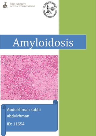
- 7. 6 Macroscopic Microscopic Amyloid appears first in the space of Disse and then progressively encroaches on adjacent hepatic parenchymal cells and sinusoids. In time, deformity, pressure atrophy, and disappearance of hepatocytes occur, causing total replacement of large areas of liver parenchyma.
- 8. 7 Microscopic Kidney Macroscopic pale and shrunken because of ischemia caused by vascular narrowing induced by the deposition of amyloid within capillaries wall. Microscopic the amyloid is deposited primarily in the glomeruli, but the interstitial peritubular capillaries are also affected.
- 10. 9 -Vetreninary pathology course (302) http://en.wikipedia.org/wiki/Amyloidosis- -http://research.vet.upenn.edu/ -http://www.unckidneycenter.org/ -http://webpathology.com References: