Pathologies of the gastrointestinal tract
•Als PPTX, PDF herunterladen•
21 gefällt mir•10,703 views

Pathologies of the gastrointestinal tract

Empfohlen
Weitere ähnliche Inhalte
Was ist angesagt?
Was ist angesagt? (20)
Ähnlich wie Pathologies of the gastrointestinal tract
Ähnlich wie Pathologies of the gastrointestinal tract (20)
Mehr von Dr. Varughese George
Mehr von Dr. Varughese George (20)
Kürzlich hochgeladen
Kürzlich hochgeladen (20)
Pathologies of the gastrointestinal tract
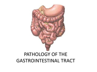
- 1. PATHOLOGY OF THE GASTROINTESTINAL TRACT
- 2. OBJECTIVES • At the end of the session, you should know about • Peptic ulcer – Gross and microscopic features • Carcinoma stomach – Gross and microscopic features • Ulcerative lesions of intestine • Carcinoma colon – Gross and microscopic features
- 3. BENIGN ULCER V/S MALIGNANT ULCER Benign ulcer Malignant ulcerFeatures
- 4. PEPTIC ULCER
- 5. PEPTIC ULCER GROSS FEATURES • Gastric ulcers are found predominantly along the lesser curvature in the region of pyloric antrum, more commonly on the posterior wall. • Duodenal ulcers are commonly found in first part of the duodenum, more commonly on the anterior wall. • Peptic ulcers of either gastric or duodenal mucosa are small (1-2.5 cm in diameter), round to oval and punched out. • The mucosal folds converge towards the ulcer. Benign chronic gastric ulcer •Partial gastrectomy specimen is identified by thick muscular wall and irregular mucosal folds. •The luminal surface shows a punched out round to oval ulcer, about 1 cm in diameter (arrow) penetrating into muscularis layer.
- 6. PEPTIC ULCER MICROSCOPIC FEATURES 4 histologic zones 1. Necrotic zone lies in the floor of the ulcer • The tissue elements show coagulative necrosis giving eosinophilic smudgy appearance with nuclear debris. 2. Superficial exudative zone • lies underneath the necrotic zone and is composed of fibrinous exudate containing necrotic debris and a few leucocytes, predominantly neutrophils. Chronic peptic ulcer Histologic zones of the ulcer are illustrated. The mucosal surface shows necrosis, ulceration, and inflammation.
- 7. PEPTIC ULCER MICROSCOPIC FEATURES 4 histologic zones 3. Granulation tissue zone is seen merging into the necrotic zone. • Composed of nonspecific chronic inflammatory infiltrate and proliferating capillaries. 4. Zone of cicatrisation is seen outer to the layer of granulation tissue • Composed of dense fibrocollagenic scar tissue. Chronic peptic ulcer Histologic zones of the ulcer are illustrated. The mucosal surface shows necrosis, ulceration, and inflammation.
- 8. CARCINOMA STOMACH GROSS FEATURES • Most common pattern is flat, infiltrating and ulcerative growth with irregular necrotic base and raised margin. • Other gross patterns include – fungating (polypoid) – scirrhous (linitis plastica) – colloid (mucoid) – ulcer cancer. Ulcerative carcinoma stomach. The luminal surface of the stomach in the region of the pyloric canal shows an elevated irregular growth with ulcerated surface and raised margins (arrow).
- 9. CARCINOMA STOMACH MICROSCOPIC FEATURES • Tubular and acinar pattern of growth is seen infiltrating the stomach wall. • The tumour invades into the wall of stomach for variable • depth. • The tumour cells show varying degree of anaplasia but is more often poorly-differentiated with high degree of anaplasia
- 10. ULCERATIVE LESIONS OF GIT
- 11. ULCERATIVE LESIONS OF GIT Cushing ulcer • seen in esophagus, stomach or the duodenum. • associated with intracranial disease or head injury. • caused by gastric acid hypersecretion due to vagal nuclei stimulation. Curling ulcer • seen in proximal duodenum. • associated with burns or trauma. • Caused due to reduced blood supply and systemic acidosis in burns or trauma.
- 12. CARCINOMA COLON GROSS FEATURES The right-sided growth, tends to be fungating, large, cauliflower-like, soft and friable mass projecting into the lumen. The left-sided growth has napkin-ring configuration it encircles the bowel wall circumferentially with increased fibrous tissue forming annular ring with central mucosal ulceration.
- 13. CARCINOMA COLON MICROSCOPIC FEATURES The tumour has infiltrating glandular pattern in the colonic wall with varying grades of differentiation of tumour cells. About 10% cases show mucin-secreting colloid carcinoma with pools of mucin