SlideShare ist ein Scribd-Unternehmen logo
1 von 56
White Blood Cell (WBC) Disorders
By
Dr. Varughese George
Introduction
• Disorders of white cells include
– Proliferations (leukocytosis)
• reactive
• neoplastic.
– Deficiencies (leukopenias)
• Reactive proliferation of WBCs in response to an underlying
primary, often microbial, disease is fairly common.
• Neoplastic disorders, though less common, are more
ominous & cause approximately 9% of all cancer deaths in
adults and a staggering 40% in children younger than 15
years.
• In this session, we ll discuss about non-neoplastic disorders
of white cells.
Quantitative WBC Disorders
Leukocytosis
• An increase in the number of white blood cells is common in a
variety of inflammatory states caused by microbial & non-
microbial stimuli.
• Leukocytoses are relatively non-specific & are classified
according to the particular white cell series that is affected.
• In some cases reactive leukocytosis may mimic leukemia.
– Such “leukemoid” reactions must be distinguished from true
white cell malignancies.
– Infectious mononucleosis merits separate consideration
because it gives rise to distinctive syndrome associated with
lymphocytosis.
Quantitative WBC Disorders
• Refers to an absolute neutrophil count >7500/mm³.
• Causes include:
– Bacterial infections.
– Tissue damage or necrosis: e.g. surgery, burns, trauma, hyperthermia,
myocardial infarction and other tissue necrosis.
– Inflammatory disorders: e.g. acute appendicitis, acute pancreatitis,
colitis, vasculitis, rheumatoid arthritis.
– Acute stress or physical exertion, pregnancy, seizures
– Acute hemorrhage.
– Hemolysis: acute or chronic
– Metabolic disorders: diabetic ketoacidosis, uraemia, toxins, gout,
– Medications: e.g. lithium, corticosteroids, epinephrine, hematopoietic
growth factors.
– Physiological: newborns, pregnancy, stress, exercise
– Haematological malignancies: myeloproliferative neoplasms, chronic
myelomonocytic leukaemia, acute myeloid leukaemia
– Conditions like chronic idiopathic neutrophilia & hereditary neutrophilia.
Neutrophilic leukocytosis
Quantitative WBC Disorders
Pathogenesis
• Cytokines stimulate increased bone marrow production and/or early release of
segmented neutrophils/bands in response to inflammation/infections.
• Decreased activation of neutrophil adhesion molecules
a. Fewer neutrophils adhere to endothelial cells and become part of the
circulating pool of neutrophils.
b. Agents that decrease activation of neutrophil adhesion molecules include
corticosteroids, catecholamines & lithium
Neutrophilic leukocytosis
Quantitative WBC Disorders
• The most common cause of neutrophilic leucocytosis is bacterial
infections particularly by Gram-positive cocci (Staphylococci)
• Bacterial infections are frequently associated with following
alterations in peripheral blood—
i. Neutrophilic leucocytosis with shift to left
ii. Toxic granules
iii. Döhle inclusion bodies:
iv. Cytoplasmic vacuoles:
Neutrophilic leukocytosis
Quantitative WBC Disorders
i. Neutrophilic leucocytosis with shift to left
• Normally neutrophils with 3 lobes predominate, while some have 4 lobes & only
a few have 2 or 5 lobes.
• In mild to moderate left shift, immature cells are limited to band forms &
metamyelocytes.
• In severe left shift, immature cells like myeloblast, promyelocytes &myelocytes
are also seen
Neutrophilic leukocytosis
Quantitative WBC Disorders
ii. Toxic granules
• These are dark blue or purple granules in the cytoplasm of segmented neutrophils,
band forms,and metamyelocytes.
• They represent azurophil granules
• Toxic granules probably result from impaired cytoplasmic maturation while generating
large number of neutrophils.
Neutrophilic leukocytosis
Quantitative WBC Disorders
iii. Döhle inclusion bodies:
• These are small, pale blue inclusion bodies in the periphery of
cytoplasm of neutrophils.
• They represent rows of rough endoplasmic reticulum
Neutrophilic leukocytosis
Quantitative WBC Disorders
Neutrophilic leukocytosis
iv. Cytoplasmic vacuoles:
Cytoplasmic vacuoles are indicative of phagocytosis.
Quantitative WBC Disorders
Neutrophilic leukocytosis
• There is neutrophilia with neutrophils
• constituting 88% of the differential
leucocyte count.
• The patient had bronchopneumonia
Band forms in peripheral blood
• Band forms of neutrophils are
preponderant in children with
bacterial infections.
• Note the cytoplasmic vacuoles and toxic
granules in these cells.
Quantitative WBC Disorders
Leukopenia
• Results most commonly from a decrease in granulocytes.
• Neutropenia
– For children from 1 month - 10 years old, neutropenia is defined as a
absolute neutrophil count < 1500/mm³.
– For individuals older than age 10 years, neutropenia is defined as a
absolute neutrophil count ≲ 1800/mm³.
• Agranulocytosis
– literally means a complete absence of blood granulocytes.
– indicates severe neutropenia, i.e, counts < 500/mm³.
– serious consequence of making individuals susceptible to bacterial and
fungal infections.
Causes of neutropenia
I. Inadequate or ineffective granulopoiesis
– Suppression of Hematopoietic stem cells (e.g. aplastic anemia)
– Infiltrative marrow disorders (tumors, granulomatous disease).
– Suppression of committed granulocytic precursors (e.g., after drug exposure).
– Disease states characterized by ineffective granulopoiesis in conditions such as
megaloblastic anemias [vitamin B12 deficiency] & myelodysplastic syndromes
[MDSs]
– Rare inherited conditions (e.g. Kostmann syndrome) impairing differentiation.
II. Accelerated removal or destruction of neutrophils
– Neutrophil injury caused by immunologic disorders (e.g. systemic lupus
erythematosus) or drug exposures.
– Splenic sequestration as a result of splenomegaly.
– Increased peripheral use in overwhelming infections
Causes of neutropenia
III. Drug toxicity
– Immune mediated destruction of neutrophils by drugs like aminopyrine, penicillin,
gold and antithyroid drugs.
– Circulating immune complexes are formed in response to the inducing drug, which
in turn bind to the neutrophils destroying them.
– Dose dependent direct toxic effect on granulopoiesis by drugs such as β lactam
antibiotics, chlorpromazine,procainamide, clozapine, dapsone, sulfonamides,
– phenytoin, indomethacin & diclofenac, which are toxic to neutrophils,
microenvironment of the bone marrow & myeloid precursors.
Morphology in neutropenia
• Marrow anatomic alterations depend on the underlying cause.
• Hypocellularity occurs with agents that suppress granulocyte progenitor cell growth
and survival.
– these may be granulocyte specific or
– can potentially affect erythroid and megakaryocytic progenitors, leading to
pancytopenia and aplastic anemia (empty marrow).
• Hypercellularity occurs in conditions
– with ineffective granulopoiesis (MDSs) or
– when there is increased peripheral destruction of neutrophils.
• Need to rule out other causes of neutropenia like subleukemic/aleukemic leukemia,
megaloblastic anemia & aplastic anemia.
Morphology in neutropenia
(A) Note the characteristic maturation arrest at the stage of promyelocyte/myelocyte
in the bone marrow smear from the severe congenital neutropenia patient.
(B) In contrast, immature and mature forms of neutrophils are found in a bone
marrow smear from a healthy control.
Bone marrow smear from a patient with neutropenia & a healthy person
Clinical features in neutropenia
• Symptoms & signs relate to intercurrent infections include malaise, chills &
fever, often with marked weakness & fatigability.
• Serious infections are most likely when the neutrophil count ≤ 500cells/mm³.
• Ulcerating necrotizing lesions of the gingiva, buccal mucosa, or pharynx are
characteristic.
• Severe, life-threatening, invasive bacterial or fungal infections can occur in the
lungs, kidney or urinary tract.
• Neutropenic patients are at high risk for deep Candida/Aspergillus infections.
• Infections are often fulminant.
• Neutropenic patients are therefore treated with broad-spectrum antibiotics at
the first sign of infection.
• Granulocyte colony–stimulating factor therapy decreases the duration &
severity of the neutrophil nadir caused by chemotherapeutic drugs.
Quantitative WBC Disorders
Lymphocytosis
• Refers to an absolute lymphocyte count >5000 cells/mm³ in
adults or >8000 cells/mm³ in children.
• Causes include:
• Infections
• Predominantly viral (e.g. infectious mononucleosis, dengue)
• Occasionally bacterial (e.g. pertussis and chronic infections like tuberculosis)
• Unusually parasites (e.g. babesiosis)
• Stress and Postsplenectomy
• Hypersensitivity Reactions
• Autoimmune Disorders (e.g. Grave’s disease)
• Thymoma
• Clonal
• Monoclonal B cell lymphocytosis
• Lymphoproliferative disorders especially chronic lymphocytic leukaemia &
lymphomas
Quantitative WBC Disorders
Lymphocytosis
Pathogenesis
• Decreased entry of lymphocytes into the lymph nodes.
e.g. lymphocytosis-promoting factor produced by Bordetella
pertussis (whooping cough)
• Increased production of lymphocytes (e.g. CLL).
Quantitative WBC Disorders
Lymphocytosis
Mature lymphocytes are increased in a case of
tuberculosis
Quantitative WBC Disorders
Lymphopenia
• Refers to absolute lymphocyte count of <1500 cells/mm³ in adults or <3000 cells/mm³
in children
• Causes include:
• Human immunodeficiency virus (HIV)- AIDS caused by deficiency of CD4 T helper
cells.
• Immunodeficiency disorders
e.g. DiGeorge syndrome (T-cell deficiency), Severe combined immunodeficiency
(SCID; B- and T-cell deficiency), Bruton agammaglobulinemia (B-cell deficiency),
Wiskott Aldrich Syndrome.
• Infectious
e.g. viral hepatitis, influenza, typhoid fever.
• Drugs
e.g. Corticosteroids ,Cyclophosphamide, Cytotoxic chemotherapy
• Ionizing radiation.
• Lymphocytes are the most sensitive cells to destruction by radiation
Quantitative WBC Disorders
Eosinophilia
• Refers to an absolute eosinophil count of >400 cells/mm³ in the peripheral blood.
• Causes include:
– Allergic Disorders
• Allergic rhinitis, asthma and atopic dermatitis.
• Gastrointestinal disorders – may be associated with tissue eosinophilia rather than peripheral
blood eosinophilia.
• Drug reactions including the DRESS syndrome (drug reaction with eosinophilia and systemic
symptoms).
– Parasitic Infections – especially with helminthic infestations
– Immunodeficiency Disorders
• Hyper IgE (Job) syndrome.
• Autoimmune lymphoproliferative syndrome.
• Graft-versus-host disease.
• Connective tissue/rheumatology disorders
– Neoplastic Diseases
• Primary/neoplastic hypereosinophilia, e.g. associated with FIP1L1-PDGFRA fusion gene.
• Myeloproliferative neoplasms such as chronic myeloid leukaemia & systemic mastocytosis
• Acute or chronic eosinophilic leukaemia.
• Reactive to other neoplasms, e.g. to B- or T-cell lymphoma or leukaemia or solid tumor.
Quantitative WBC Disorders
Eosinophilia
Pathogenesis :
• Caused by the release of eosinophil chemotactic factor (ECF) from
mast cells
– Type I Hypersensitivity reactions
• Eosinophils in lymph nodes are not sequestered; hence, more
eosinophils are circulating in the blood.
– e.g. hypocortisolism
Quantitative WBC Disorders
Eosinophilia
Eosinophilia in a case of tropical eosinophilia
Some of the eosinophils show degranuation
which are visible as clear vacuole like spaces
(arrow )
Eosinophifia bone marrow biopsy shows
eosinophils & their precursors are
prominent in a case of eosinophilia
(arrow )
Quantitative WBC Disorders
Eosinopenia
• Refers to a decrease in the total number of circulating
eosinophils in the peripheral blood.
• Hypercortisolism (Cushing syndrome, corticosteroids) is the
most common cause of eosinopenia.
– Corticosteroids sequester eosinophils in lymph nodes and
trigger apoptosis.
Quantitative WBC Disorders
Basophilia
• Refers to an absolute basophil count > 110 cells/ mm³
• Causes include:
– Chronic myeloproliferative disorders
• Chronic myelogenous leukemia (CML)
• Polycythemia vera
– Allergic reactions
– Varicella infections.
– Rare cases like Acute basophilic leukemia
Quantitative WBC Disorders
Basophilia
• Peripheral blood film from a case of CML shows immature myeloid precursors with 2
basophils (arrows) having coarse basophilic granules covering the nuclei.
• Pericellular pinkish halo is useful In identifying basophils in the peripheral smear and in
bone marrow smears.
Quantitative WBC Disorders
Monocytosis
• Refers to an absolute monocyte count >800 cells / mm³.
• Causes include:
a) Chronic infections/inflammation:
e.g. Tuberculosis, subacute bacterial endocarditis, syphilis, rickettsia,
malaria, typhoid, kala-azar
b) Haematological malignancies:
e.g. Acute myelomonocytic leukaemia (AML M4), acute monocytic
leukaemia (AML M5), myeloproliferative neoplasms, chronic
myelomonocytic leukaemia, myelodysplastic syndrome,
c) Others:
e.g. Sarcoidosis, ulcerative colitis, regional enteritis, carcinomas
• Pathogenesis:
• Immune response to chronic inflammation, autoimmune or malignancy
Quantitative WBC Disorders
Monocytosis
Peripheral blood film demonstrates increased monocytes
Quantitative WBC Disorders
Unusual benign leukocyte reactions
Leukemoid reaction
• Refers to an absolute leukocyte count that is usually > 50,000 – 1,00,000
cells/mm³.
• It is caused by normal bone marrow response to cytokines released by
cells (e.g., lymphocytes, stromal cells, macrophages) to severe infection
(most common), trauma, burns, malignancies (especially with bone
marrow metastases), myelofibrosis, hemorrhage/hemolysis & eclampsia.
• The triggering factors induces proliferation of precursors in the bone
marrow caused by increased production of colony-stimulating factors
and thus compensates for the loss of these cells in the inflammatory
reaction.
• The reaction can be predominantly granulocytic that includes
prdominantly mature/segmented neutrophils followed by few bands &
metamyelocyte ; lymphocytic as seen in children with e.g. whooping
cough & occasionally monocytic.
Quantitative WBC Disorders
Unusual benign leukocyte reactions
Leukemoid reaction
Neutrophil leukemoid reaction in the peripheral blood.
• Note the great number of segmented and band neutrophils
as well as metamyelocytes (red arrow).
• This smear would be difficult to distinguish from chronic
myelogenous leukemia without chromosome studies.
Parameters Leukemoid reaction Leukemia
Clinical features As per underlying disease Splenomegaly
Total white cell count Increase usually only
moderate; seldom
exceeds 1,00,000 cells/mm³
Can exceed 1,00,000 cells/mm³
Proportion of immature cells Usually small or moderate.
Myelocytes seldom exceed
5-15% & blasts 5 %
Usually numerous
White cell morphology Toxic changes may be seen
in infective cases
Cells often atypical & immature.
Toxic changes uncommon.
Bone marrow White cell hyperplasia may
be present but seldom to
same degree as in
leukaemia
Hyperplastic with potentially
large proportion of immature
cells
Neutrophil Alkaline
Phosphatase Score
Normal or increased Low
Genetic analysis Normal Abnormal
(eg Philadelphia Chromosome)
LEUKEMOID REACTION V/S LEUKEMIA
Quantitative WBC Disorders
Unusual benign leukocyte reactions
Leukoerythroblastosis (leukoerythroblastic reaction)
• Refers to the presence of immature myeloid & nucleated red
blood cells (normoblasts) in the peripheral blood, irrespective of
the total leukocyte count.
• It often occurs as a consequence of disturbance of the bone
marrow architecture by abnormal tissue.
• Example of a leukoerythroblastic reaction with a normal bone
marrow
– severe acute hemolytic anemia (e.g., sickle cell disease).
Quantitative WBC Disorders
Leukoerythroblastosis (leukoerythroblastic reaction)
• Examples of a leukoerythroblastic reaction with an abnormal
bone marrow include:
– bone marrow infiltrative disease (e.g., amyloidosis),
– metastatic malignancy (e.g., breast cancer).
– granulomatous disease (e.g., tuberculosis [TB], systemic fungal
disease, sarcoidosis).
– hematologic malignancies (e.g., acute leukemia, multiple myeloma)
chronic
– myeloproliferative diseases (e.g., primary myelofibrosis).
– Other causes include massive trauma with multiple fractures, Paget
disease of bone & extramedullary hematopoiesis (EMH).
Unusual benign leukocyte reactions
Quantitative WBC Disorders
Leukoerythroblastosis (leukoerythroblastic reaction)
• The blue arrow shows a teardrop red blood cell (RBC).
• Immature myeloid cells are also present .
• A nucleated RBC (red arrow).
Unusual benign leukocyte reactions
Quantitative WBC Disorders
Unusual benign leukocyte conditions
Infectious mononucleosis
• Disease of the young (15-25 years of age) caused by Epstein-Barr
Virus (EBV).
• Characterized by fever, sore throat ,cervical lymphadenopathy &
splenomegaly.
• There is a response of cytotoxic T-lymphocytes (CD8+) subset
reflected by the presence of atypical/reactive (transformed)
lymphocytes in the peripheral blood to infected B-lymphocytes
present especially in the tonsils.
Atypical / Reactive lymphocytes
Quantitative WBC Disorders
Atypical / Reactive lymphocytes
Unusual benign leukocyte conditions
Infectious mononucleosis
• There is moderate degree of leucocytosis with a TLC of 10000 -
25000 cells/mm³ .
• Reactive lymphocytosis > 50% is present.
• Besides the presence of mature lymphocytes, there are large
number of atypical lymphocytes and should be > 10%.
• These cells appear on 3rd -4th day of fever and persist for
several months.
Quantitative WBC Disorders
Atypical / Reactive lymphocytes
Unusual benign leukocyte conditions
• Transformed benign lymphocytes as a result of antigen stimulation.
• There is marked variation in cell size and morphology.
• Cells are large with abundant pale blue cytoplasm.
• The nuclei may be round,oval, tabulated or eccentric and few may
have open nuclear chromatin ( blastoid).
• Some cells with foamy basophilic cytoplasm and kidney shaped
nucleus (monocytoid cells).
• Some cells have eccentric nuclei with pale basophilic cytoplasm
(plasmacytoid cells).
• Some cells have irregular nuclei with cytoplasmic vacuulation.
• These are not pathognomonic for IM since similar cells are
observed in other viral infections also.
Quantitative WBC Disorders
Atypical/Reactive Lymphocyte
Peripheral blood smear showing monocytoid
atypical/reactive lymphocytes.
• The lymphocytes are large and have
abundant blue-gray cytoplasm.
• Nuclei are irregular and have dark
chromatin with inconspicuous nucleoli.
Peripheral blood smear showing
plasmacytoid atypical/reactive lymphocytes
which are T cells in response to B cells
infected by Epstein-Barr Virus.
There is variation in the cell size and
morphology.
Quantitative WBC Disorders
Atypical/Reactive Lymphocyte
Reactive lymphocytes are also referred to as Downey cells.
There are three types of Downey cells:
■ Type I: Small cells with minimum cytoplasm,
indented nucleus/ irregular nuclear membrane,
and condensed chromatin.
■ Type II: Larger cells with abundant cytoplasm;
the lymphocyte
cytoplasm seems to hug the red cells. Type II is
the most common type of Downey cell.
■ Type III: Cells with large moderate basophilic
cytoplasm and nucleus with coarse chromatin.
Nucleoli are apparent
Type I
Type II
Type III
Quantitative WBC Disorders
Atypical/Reactive Lymphocyte
• Ballerina Skirt Cells - Characterisitic appearance of atypical lymphocytes in Infectious
Mononucleosis.
• These are Downey Type II cells which are large cells with abundant agranular pale
cytoplasm and indented by surrounding red blood cells.
Qualitative WBC Disorders
• Rare familial disorders which manifest morphologic and
functional changes in the leucocytes.
• Few disorders are picked up while examining the peripheral
smear and/or bone marrow.
1. Pelger-Huet Anomaly
2. Alder-Reilly Anomaly
3. May-Hegglin Anomaly.
4. Chediak- Higashi Anomaly
5. Chronic Granulomatous Disease of
Childhood
6. Myeloperoxidase deficiency
6. Leucocyte adhesion
defect
7. Hyper IgE/Jotfs
syndrome
8. Catalase deficiency
9. CARD 9 deficiency
10.Giant neutrophils
PELGER-HUET ANOMALY
• A benign autosomal dominant anomaly of leukocytes.
• Neutrophils demonstrate lack of segmentation of nuclei with bilobed/band
forms/dumb-bell shaped/smooth rounded small nuclei in > 70 % of neutrophils.
• < 25% band forms are present in peripheral blood.
• The nuclear chromatin is coarse.
• Confused with leukemoid reaction.
• A similar morphological appearance of neutrophils (Acquired or pseudo-Felger
Huet anomaly) is seen in MDS. AML, CML and multiple myeloma.
ALDER REILLY ANOMALY
• An autosomal recessive disorder
• Neutrophils demonstrate large, darkly staining metachromatic cytoplasmic
granules composed primarily of partially digested mucopolysaccharides referred
to as Alder-Reilly bodies.
• Similar inclusions are also seen in lymphocytes and monocytes.
• Observed in Hurler syndrome & Hunter syndrome and the cell function is normal.
Note the dark granules present in both cells in two neutrophils from a patient with
Alder-Reilly anomaly.
MAY-HEGGLIN ANOMALY
• A rare autosomal dominant disorder
• characterised by the presence of basophilic inclusions in neutrophils,
eosinophils, basophils and monocytes.
• This is associated with giant platelets and variable thrombocytopenia.
A neutrophil and a giant platelet from a patient with May-Hegglin anomaly.
Note the large, elongated, bluish inclusion in the neutrophil cytoplasm (arrow)
CHEDIAK HIGASHI ANOMALY
• This as an autosomal recessive disease characterized by partial cutaneous and ocular
albinism, increased susceptibility to pyogenic infections and bleeding tendency due to
abnormal platelet function.
• It is due to the defect in the leukocyte NADPH oxidase-respiratory burst, oxidase.
• Neutrophils and their precursors demonstrate inclusion like large single/multiple
lysosomal granules (arrows).
• As the disease progresses, there is progressive anemia, neutropenia &
thrombocytopenia.
CHEDIAK HIGASHI ANOMALY
Large rounded granules are also seen in lymphocytes,
monocytes & eosinophils.
These granules are
myeloperoxidase positive
CHRONIC GRANULOMATOUS DISEASE
OF CHILDHOOD
• characterized by morphologically normal leucocytes which fail to generate
superoxide and hydrogen peroxide & therefore phagocytosis and killing of
organisms is impaired.
• These cases are susceptible to infections by organisms of low pathogenicity.
• A second hallmark feature is the development of inflammatory granulomas.
• Clinical features include recurrent, infections of skin, lungs and epithelial
surfaces and hepatosplenomegaly.
• Management includes bone marrow transplantation & gene therapy.
CHRONIC GRANULOMATOUS DISEASE
OF CHILDHOOD
Normal neutrophils, when stimulated
reduce the yellow water-soluble
nitroblue tetrazolium to a
dark blue insoluble formazan.
In the nitroblue tetrazolium reduction test
Neutrophils in
chronic granulomatous disease
CANNOT
perform this reduction
MYELOPEROXIDASE DEFICIENCY
• It is the most common hereditary neutrophil function defect.
• There is absence of myeloperoxidase (MPO) enzyme in the neutrophils &
monocytes, however, the cell morphology is normal on peripheral smear.
• The intracellular killing of microorganisms is slower than normal PMNs but it is
eventually achieved  so infections are not a major complication.
• But there is an increased susceptibility to Candida & staphylococcal infections.
• Diagnosis is confirmed by MPO stain of the blood smear that demonstrates a
lack of peroxidase in the neutrophils.
Peroxidase activity in polymorphonuclear neutrophils (PMN) from a healthy subject (Normal PMN) & an individual with
myeloperoxidase (MPO) deficiency (as indicated).
Panel at far right indicates normal peroxidase activity in an eosinophil isolated from an individual with MPO deficiency as
eosinophil peroxidase is encoded by another gene than MPO.
LEUCOCYTE ADHESION DEFICIENCY
• It is a rare autosomal recessive disorder, characterised by absence of leucocyte
cel! surface adhesion proteins CD11/CD18.
These surface glycoproteins play an important role in the cell's ability to kill
microorganisms.
• Neutrophils with LAD are morphologically normal, but have multiple functional
abnormalities related to adhesion, motility (chemotaxis), phagocytosis,
degranulation and respiratory burst activation.
• Patients present with recurrent bacterial and fungal infections with persistent
leucocytosis.
• Diagnosis can be established by flow cytometric analysis of CD11/GD18 levels in
neutrophils using monoclonal antibodies.
• These patients require prophylactic antibiotics and prompt treatment of
infections.
HYPER IgE/JOB SYNDROME
• It is a rare congenital disorder, characterized by defective neutrophil chemotaxis.
Patients present, with recurrent bacterial, fungal and viral infections.
• The most common pathogens are Staphylococcus aureus and Candida species.
• The most common laboratory findings are blood eosinophilia & raised IgE levels.
• This infant with markedly elevated IgE experienced early onset of a pruritic
dermatitis that shared clinical features with both seborrhea and atopic
dermatitis. The perinasal crusting caused by Staphylococcus aureus infection.
CATALASE DEFICIENCY
• The catalase enzyme plays a crucial role in protecting
neutrophils from products of NADPH oxidase by converting
H202 to H20 and 02.
• In catalase deficiency, the surface of neutrophil becomes very
susceptible to damage by H202.
• However, these patients do not manifest with very severe
infections.
CARD 9 (CASPASE ASSOCIATED
RECRUITMENT DOMAIN) DEFICIENCY
• Caspase recruitment domain-containing protein 9 deficiency is a
rare immunologic disorder because of mutation in CARD 9 gene
&results in defective defence against pathogen fungi
• like Candida.
• CARD 9 protein plays a role in apoptosis.
• Mucocutaneous candidiasis is associated with CARD9 deficiency.
GIANT/HYPERSEGMENTED NEUTROPHILS
• Giant neutrophils are larger & hypersegmented (macropolycytes) found in the
peripheral blood of individuals with Vitamin B12 & folic acid deficiency.
• Seen rarely in families as an inherited condition & observed in AIDS cases.
• Giant neutrophils, large myeloid precursors and few binucleate promyelocytes are
also seen in bone marrow and peripheral blood following G-CSF therapy.
• The granulopoiesis is ineffective, accelerated & skipping of 1 or 2 mitotic divisions
without cytoplasmic division may result in morphologically abnormal giant
neutrophils.
Neutrophils have 6 to 10 nuclear segments
& are > 5% of all the peripheral blood
neutrophils.
GIANT/HYPERSEGMENTED NEUTROPHILS
• These are larger & hypersegmented neutrophils (macropolycytes).
• They are found in peripheral smear of patients with Vitamin B12 & folic acid
deficiency.
• These are also seen rarely in families as an inherited condition & in AIDS.
• Giant neutrophils, large myeloid precursors & few binudeate promyelocytes are
also seen in bone marrow & peripheral blood following G-CSF therapy.
• The granulopoiesis is ineffective but accelerated & skipping of one or two
mitotic divisions without cytoplasmic division may result in morphologically
abnormal giant neutrophils.
Neutrophil hypersegmentation can be defined
as the presence of neutrophils ≥ 6 lobes
or
the presence of more than 3% of neutrophils
with at least 5 lobes.

Weitere ähnliche Inhalte

Was ist angesagt? (20)

Peripheral blood smear examination
Peripheral blood smear examinationPeripheral blood smear examination
Peripheral blood smear examination
 
Coombs test
Coombs testCoombs test
Coombs test
 
Haemolytic anaemias
Haemolytic anaemiasHaemolytic anaemias
Haemolytic anaemias
 
Acute leukemia
Acute leukemia Acute leukemia
Acute leukemia
 
Leukemia
LeukemiaLeukemia
Leukemia
 
Platelet Function Tests
Platelet Function TestsPlatelet Function Tests
Platelet Function Tests
 
Osmotic fragility test
Osmotic fragility testOsmotic fragility test
Osmotic fragility test
 
Sickle cell Anemia
Sickle cell AnemiaSickle cell Anemia
Sickle cell Anemia
 
stains
stainsstains
stains
 
Blood indices
Blood indicesBlood indices
Blood indices
 
Laboratory diagnosis of anemia
Laboratory diagnosis of anemiaLaboratory diagnosis of anemia
Laboratory diagnosis of anemia
 
Leukocytosis. Leukopenia. Leukosis
Leukocytosis. Leukopenia. LeukosisLeukocytosis. Leukopenia. Leukosis
Leukocytosis. Leukopenia. Leukosis
 
Abnormalities of WBC
Abnormalities of WBCAbnormalities of WBC
Abnormalities of WBC
 
Chronic leukemias
Chronic leukemiasChronic leukemias
Chronic leukemias
 
Interpretation of histograms
Interpretation of histogramsInterpretation of histograms
Interpretation of histograms
 
Disorders of white blood cells
Disorders of white blood cellsDisorders of white blood cells
Disorders of white blood cells
 
Peripheral Blood Smear Examination
Peripheral Blood Smear ExaminationPeripheral Blood Smear Examination
Peripheral Blood Smear Examination
 
Automation in hematology part 1
Automation in hematology part 1Automation in hematology part 1
Automation in hematology part 1
 
Hemolytic anemia ppt presentation
Hemolytic anemia ppt presentationHemolytic anemia ppt presentation
Hemolytic anemia ppt presentation
 
Introduction to hematology
Introduction to hematologyIntroduction to hematology
Introduction to hematology
 

Ähnlich wie Benign White blood cell (WBC) Disorders

Pathology of WBC
Pathology of WBCPathology of WBC
Pathology of WBCshaimaaf12
 
Multiple myeloma 06-11-2022.pptx
Multiple myeloma 06-11-2022.pptxMultiple myeloma 06-11-2022.pptx
Multiple myeloma 06-11-2022.pptxmanjujanhavi
 
White Blood Cell Disorders (ramkumaradhikari)
White Blood Cell Disorders (ramkumaradhikari)White Blood Cell Disorders (ramkumaradhikari)
White Blood Cell Disorders (ramkumaradhikari)Ramkumar Adhikari
 
Causes of Leukocytosis, Leokopenia, Lymphocytosis and Leukemoid reactions..pptx
Causes of Leukocytosis, Leokopenia, Lymphocytosis and Leukemoid reactions..pptxCauses of Leukocytosis, Leokopenia, Lymphocytosis and Leukemoid reactions..pptx
Causes of Leukocytosis, Leokopenia, Lymphocytosis and Leukemoid reactions..pptxUtkarsh Sharma
 
8. White Cell Disorders.pptx
8. White Cell Disorders.pptx8. White Cell Disorders.pptx
8. White Cell Disorders.pptxmariaidrees3
 
WBC normal and abnormal final.pptx
WBC normal and abnormal final.pptxWBC normal and abnormal final.pptx
WBC normal and abnormal final.pptxKaushik Bhuva
 
Blood and its components Part 2
Blood and its components Part 2Blood and its components Part 2
Blood and its components Part 2Arun Panwar
 
medicine.Acute leukemias.(dr.sabir)
medicine.Acute leukemias.(dr.sabir)medicine.Acute leukemias.(dr.sabir)
medicine.Acute leukemias.(dr.sabir)student
 
Approach to thrombocytopenia
Approach to thrombocytopeniaApproach to thrombocytopenia
Approach to thrombocytopeniaSukritiAzad1
 
NON-MALIGNANT REACTIVE DISORDERS OF LYMPHOCYTES
NON-MALIGNANT REACTIVE DISORDERS OF LYMPHOCYTESNON-MALIGNANT REACTIVE DISORDERS OF LYMPHOCYTES
NON-MALIGNANT REACTIVE DISORDERS OF LYMPHOCYTEShm alumia
 
Haematological malignancies-1.pptx
Haematological malignancies-1.pptxHaematological malignancies-1.pptx
Haematological malignancies-1.pptxHassan25409
 
Periodontitis as a manifestation of systemic diseases
Periodontitis as a manifestation of systemic diseasesPeriodontitis as a manifestation of systemic diseases
Periodontitis as a manifestation of systemic diseasesDr. vasavi reddy
 
9. Chronic Myeloid Leukaemia.ppt
9. Chronic Myeloid Leukaemia.ppt9. Chronic Myeloid Leukaemia.ppt
9. Chronic Myeloid Leukaemia.pptAnayaKhan992603
 

Ähnlich wie Benign White blood cell (WBC) Disorders (20)

Pathology of WBC
Pathology of WBCPathology of WBC
Pathology of WBC
 
Multiple myeloma 06-11-2022.pptx
Multiple myeloma 06-11-2022.pptxMultiple myeloma 06-11-2022.pptx
Multiple myeloma 06-11-2022.pptx
 
White Blood Cell Disorders (ramkumaradhikari)
White Blood Cell Disorders (ramkumaradhikari)White Blood Cell Disorders (ramkumaradhikari)
White Blood Cell Disorders (ramkumaradhikari)
 
Causes of Leukocytosis, Leokopenia, Lymphocytosis and Leukemoid reactions..pptx
Causes of Leukocytosis, Leokopenia, Lymphocytosis and Leukemoid reactions..pptxCauses of Leukocytosis, Leokopenia, Lymphocytosis and Leukemoid reactions..pptx
Causes of Leukocytosis, Leokopenia, Lymphocytosis and Leukemoid reactions..pptx
 
8. White Cell Disorders.pptx
8. White Cell Disorders.pptx8. White Cell Disorders.pptx
8. White Cell Disorders.pptx
 
WBC normal and abnormal final.pptx
WBC normal and abnormal final.pptxWBC normal and abnormal final.pptx
WBC normal and abnormal final.pptx
 
Medicine 5th year, 3rd lecture (Dr. Abdulla Sharief)
Medicine 5th year, 3rd lecture (Dr. Abdulla Sharief)Medicine 5th year, 3rd lecture (Dr. Abdulla Sharief)
Medicine 5th year, 3rd lecture (Dr. Abdulla Sharief)
 
blood diseases.pptx
blood diseases.pptxblood diseases.pptx
blood diseases.pptx
 
Blood and its components Part 2
Blood and its components Part 2Blood and its components Part 2
Blood and its components Part 2
 
medicine.Acute leukemias.(dr.sabir)
medicine.Acute leukemias.(dr.sabir)medicine.Acute leukemias.(dr.sabir)
medicine.Acute leukemias.(dr.sabir)
 
Acute leukemias
Acute leukemiasAcute leukemias
Acute leukemias
 
Approach to thrombocytopenia
Approach to thrombocytopeniaApproach to thrombocytopenia
Approach to thrombocytopenia
 
G.n
G.nG.n
G.n
 
NON-MALIGNANT REACTIVE DISORDERS OF LYMPHOCYTES
NON-MALIGNANT REACTIVE DISORDERS OF LYMPHOCYTESNON-MALIGNANT REACTIVE DISORDERS OF LYMPHOCYTES
NON-MALIGNANT REACTIVE DISORDERS OF LYMPHOCYTES
 
Haematological malignancies-1.pptx
Haematological malignancies-1.pptxHaematological malignancies-1.pptx
Haematological malignancies-1.pptx
 
Leukemia
LeukemiaLeukemia
Leukemia
 
Periodontitis as a manifestation of systemic diseases
Periodontitis as a manifestation of systemic diseasesPeriodontitis as a manifestation of systemic diseases
Periodontitis as a manifestation of systemic diseases
 
Leukemias
LeukemiasLeukemias
Leukemias
 
9. Chronic Myeloid Leukaemia.ppt
9. Chronic Myeloid Leukaemia.ppt9. Chronic Myeloid Leukaemia.ppt
9. Chronic Myeloid Leukaemia.ppt
 
Leukemia.pptx
Leukemia.pptxLeukemia.pptx
Leukemia.pptx
 

Mehr von Dr. Varughese George (20)

Urine analysis Class I
Urine analysis   Class IUrine analysis   Class I
Urine analysis Class I
 
Urine analysis Class II
Urine analysis   Class IIUrine analysis   Class II
Urine analysis Class II
 
Stool Examination
Stool ExaminationStool Examination
Stool Examination
 
Stool Examination Abridged What A Medical Graduate Should Know
Stool Examination Abridged   What A Medical Graduate Should KnowStool Examination Abridged   What A Medical Graduate Should Know
Stool Examination Abridged What A Medical Graduate Should Know
 
Muscle biopsy
Muscle biopsyMuscle biopsy
Muscle biopsy
 
Cpc meeting 12032020
Cpc meeting 12032020Cpc meeting 12032020
Cpc meeting 12032020
 
Cpc meeting 17022020
Cpc meeting 17022020Cpc meeting 17022020
Cpc meeting 17022020
 
Urine analysis
Urine analysisUrine analysis
Urine analysis
 
MOSCMC CPC 28102019 - Follicular Lymphoma
MOSCMC CPC 28102019 - Follicular LymphomaMOSCMC CPC 28102019 - Follicular Lymphoma
MOSCMC CPC 28102019 - Follicular Lymphoma
 
Infectious diseases
Infectious diseasesInfectious diseases
Infectious diseases
 
Diseases of the kidney
Diseases of the kidneyDiseases of the kidney
Diseases of the kidney
 
Aneurysms
AneurysmsAneurysms
Aneurysms
 
Tumors of the breast
Tumors of the breastTumors of the breast
Tumors of the breast
 
Tumor board Ca stomach
Tumor board Ca stomachTumor board Ca stomach
Tumor board Ca stomach
 
Tumor board soft tissue sarcoma
Tumor board  soft tissue sarcomaTumor board  soft tissue sarcoma
Tumor board soft tissue sarcoma
 
Approach to infertility
Approach to infertilityApproach to infertility
Approach to infertility
 
Tumor board melanoma 28082018
Tumor board melanoma 28082018Tumor board melanoma 28082018
Tumor board melanoma 28082018
 
Tumor board ca stomach 28082018
Tumor board ca stomach 28082018Tumor board ca stomach 28082018
Tumor board ca stomach 28082018
 
Dermatitis herpetiformis
Dermatitis herpetiformisDermatitis herpetiformis
Dermatitis herpetiformis
 
Pathologies of the gastrointestinal tract
Pathologies of the gastrointestinal tractPathologies of the gastrointestinal tract
Pathologies of the gastrointestinal tract
 

Kürzlich hochgeladen

High Profile Call Girls Jaipur Vani 8445551418 Independent Escort Service Jaipur
High Profile Call Girls Jaipur Vani 8445551418 Independent Escort Service JaipurHigh Profile Call Girls Jaipur Vani 8445551418 Independent Escort Service Jaipur
High Profile Call Girls Jaipur Vani 8445551418 Independent Escort Service Jaipurparulsinha
 
Call Girls ITPL Just Call 7001305949 Top Class Call Girl Service Available
Call Girls ITPL Just Call 7001305949 Top Class Call Girl Service AvailableCall Girls ITPL Just Call 7001305949 Top Class Call Girl Service Available
Call Girls ITPL Just Call 7001305949 Top Class Call Girl Service Availablenarwatsonia7
 
Ahmedabad Call Girls CG Road 🔝9907093804 Short 1500 💋 Night 6000
Ahmedabad Call Girls CG Road 🔝9907093804  Short 1500  💋 Night 6000Ahmedabad Call Girls CG Road 🔝9907093804  Short 1500  💋 Night 6000
Ahmedabad Call Girls CG Road 🔝9907093804 Short 1500 💋 Night 6000aliya bhat
 
Call Girls Jp Nagar Just Call 7001305949 Top Class Call Girl Service Available
Call Girls Jp Nagar Just Call 7001305949 Top Class Call Girl Service AvailableCall Girls Jp Nagar Just Call 7001305949 Top Class Call Girl Service Available
Call Girls Jp Nagar Just Call 7001305949 Top Class Call Girl Service Availablenarwatsonia7
 
See the 2,456 pharmacies on the National E-Pharmacy Platform
See the 2,456 pharmacies on the National E-Pharmacy PlatformSee the 2,456 pharmacies on the National E-Pharmacy Platform
See the 2,456 pharmacies on the National E-Pharmacy PlatformKweku Zurek
 
Call Girl Service Bidadi - For 7001305949 Cheap & Best with original Photos
Call Girl Service Bidadi - For 7001305949 Cheap & Best with original PhotosCall Girl Service Bidadi - For 7001305949 Cheap & Best with original Photos
Call Girl Service Bidadi - For 7001305949 Cheap & Best with original Photosnarwatsonia7
 
Call Girls Kanakapura Road Just Call 7001305949 Top Class Call Girl Service A...
Call Girls Kanakapura Road Just Call 7001305949 Top Class Call Girl Service A...Call Girls Kanakapura Road Just Call 7001305949 Top Class Call Girl Service A...
Call Girls Kanakapura Road Just Call 7001305949 Top Class Call Girl Service A...narwatsonia7
 
call girls in Connaught Place DELHI 🔝 >༒9540349809 🔝 genuine Escort Service ...
call girls in Connaught Place  DELHI 🔝 >༒9540349809 🔝 genuine Escort Service ...call girls in Connaught Place  DELHI 🔝 >༒9540349809 🔝 genuine Escort Service ...
call girls in Connaught Place DELHI 🔝 >༒9540349809 🔝 genuine Escort Service ...saminamagar
 
Call Girls Whitefield Just Call 7001305949 Top Class Call Girl Service Available
Call Girls Whitefield Just Call 7001305949 Top Class Call Girl Service AvailableCall Girls Whitefield Just Call 7001305949 Top Class Call Girl Service Available
Call Girls Whitefield Just Call 7001305949 Top Class Call Girl Service Availablenarwatsonia7
 
Book Call Girls in Kasavanahalli - 7001305949 with real photos and phone numbers
Book Call Girls in Kasavanahalli - 7001305949 with real photos and phone numbersBook Call Girls in Kasavanahalli - 7001305949 with real photos and phone numbers
Book Call Girls in Kasavanahalli - 7001305949 with real photos and phone numbersnarwatsonia7
 
Housewife Call Girls Hoskote | 7001305949 At Low Cost Cash Payment Booking
Housewife Call Girls Hoskote | 7001305949 At Low Cost Cash Payment BookingHousewife Call Girls Hoskote | 7001305949 At Low Cost Cash Payment Booking
Housewife Call Girls Hoskote | 7001305949 At Low Cost Cash Payment Bookingnarwatsonia7
 
Artifacts in Nuclear Medicine with Identifying and resolving artifacts.
Artifacts in Nuclear Medicine with Identifying and resolving artifacts.Artifacts in Nuclear Medicine with Identifying and resolving artifacts.
Artifacts in Nuclear Medicine with Identifying and resolving artifacts.MiadAlsulami
 
College Call Girls Vyasarpadi Whatsapp 7001305949 Independent Escort Service
College Call Girls Vyasarpadi Whatsapp 7001305949 Independent Escort ServiceCollege Call Girls Vyasarpadi Whatsapp 7001305949 Independent Escort Service
College Call Girls Vyasarpadi Whatsapp 7001305949 Independent Escort ServiceNehru place Escorts
 
VIP Call Girls Pune Vrinda 9907093804 Short 1500 Night 6000 Best call girls S...
VIP Call Girls Pune Vrinda 9907093804 Short 1500 Night 6000 Best call girls S...VIP Call Girls Pune Vrinda 9907093804 Short 1500 Night 6000 Best call girls S...
VIP Call Girls Pune Vrinda 9907093804 Short 1500 Night 6000 Best call girls S...Miss joya
 
Glomerular Filtration and determinants of glomerular filtration .pptx
Glomerular Filtration and  determinants of glomerular filtration .pptxGlomerular Filtration and  determinants of glomerular filtration .pptx
Glomerular Filtration and determinants of glomerular filtration .pptxDr.Nusrat Tariq
 
Call Girls Service in Bommanahalli - 7001305949 with real photos and phone nu...
Call Girls Service in Bommanahalli - 7001305949 with real photos and phone nu...Call Girls Service in Bommanahalli - 7001305949 with real photos and phone nu...
Call Girls Service in Bommanahalli - 7001305949 with real photos and phone nu...narwatsonia7
 
Call Girls In Andheri East Call 9920874524 Book Hot And Sexy Girls
Call Girls In Andheri East Call 9920874524 Book Hot And Sexy GirlsCall Girls In Andheri East Call 9920874524 Book Hot And Sexy Girls
Call Girls In Andheri East Call 9920874524 Book Hot And Sexy Girlsnehamumbai
 
Call Girls Hsr Layout Just Call 7001305949 Top Class Call Girl Service Available
Call Girls Hsr Layout Just Call 7001305949 Top Class Call Girl Service AvailableCall Girls Hsr Layout Just Call 7001305949 Top Class Call Girl Service Available
Call Girls Hsr Layout Just Call 7001305949 Top Class Call Girl Service Availablenarwatsonia7
 
Call Girls Jayanagar Just Call 7001305949 Top Class Call Girl Service Available
Call Girls Jayanagar Just Call 7001305949 Top Class Call Girl Service AvailableCall Girls Jayanagar Just Call 7001305949 Top Class Call Girl Service Available
Call Girls Jayanagar Just Call 7001305949 Top Class Call Girl Service Availablenarwatsonia7
 

Kürzlich hochgeladen (20)

High Profile Call Girls Jaipur Vani 8445551418 Independent Escort Service Jaipur
High Profile Call Girls Jaipur Vani 8445551418 Independent Escort Service JaipurHigh Profile Call Girls Jaipur Vani 8445551418 Independent Escort Service Jaipur
High Profile Call Girls Jaipur Vani 8445551418 Independent Escort Service Jaipur
 
Call Girls ITPL Just Call 7001305949 Top Class Call Girl Service Available
Call Girls ITPL Just Call 7001305949 Top Class Call Girl Service AvailableCall Girls ITPL Just Call 7001305949 Top Class Call Girl Service Available
Call Girls ITPL Just Call 7001305949 Top Class Call Girl Service Available
 
Ahmedabad Call Girls CG Road 🔝9907093804 Short 1500 💋 Night 6000
Ahmedabad Call Girls CG Road 🔝9907093804  Short 1500  💋 Night 6000Ahmedabad Call Girls CG Road 🔝9907093804  Short 1500  💋 Night 6000
Ahmedabad Call Girls CG Road 🔝9907093804 Short 1500 💋 Night 6000
 
Call Girls Jp Nagar Just Call 7001305949 Top Class Call Girl Service Available
Call Girls Jp Nagar Just Call 7001305949 Top Class Call Girl Service AvailableCall Girls Jp Nagar Just Call 7001305949 Top Class Call Girl Service Available
Call Girls Jp Nagar Just Call 7001305949 Top Class Call Girl Service Available
 
See the 2,456 pharmacies on the National E-Pharmacy Platform
See the 2,456 pharmacies on the National E-Pharmacy PlatformSee the 2,456 pharmacies on the National E-Pharmacy Platform
See the 2,456 pharmacies on the National E-Pharmacy Platform
 
Escort Service Call Girls In Sarita Vihar,, 99530°56974 Delhi NCR
Escort Service Call Girls In Sarita Vihar,, 99530°56974 Delhi NCREscort Service Call Girls In Sarita Vihar,, 99530°56974 Delhi NCR
Escort Service Call Girls In Sarita Vihar,, 99530°56974 Delhi NCR
 
Call Girl Service Bidadi - For 7001305949 Cheap & Best with original Photos
Call Girl Service Bidadi - For 7001305949 Cheap & Best with original PhotosCall Girl Service Bidadi - For 7001305949 Cheap & Best with original Photos
Call Girl Service Bidadi - For 7001305949 Cheap & Best with original Photos
 
Call Girls Kanakapura Road Just Call 7001305949 Top Class Call Girl Service A...
Call Girls Kanakapura Road Just Call 7001305949 Top Class Call Girl Service A...Call Girls Kanakapura Road Just Call 7001305949 Top Class Call Girl Service A...
Call Girls Kanakapura Road Just Call 7001305949 Top Class Call Girl Service A...
 
call girls in Connaught Place DELHI 🔝 >༒9540349809 🔝 genuine Escort Service ...
call girls in Connaught Place  DELHI 🔝 >༒9540349809 🔝 genuine Escort Service ...call girls in Connaught Place  DELHI 🔝 >༒9540349809 🔝 genuine Escort Service ...
call girls in Connaught Place DELHI 🔝 >༒9540349809 🔝 genuine Escort Service ...
 
Call Girls Whitefield Just Call 7001305949 Top Class Call Girl Service Available
Call Girls Whitefield Just Call 7001305949 Top Class Call Girl Service AvailableCall Girls Whitefield Just Call 7001305949 Top Class Call Girl Service Available
Call Girls Whitefield Just Call 7001305949 Top Class Call Girl Service Available
 
Book Call Girls in Kasavanahalli - 7001305949 with real photos and phone numbers
Book Call Girls in Kasavanahalli - 7001305949 with real photos and phone numbersBook Call Girls in Kasavanahalli - 7001305949 with real photos and phone numbers
Book Call Girls in Kasavanahalli - 7001305949 with real photos and phone numbers
 
Housewife Call Girls Hoskote | 7001305949 At Low Cost Cash Payment Booking
Housewife Call Girls Hoskote | 7001305949 At Low Cost Cash Payment BookingHousewife Call Girls Hoskote | 7001305949 At Low Cost Cash Payment Booking
Housewife Call Girls Hoskote | 7001305949 At Low Cost Cash Payment Booking
 
Artifacts in Nuclear Medicine with Identifying and resolving artifacts.
Artifacts in Nuclear Medicine with Identifying and resolving artifacts.Artifacts in Nuclear Medicine with Identifying and resolving artifacts.
Artifacts in Nuclear Medicine with Identifying and resolving artifacts.
 
College Call Girls Vyasarpadi Whatsapp 7001305949 Independent Escort Service
College Call Girls Vyasarpadi Whatsapp 7001305949 Independent Escort ServiceCollege Call Girls Vyasarpadi Whatsapp 7001305949 Independent Escort Service
College Call Girls Vyasarpadi Whatsapp 7001305949 Independent Escort Service
 
VIP Call Girls Pune Vrinda 9907093804 Short 1500 Night 6000 Best call girls S...
VIP Call Girls Pune Vrinda 9907093804 Short 1500 Night 6000 Best call girls S...VIP Call Girls Pune Vrinda 9907093804 Short 1500 Night 6000 Best call girls S...
VIP Call Girls Pune Vrinda 9907093804 Short 1500 Night 6000 Best call girls S...
 
Glomerular Filtration and determinants of glomerular filtration .pptx
Glomerular Filtration and  determinants of glomerular filtration .pptxGlomerular Filtration and  determinants of glomerular filtration .pptx
Glomerular Filtration and determinants of glomerular filtration .pptx
 
Call Girls Service in Bommanahalli - 7001305949 with real photos and phone nu...
Call Girls Service in Bommanahalli - 7001305949 with real photos and phone nu...Call Girls Service in Bommanahalli - 7001305949 with real photos and phone nu...
Call Girls Service in Bommanahalli - 7001305949 with real photos and phone nu...
 
Call Girls In Andheri East Call 9920874524 Book Hot And Sexy Girls
Call Girls In Andheri East Call 9920874524 Book Hot And Sexy GirlsCall Girls In Andheri East Call 9920874524 Book Hot And Sexy Girls
Call Girls In Andheri East Call 9920874524 Book Hot And Sexy Girls
 
Call Girls Hsr Layout Just Call 7001305949 Top Class Call Girl Service Available
Call Girls Hsr Layout Just Call 7001305949 Top Class Call Girl Service AvailableCall Girls Hsr Layout Just Call 7001305949 Top Class Call Girl Service Available
Call Girls Hsr Layout Just Call 7001305949 Top Class Call Girl Service Available
 
Call Girls Jayanagar Just Call 7001305949 Top Class Call Girl Service Available
Call Girls Jayanagar Just Call 7001305949 Top Class Call Girl Service AvailableCall Girls Jayanagar Just Call 7001305949 Top Class Call Girl Service Available
Call Girls Jayanagar Just Call 7001305949 Top Class Call Girl Service Available
 

Benign White blood cell (WBC) Disorders

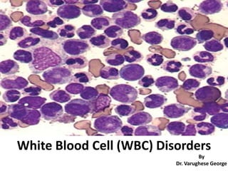
  • 1. White Blood Cell (WBC) Disorders By Dr. Varughese George
  • 2. Introduction • Disorders of white cells include – Proliferations (leukocytosis) • reactive • neoplastic. – Deficiencies (leukopenias) • Reactive proliferation of WBCs in response to an underlying primary, often microbial, disease is fairly common. • Neoplastic disorders, though less common, are more ominous & cause approximately 9% of all cancer deaths in adults and a staggering 40% in children younger than 15 years. • In this session, we ll discuss about non-neoplastic disorders of white cells.
  • 3. Quantitative WBC Disorders Leukocytosis • An increase in the number of white blood cells is common in a variety of inflammatory states caused by microbial & non- microbial stimuli. • Leukocytoses are relatively non-specific & are classified according to the particular white cell series that is affected. • In some cases reactive leukocytosis may mimic leukemia. – Such “leukemoid” reactions must be distinguished from true white cell malignancies. – Infectious mononucleosis merits separate consideration because it gives rise to distinctive syndrome associated with lymphocytosis.
  • 4. Quantitative WBC Disorders • Refers to an absolute neutrophil count >7500/mm³. • Causes include: – Bacterial infections. – Tissue damage or necrosis: e.g. surgery, burns, trauma, hyperthermia, myocardial infarction and other tissue necrosis. – Inflammatory disorders: e.g. acute appendicitis, acute pancreatitis, colitis, vasculitis, rheumatoid arthritis. – Acute stress or physical exertion, pregnancy, seizures – Acute hemorrhage. – Hemolysis: acute or chronic – Metabolic disorders: diabetic ketoacidosis, uraemia, toxins, gout, – Medications: e.g. lithium, corticosteroids, epinephrine, hematopoietic growth factors. – Physiological: newborns, pregnancy, stress, exercise – Haematological malignancies: myeloproliferative neoplasms, chronic myelomonocytic leukaemia, acute myeloid leukaemia – Conditions like chronic idiopathic neutrophilia & hereditary neutrophilia. Neutrophilic leukocytosis
  • 5. Quantitative WBC Disorders Pathogenesis • Cytokines stimulate increased bone marrow production and/or early release of segmented neutrophils/bands in response to inflammation/infections. • Decreased activation of neutrophil adhesion molecules a. Fewer neutrophils adhere to endothelial cells and become part of the circulating pool of neutrophils. b. Agents that decrease activation of neutrophil adhesion molecules include corticosteroids, catecholamines & lithium Neutrophilic leukocytosis
  • 6. Quantitative WBC Disorders • The most common cause of neutrophilic leucocytosis is bacterial infections particularly by Gram-positive cocci (Staphylococci) • Bacterial infections are frequently associated with following alterations in peripheral blood— i. Neutrophilic leucocytosis with shift to left ii. Toxic granules iii. Döhle inclusion bodies: iv. Cytoplasmic vacuoles: Neutrophilic leukocytosis
  • 7. Quantitative WBC Disorders i. Neutrophilic leucocytosis with shift to left • Normally neutrophils with 3 lobes predominate, while some have 4 lobes & only a few have 2 or 5 lobes. • In mild to moderate left shift, immature cells are limited to band forms & metamyelocytes. • In severe left shift, immature cells like myeloblast, promyelocytes &myelocytes are also seen Neutrophilic leukocytosis
  • 8. Quantitative WBC Disorders ii. Toxic granules • These are dark blue or purple granules in the cytoplasm of segmented neutrophils, band forms,and metamyelocytes. • They represent azurophil granules • Toxic granules probably result from impaired cytoplasmic maturation while generating large number of neutrophils. Neutrophilic leukocytosis
  • 9. Quantitative WBC Disorders iii. Döhle inclusion bodies: • These are small, pale blue inclusion bodies in the periphery of cytoplasm of neutrophils. • They represent rows of rough endoplasmic reticulum Neutrophilic leukocytosis
  • 10. Quantitative WBC Disorders Neutrophilic leukocytosis iv. Cytoplasmic vacuoles: Cytoplasmic vacuoles are indicative of phagocytosis.
  • 11. Quantitative WBC Disorders Neutrophilic leukocytosis • There is neutrophilia with neutrophils • constituting 88% of the differential leucocyte count. • The patient had bronchopneumonia Band forms in peripheral blood • Band forms of neutrophils are preponderant in children with bacterial infections. • Note the cytoplasmic vacuoles and toxic granules in these cells.
  • 12. Quantitative WBC Disorders Leukopenia • Results most commonly from a decrease in granulocytes. • Neutropenia – For children from 1 month - 10 years old, neutropenia is defined as a absolute neutrophil count < 1500/mm³. – For individuals older than age 10 years, neutropenia is defined as a absolute neutrophil count ≲ 1800/mm³. • Agranulocytosis – literally means a complete absence of blood granulocytes. – indicates severe neutropenia, i.e, counts < 500/mm³. – serious consequence of making individuals susceptible to bacterial and fungal infections.
  • 13. Causes of neutropenia I. Inadequate or ineffective granulopoiesis – Suppression of Hematopoietic stem cells (e.g. aplastic anemia) – Infiltrative marrow disorders (tumors, granulomatous disease). – Suppression of committed granulocytic precursors (e.g., after drug exposure). – Disease states characterized by ineffective granulopoiesis in conditions such as megaloblastic anemias [vitamin B12 deficiency] & myelodysplastic syndromes [MDSs] – Rare inherited conditions (e.g. Kostmann syndrome) impairing differentiation. II. Accelerated removal or destruction of neutrophils – Neutrophil injury caused by immunologic disorders (e.g. systemic lupus erythematosus) or drug exposures. – Splenic sequestration as a result of splenomegaly. – Increased peripheral use in overwhelming infections
  • 14. Causes of neutropenia III. Drug toxicity – Immune mediated destruction of neutrophils by drugs like aminopyrine, penicillin, gold and antithyroid drugs. – Circulating immune complexes are formed in response to the inducing drug, which in turn bind to the neutrophils destroying them. – Dose dependent direct toxic effect on granulopoiesis by drugs such as β lactam antibiotics, chlorpromazine,procainamide, clozapine, dapsone, sulfonamides, – phenytoin, indomethacin & diclofenac, which are toxic to neutrophils, microenvironment of the bone marrow & myeloid precursors.
  • 15. Morphology in neutropenia • Marrow anatomic alterations depend on the underlying cause. • Hypocellularity occurs with agents that suppress granulocyte progenitor cell growth and survival. – these may be granulocyte specific or – can potentially affect erythroid and megakaryocytic progenitors, leading to pancytopenia and aplastic anemia (empty marrow). • Hypercellularity occurs in conditions – with ineffective granulopoiesis (MDSs) or – when there is increased peripheral destruction of neutrophils. • Need to rule out other causes of neutropenia like subleukemic/aleukemic leukemia, megaloblastic anemia & aplastic anemia.
  • 16. Morphology in neutropenia (A) Note the characteristic maturation arrest at the stage of promyelocyte/myelocyte in the bone marrow smear from the severe congenital neutropenia patient. (B) In contrast, immature and mature forms of neutrophils are found in a bone marrow smear from a healthy control. Bone marrow smear from a patient with neutropenia & a healthy person
  • 17. Clinical features in neutropenia • Symptoms & signs relate to intercurrent infections include malaise, chills & fever, often with marked weakness & fatigability. • Serious infections are most likely when the neutrophil count ≤ 500cells/mm³. • Ulcerating necrotizing lesions of the gingiva, buccal mucosa, or pharynx are characteristic. • Severe, life-threatening, invasive bacterial or fungal infections can occur in the lungs, kidney or urinary tract. • Neutropenic patients are at high risk for deep Candida/Aspergillus infections. • Infections are often fulminant. • Neutropenic patients are therefore treated with broad-spectrum antibiotics at the first sign of infection. • Granulocyte colony–stimulating factor therapy decreases the duration & severity of the neutrophil nadir caused by chemotherapeutic drugs.
  • 18. Quantitative WBC Disorders Lymphocytosis • Refers to an absolute lymphocyte count >5000 cells/mm³ in adults or >8000 cells/mm³ in children. • Causes include: • Infections • Predominantly viral (e.g. infectious mononucleosis, dengue) • Occasionally bacterial (e.g. pertussis and chronic infections like tuberculosis) • Unusually parasites (e.g. babesiosis) • Stress and Postsplenectomy • Hypersensitivity Reactions • Autoimmune Disorders (e.g. Grave’s disease) • Thymoma • Clonal • Monoclonal B cell lymphocytosis • Lymphoproliferative disorders especially chronic lymphocytic leukaemia & lymphomas
  • 19. Quantitative WBC Disorders Lymphocytosis Pathogenesis • Decreased entry of lymphocytes into the lymph nodes. e.g. lymphocytosis-promoting factor produced by Bordetella pertussis (whooping cough) • Increased production of lymphocytes (e.g. CLL).
  • 20. Quantitative WBC Disorders Lymphocytosis Mature lymphocytes are increased in a case of tuberculosis
  • 21. Quantitative WBC Disorders Lymphopenia • Refers to absolute lymphocyte count of <1500 cells/mm³ in adults or <3000 cells/mm³ in children • Causes include: • Human immunodeficiency virus (HIV)- AIDS caused by deficiency of CD4 T helper cells. • Immunodeficiency disorders e.g. DiGeorge syndrome (T-cell deficiency), Severe combined immunodeficiency (SCID; B- and T-cell deficiency), Bruton agammaglobulinemia (B-cell deficiency), Wiskott Aldrich Syndrome. • Infectious e.g. viral hepatitis, influenza, typhoid fever. • Drugs e.g. Corticosteroids ,Cyclophosphamide, Cytotoxic chemotherapy • Ionizing radiation. • Lymphocytes are the most sensitive cells to destruction by radiation
  • 22. Quantitative WBC Disorders Eosinophilia • Refers to an absolute eosinophil count of >400 cells/mm³ in the peripheral blood. • Causes include: – Allergic Disorders • Allergic rhinitis, asthma and atopic dermatitis. • Gastrointestinal disorders – may be associated with tissue eosinophilia rather than peripheral blood eosinophilia. • Drug reactions including the DRESS syndrome (drug reaction with eosinophilia and systemic symptoms). – Parasitic Infections – especially with helminthic infestations – Immunodeficiency Disorders • Hyper IgE (Job) syndrome. • Autoimmune lymphoproliferative syndrome. • Graft-versus-host disease. • Connective tissue/rheumatology disorders – Neoplastic Diseases • Primary/neoplastic hypereosinophilia, e.g. associated with FIP1L1-PDGFRA fusion gene. • Myeloproliferative neoplasms such as chronic myeloid leukaemia & systemic mastocytosis • Acute or chronic eosinophilic leukaemia. • Reactive to other neoplasms, e.g. to B- or T-cell lymphoma or leukaemia or solid tumor.
  • 23. Quantitative WBC Disorders Eosinophilia Pathogenesis : • Caused by the release of eosinophil chemotactic factor (ECF) from mast cells – Type I Hypersensitivity reactions • Eosinophils in lymph nodes are not sequestered; hence, more eosinophils are circulating in the blood. – e.g. hypocortisolism
  • 24. Quantitative WBC Disorders Eosinophilia Eosinophilia in a case of tropical eosinophilia Some of the eosinophils show degranuation which are visible as clear vacuole like spaces (arrow ) Eosinophifia bone marrow biopsy shows eosinophils & their precursors are prominent in a case of eosinophilia (arrow )
  • 25. Quantitative WBC Disorders Eosinopenia • Refers to a decrease in the total number of circulating eosinophils in the peripheral blood. • Hypercortisolism (Cushing syndrome, corticosteroids) is the most common cause of eosinopenia. – Corticosteroids sequester eosinophils in lymph nodes and trigger apoptosis.
  • 26. Quantitative WBC Disorders Basophilia • Refers to an absolute basophil count > 110 cells/ mm³ • Causes include: – Chronic myeloproliferative disorders • Chronic myelogenous leukemia (CML) • Polycythemia vera – Allergic reactions – Varicella infections. – Rare cases like Acute basophilic leukemia
  • 27. Quantitative WBC Disorders Basophilia • Peripheral blood film from a case of CML shows immature myeloid precursors with 2 basophils (arrows) having coarse basophilic granules covering the nuclei. • Pericellular pinkish halo is useful In identifying basophils in the peripheral smear and in bone marrow smears.
  • 28. Quantitative WBC Disorders Monocytosis • Refers to an absolute monocyte count >800 cells / mm³. • Causes include: a) Chronic infections/inflammation: e.g. Tuberculosis, subacute bacterial endocarditis, syphilis, rickettsia, malaria, typhoid, kala-azar b) Haematological malignancies: e.g. Acute myelomonocytic leukaemia (AML M4), acute monocytic leukaemia (AML M5), myeloproliferative neoplasms, chronic myelomonocytic leukaemia, myelodysplastic syndrome, c) Others: e.g. Sarcoidosis, ulcerative colitis, regional enteritis, carcinomas • Pathogenesis: • Immune response to chronic inflammation, autoimmune or malignancy
  • 29. Quantitative WBC Disorders Monocytosis Peripheral blood film demonstrates increased monocytes
  • 30. Quantitative WBC Disorders Unusual benign leukocyte reactions Leukemoid reaction • Refers to an absolute leukocyte count that is usually > 50,000 – 1,00,000 cells/mm³. • It is caused by normal bone marrow response to cytokines released by cells (e.g., lymphocytes, stromal cells, macrophages) to severe infection (most common), trauma, burns, malignancies (especially with bone marrow metastases), myelofibrosis, hemorrhage/hemolysis & eclampsia. • The triggering factors induces proliferation of precursors in the bone marrow caused by increased production of colony-stimulating factors and thus compensates for the loss of these cells in the inflammatory reaction. • The reaction can be predominantly granulocytic that includes prdominantly mature/segmented neutrophils followed by few bands & metamyelocyte ; lymphocytic as seen in children with e.g. whooping cough & occasionally monocytic.
  • 31. Quantitative WBC Disorders Unusual benign leukocyte reactions Leukemoid reaction Neutrophil leukemoid reaction in the peripheral blood. • Note the great number of segmented and band neutrophils as well as metamyelocytes (red arrow). • This smear would be difficult to distinguish from chronic myelogenous leukemia without chromosome studies.
  • 32. Parameters Leukemoid reaction Leukemia Clinical features As per underlying disease Splenomegaly Total white cell count Increase usually only moderate; seldom exceeds 1,00,000 cells/mm³ Can exceed 1,00,000 cells/mm³ Proportion of immature cells Usually small or moderate. Myelocytes seldom exceed 5-15% & blasts 5 % Usually numerous White cell morphology Toxic changes may be seen in infective cases Cells often atypical & immature. Toxic changes uncommon. Bone marrow White cell hyperplasia may be present but seldom to same degree as in leukaemia Hyperplastic with potentially large proportion of immature cells Neutrophil Alkaline Phosphatase Score Normal or increased Low Genetic analysis Normal Abnormal (eg Philadelphia Chromosome) LEUKEMOID REACTION V/S LEUKEMIA
  • 33. Quantitative WBC Disorders Unusual benign leukocyte reactions Leukoerythroblastosis (leukoerythroblastic reaction) • Refers to the presence of immature myeloid & nucleated red blood cells (normoblasts) in the peripheral blood, irrespective of the total leukocyte count. • It often occurs as a consequence of disturbance of the bone marrow architecture by abnormal tissue. • Example of a leukoerythroblastic reaction with a normal bone marrow – severe acute hemolytic anemia (e.g., sickle cell disease).
  • 34. Quantitative WBC Disorders Leukoerythroblastosis (leukoerythroblastic reaction) • Examples of a leukoerythroblastic reaction with an abnormal bone marrow include: – bone marrow infiltrative disease (e.g., amyloidosis), – metastatic malignancy (e.g., breast cancer). – granulomatous disease (e.g., tuberculosis [TB], systemic fungal disease, sarcoidosis). – hematologic malignancies (e.g., acute leukemia, multiple myeloma) chronic – myeloproliferative diseases (e.g., primary myelofibrosis). – Other causes include massive trauma with multiple fractures, Paget disease of bone & extramedullary hematopoiesis (EMH). Unusual benign leukocyte reactions
  • 35. Quantitative WBC Disorders Leukoerythroblastosis (leukoerythroblastic reaction) • The blue arrow shows a teardrop red blood cell (RBC). • Immature myeloid cells are also present . • A nucleated RBC (red arrow). Unusual benign leukocyte reactions
  • 36. Quantitative WBC Disorders Unusual benign leukocyte conditions Infectious mononucleosis • Disease of the young (15-25 years of age) caused by Epstein-Barr Virus (EBV). • Characterized by fever, sore throat ,cervical lymphadenopathy & splenomegaly. • There is a response of cytotoxic T-lymphocytes (CD8+) subset reflected by the presence of atypical/reactive (transformed) lymphocytes in the peripheral blood to infected B-lymphocytes present especially in the tonsils. Atypical / Reactive lymphocytes
  • 37. Quantitative WBC Disorders Atypical / Reactive lymphocytes Unusual benign leukocyte conditions Infectious mononucleosis • There is moderate degree of leucocytosis with a TLC of 10000 - 25000 cells/mm³ . • Reactive lymphocytosis > 50% is present. • Besides the presence of mature lymphocytes, there are large number of atypical lymphocytes and should be > 10%. • These cells appear on 3rd -4th day of fever and persist for several months.
  • 38. Quantitative WBC Disorders Atypical / Reactive lymphocytes Unusual benign leukocyte conditions • Transformed benign lymphocytes as a result of antigen stimulation. • There is marked variation in cell size and morphology. • Cells are large with abundant pale blue cytoplasm. • The nuclei may be round,oval, tabulated or eccentric and few may have open nuclear chromatin ( blastoid). • Some cells with foamy basophilic cytoplasm and kidney shaped nucleus (monocytoid cells). • Some cells have eccentric nuclei with pale basophilic cytoplasm (plasmacytoid cells). • Some cells have irregular nuclei with cytoplasmic vacuulation. • These are not pathognomonic for IM since similar cells are observed in other viral infections also.
  • 39. Quantitative WBC Disorders Atypical/Reactive Lymphocyte Peripheral blood smear showing monocytoid atypical/reactive lymphocytes. • The lymphocytes are large and have abundant blue-gray cytoplasm. • Nuclei are irregular and have dark chromatin with inconspicuous nucleoli. Peripheral blood smear showing plasmacytoid atypical/reactive lymphocytes which are T cells in response to B cells infected by Epstein-Barr Virus. There is variation in the cell size and morphology.
  • 40. Quantitative WBC Disorders Atypical/Reactive Lymphocyte Reactive lymphocytes are also referred to as Downey cells. There are three types of Downey cells: ■ Type I: Small cells with minimum cytoplasm, indented nucleus/ irregular nuclear membrane, and condensed chromatin. ■ Type II: Larger cells with abundant cytoplasm; the lymphocyte cytoplasm seems to hug the red cells. Type II is the most common type of Downey cell. ■ Type III: Cells with large moderate basophilic cytoplasm and nucleus with coarse chromatin. Nucleoli are apparent Type I Type II Type III
  • 41. Quantitative WBC Disorders Atypical/Reactive Lymphocyte • Ballerina Skirt Cells - Characterisitic appearance of atypical lymphocytes in Infectious Mononucleosis. • These are Downey Type II cells which are large cells with abundant agranular pale cytoplasm and indented by surrounding red blood cells.
  • 42. Qualitative WBC Disorders • Rare familial disorders which manifest morphologic and functional changes in the leucocytes. • Few disorders are picked up while examining the peripheral smear and/or bone marrow. 1. Pelger-Huet Anomaly 2. Alder-Reilly Anomaly 3. May-Hegglin Anomaly. 4. Chediak- Higashi Anomaly 5. Chronic Granulomatous Disease of Childhood 6. Myeloperoxidase deficiency 6. Leucocyte adhesion defect 7. Hyper IgE/Jotfs syndrome 8. Catalase deficiency 9. CARD 9 deficiency 10.Giant neutrophils
  • 43. PELGER-HUET ANOMALY • A benign autosomal dominant anomaly of leukocytes. • Neutrophils demonstrate lack of segmentation of nuclei with bilobed/band forms/dumb-bell shaped/smooth rounded small nuclei in > 70 % of neutrophils. • < 25% band forms are present in peripheral blood. • The nuclear chromatin is coarse. • Confused with leukemoid reaction. • A similar morphological appearance of neutrophils (Acquired or pseudo-Felger Huet anomaly) is seen in MDS. AML, CML and multiple myeloma.
  • 44. ALDER REILLY ANOMALY • An autosomal recessive disorder • Neutrophils demonstrate large, darkly staining metachromatic cytoplasmic granules composed primarily of partially digested mucopolysaccharides referred to as Alder-Reilly bodies. • Similar inclusions are also seen in lymphocytes and monocytes. • Observed in Hurler syndrome & Hunter syndrome and the cell function is normal. Note the dark granules present in both cells in two neutrophils from a patient with Alder-Reilly anomaly.
  • 45. MAY-HEGGLIN ANOMALY • A rare autosomal dominant disorder • characterised by the presence of basophilic inclusions in neutrophils, eosinophils, basophils and monocytes. • This is associated with giant platelets and variable thrombocytopenia. A neutrophil and a giant platelet from a patient with May-Hegglin anomaly. Note the large, elongated, bluish inclusion in the neutrophil cytoplasm (arrow)
  • 46. CHEDIAK HIGASHI ANOMALY • This as an autosomal recessive disease characterized by partial cutaneous and ocular albinism, increased susceptibility to pyogenic infections and bleeding tendency due to abnormal platelet function. • It is due to the defect in the leukocyte NADPH oxidase-respiratory burst, oxidase. • Neutrophils and their precursors demonstrate inclusion like large single/multiple lysosomal granules (arrows). • As the disease progresses, there is progressive anemia, neutropenia & thrombocytopenia.
  • 47. CHEDIAK HIGASHI ANOMALY Large rounded granules are also seen in lymphocytes, monocytes & eosinophils. These granules are myeloperoxidase positive
  • 48. CHRONIC GRANULOMATOUS DISEASE OF CHILDHOOD • characterized by morphologically normal leucocytes which fail to generate superoxide and hydrogen peroxide & therefore phagocytosis and killing of organisms is impaired. • These cases are susceptible to infections by organisms of low pathogenicity. • A second hallmark feature is the development of inflammatory granulomas. • Clinical features include recurrent, infections of skin, lungs and epithelial surfaces and hepatosplenomegaly. • Management includes bone marrow transplantation & gene therapy.
  • 49. CHRONIC GRANULOMATOUS DISEASE OF CHILDHOOD Normal neutrophils, when stimulated reduce the yellow water-soluble nitroblue tetrazolium to a dark blue insoluble formazan. In the nitroblue tetrazolium reduction test Neutrophils in chronic granulomatous disease CANNOT perform this reduction
  • 50. MYELOPEROXIDASE DEFICIENCY • It is the most common hereditary neutrophil function defect. • There is absence of myeloperoxidase (MPO) enzyme in the neutrophils & monocytes, however, the cell morphology is normal on peripheral smear. • The intracellular killing of microorganisms is slower than normal PMNs but it is eventually achieved  so infections are not a major complication. • But there is an increased susceptibility to Candida & staphylococcal infections. • Diagnosis is confirmed by MPO stain of the blood smear that demonstrates a lack of peroxidase in the neutrophils. Peroxidase activity in polymorphonuclear neutrophils (PMN) from a healthy subject (Normal PMN) & an individual with myeloperoxidase (MPO) deficiency (as indicated). Panel at far right indicates normal peroxidase activity in an eosinophil isolated from an individual with MPO deficiency as eosinophil peroxidase is encoded by another gene than MPO.
  • 51. LEUCOCYTE ADHESION DEFICIENCY • It is a rare autosomal recessive disorder, characterised by absence of leucocyte cel! surface adhesion proteins CD11/CD18. These surface glycoproteins play an important role in the cell's ability to kill microorganisms. • Neutrophils with LAD are morphologically normal, but have multiple functional abnormalities related to adhesion, motility (chemotaxis), phagocytosis, degranulation and respiratory burst activation. • Patients present with recurrent bacterial and fungal infections with persistent leucocytosis. • Diagnosis can be established by flow cytometric analysis of CD11/GD18 levels in neutrophils using monoclonal antibodies. • These patients require prophylactic antibiotics and prompt treatment of infections.
  • 52. HYPER IgE/JOB SYNDROME • It is a rare congenital disorder, characterized by defective neutrophil chemotaxis. Patients present, with recurrent bacterial, fungal and viral infections. • The most common pathogens are Staphylococcus aureus and Candida species. • The most common laboratory findings are blood eosinophilia & raised IgE levels. • This infant with markedly elevated IgE experienced early onset of a pruritic dermatitis that shared clinical features with both seborrhea and atopic dermatitis. The perinasal crusting caused by Staphylococcus aureus infection.
  • 53. CATALASE DEFICIENCY • The catalase enzyme plays a crucial role in protecting neutrophils from products of NADPH oxidase by converting H202 to H20 and 02. • In catalase deficiency, the surface of neutrophil becomes very susceptible to damage by H202. • However, these patients do not manifest with very severe infections.
  • 54. CARD 9 (CASPASE ASSOCIATED RECRUITMENT DOMAIN) DEFICIENCY • Caspase recruitment domain-containing protein 9 deficiency is a rare immunologic disorder because of mutation in CARD 9 gene &results in defective defence against pathogen fungi • like Candida. • CARD 9 protein plays a role in apoptosis. • Mucocutaneous candidiasis is associated with CARD9 deficiency.
  • 55. GIANT/HYPERSEGMENTED NEUTROPHILS • Giant neutrophils are larger & hypersegmented (macropolycytes) found in the peripheral blood of individuals with Vitamin B12 & folic acid deficiency. • Seen rarely in families as an inherited condition & observed in AIDS cases. • Giant neutrophils, large myeloid precursors and few binucleate promyelocytes are also seen in bone marrow and peripheral blood following G-CSF therapy. • The granulopoiesis is ineffective, accelerated & skipping of 1 or 2 mitotic divisions without cytoplasmic division may result in morphologically abnormal giant neutrophils. Neutrophils have 6 to 10 nuclear segments & are > 5% of all the peripheral blood neutrophils.
  • 56. GIANT/HYPERSEGMENTED NEUTROPHILS • These are larger & hypersegmented neutrophils (macropolycytes). • They are found in peripheral smear of patients with Vitamin B12 & folic acid deficiency. • These are also seen rarely in families as an inherited condition & in AIDS. • Giant neutrophils, large myeloid precursors & few binudeate promyelocytes are also seen in bone marrow & peripheral blood following G-CSF therapy. • The granulopoiesis is ineffective but accelerated & skipping of one or two mitotic divisions without cytoplasmic division may result in morphologically abnormal giant neutrophils. Neutrophil hypersegmentation can be defined as the presence of neutrophils ≥ 6 lobes or the presence of more than 3% of neutrophils with at least 5 lobes.

Hinweis der Redaktion

  1. It is noteworthy that eosinophils are never involved in MPO deficiency since eosinophil peroxidase is encoded by another gene than MPO.