SlideShare ist ein Scribd-Unternehmen logo
1 von 46
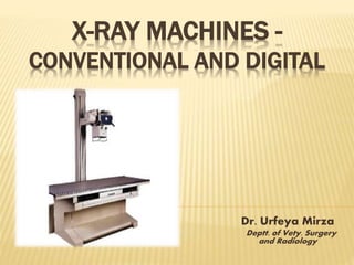
X-RAY MACHINES -
CONVENTIONAL AND DIGITAL
Dr. Urfeya Mirza
Deptt. of Vety. Surgery
and Radiology
INTRODUCTION
• X rays are the ionizing electromagnetic radiation emitted
from a highly evacuated high-voltage tube. Inner orbital
electrons in the target anode are stimulated to emit x-
radiation via bombardment by a stream of electrons from a
heated cathode
• X-rays, like gamma rays, are penetrating and carry
enough energy to ionize atoms in their path. Nearly
identical to gamma rays, x-rays require shielding to reduce
their intensity and minimize the danger of tissue damage to
personnel. Mishaps with x-rays can cause severe
radiation burns and deep tissue damage and can lead to
various cancers
X-rays were discovered in 1895 when Wilhelm Conrad
Roentgen observed that a screen coated with a barium salt
fluoresced when placed near a cathode ray tube. Roentgen
concluded that a form of penetrating radiation was being
emitted by the cathode ray tube and called the unknown
rays, X-rays .
First X-ray Image
X-RAY MACHINE
An x ray machine is a complex device used in
variety of applications around the world. With the
ability to penetrate hard objects, they are used for
purposes such as to look for broken bones or
problems within the body in the medical
community, air port security check points, in the
industrial QC applications and for research
purposes.
PRINCIPLES OF OPERATION
 An x-ray machine is essentially a camera. Instead of
visible light, however, it uses X-rays to expose the film. X-
rays are like light in that they are electromagnetic waves,
but they are more energetic so they can penetrate many
materials to varying degrees. When the X-rays hit the film,
they expose it just as light would
 Since bone, fat, muscle, tumors and other masses all
absorb X-rays at different levels, the image on the film lets
you see different (distinct) structures inside the body
because of the different levels of exposure on the film
PRODUCTION OF X-RAY
An x-ray tube requires a source of electrons, a means to accelerate
the electrons, and a target to stop the high-speed electrons.
 The filament is heated to boil off electrons which are then accelerated
to the anode
 The filament is contained within the cathode which is cup shaped to
focus the electrons onto the focus spot on the anode
 Tube currents of 50-800 milliamperes are used whereas filament
currents are in the range of 2-5 amperes
 The anode is bevelled at an angle of 12 to 17 degrees in order to
maximise the contact area while focussing the resultant beam
 The anode is usually composed of tungsten or molybdenum as it must
withstand very high temperatures (>3000 degrees C)
 Correct warm up and stand by procedures are essential to maximise
tube and filament life
 When the electrons from the cathode are accelerated at
high voltage to the anode:
 99% of the energy is dissipated as heat
(anode materials are selected to withstand the high temperatures they are able
to withstand)
 1% is given off as x-rays.
 The energy of the x-rays (keV) is determined by the
voltage applied (kVp) while,
 The amount of x-rays is determined by the current (mA).
Block Diagram of the X-ray
Machine
PARTS OF X-RAY MACHINE
X-ray has three main components:
 Operating Console
 High Frequency Generator
 X-ray Tube
Internal
External
Other Parts include:
 Collimator and
 Grid Bucky
 X-ray Film
 X-Ray Generator: High voltage generator:
modifies incoming voltage and current to
provide an x-ray tube with the power needed
to produce an x-ray beam of the desired
peak-kilo-voltage (k V p) and current (mA)
and duration (Time).
 Control panel: Permits the selection of
technique factors and initiation of
radiographic exposures mA, kV, Time
 Transformer: Transformers modify the
voltage of incoming alternating-current
(AC) electrical signals to increase or
decrease the voltage in a circuit.
…CONT
 Step-up transformer: Supplies the high
voltage to the x-ray tube (voltage increases and
current decreases)
 Step-down transformer: Supplies power to
heat the filament of the x-ray tube (voltage
decreases and current increases)
 Autotransformer: Supplies the voltage for the
two circuits and provide a location for the K v
p meter (indicates the voltage applied across
the x-ray tube)
 Rectifiers: Convert AC into the direct current
(DC) required by the x-ray tube. A rectifier
restricts current flow in an x-ray tube to one
direction (from cathode to anode), thereby
preventing damage to the x-ray tube filament.
Two types: Half wave and Full wave.
…CONT
 X-RAY TUBE: It is an
expensive wearing
element in medical
radiological equipment.
It consists of :
 Anode
 Expansion bellows
(provide space for oil
to expand)
 Cathode (and heating-
coil)
 Tube envelope
(evacuated)
 Tube housing
 Cooling dielectric oil
 Rotor
vacuum
Protection
housing
Projectile
PROTECTING HOUSING
…CONT
 High Tension Cable: Special highly insulated
cables Considered are the cable capacitance (130-
230 pF/m) because it affects the average value of
the voltage and current across the x-ray tube
(increases the power delivered to the tube.
 Collimators and Grids: They are used to increase
the image contrast and to reduce the dose to the
patient by mean limiting the x-ray beam to the area
of interest.
 Collimator: It is placed between the x-ray tube
and the patient and Usually provided with an
optical device, by which the x-ray filed can be
exactly simulated by a light filed.
 Grid: It is inserted between the patient and the
film cassette in order to reduce the loss of contrast
due to scattered radiation.
…CONT
 X-ray film: X-ray film is a sensitive material (sheet) for the x-ray. A
film that has been exposed to x-rays shows an image of the x-ray
intensity.
SCHEMATIC DIAGRAM (EXTERNAL COMPONENTS)
11. Cassette holder 12. Cable 13. Imaging hand switch 14. Control panel 15. Generator
16. Display screen 17. Stretcher 18. X-ray tube 19. Collimator 20. Cassette 21. Interface
cable 22. trolley 46. Hand grip 66.tube
CONVENTIONAL X-RAY MACHINE:
 A conventional system uses an intensifying
screen to create a latent image on x-ray film.
The film is then processed, creating a
manifest image that can be interpreted by a
physician. It is later stored in the file room.
LIMITATIONS
Conventional radiography (also known as screen film
radiography SFR) is still used more widely than digital
radiography but this dominance is fast dwindling. The
reasons behind the declining popularity of SFR are —
 Diagnostic image quality is poor
 Fixed non-linear Grey scale response
 Limited potential for reducing dose to the patient
 The images cannot be changed in contrast once they
have been processed
 Film is expensive, uses hazardous materials for
processing
 This method is labour intensive
 Long term storage and retrieval of film is difficult
 SFR is not compatible with the Picture Archiving And
Communication Systems (PACS)
DIGITAL RADIOGRAPHY
 X-ray tube is coupled to a
specialized reciever that
changes x-rays into electrical
signals
 Analog image is digitalized &
displayed on integrated
computer screen
 Data is stored in magnetic
optical
discs(MODs),CDs,DVDs
ADVANTAGES :
 No films are required
 No screens are required
 No processing is required
 Brightness & contrast of images
Digital systems are traditionally split into two broadly defined
categories:
 Direct radiography(DR)
 Computed radiography (CR)
The detector classification is related with the conversion
process of X-ray energy to electric charge:
 DR technology converts X-rays into electrical charges by
means of a direct readout process using thin-film transistor
(TFT) arrays
 Concerning CR systems they use storage-phosphor image
plates with a separate image readout process, which means
an indirect conversion process
DIRECT RADIOGRAPHY
Referred to as “cassette-less” because the detector
is incorporated into the x-ray table or upright wall
unit
 Equipment may be indirect or direct conversion
 Images are ready for viewing within seconds
In a system of direct radiography, also called direct capture
radiography, the image receptor is composed of an array of
electronic sensors that respond to the radiation exiting the
patient. These sensors send that information in digital
format to a computer
DR / CHARGE COUPLED DEVICE
CCD
Lens
Scintillator
X-ray tube
DR / CHARGE COUPLED DEVICE
High Radiation / High Noise Zone
Low Radiation / Low Noise Zone
COMPUTED RADIOGRAPHY
 Image obtained using cassettes containing
photostimulable phosphor plates
 CR systems equipment includes reader for image
processing
 Note:
The phosphor plates used in computed radiography
are not as sensitive to light as x-ray, but are
extremely sensitive to scatter radiation.
COMPUTED RADIOGRAPHIC MACHINE AT TVCC SKUAST-K
X-Ray Digitizer – A scanner used to convert
existing analog images into a DICOM (Digital
Imaging and Communications in Medicine) format.
IMAGE MATRIX
VIEWING THE IMAGE
 The computer-
processed image can
be viewed on a
computer monitor or
printed on film or
paper
 For an image on a
screen to have the
quality approaching
that of a film image, a
special monitor must
be used with a
resolution of 1024 x
1024 pixels.
IMAGE PROCESSING AND POST-PROCESSING
Both allow image manipulation of :
 Density
 Structures demonstrated
 Subtraction permits viewing of bone only or
tissues only
 Contrast enhancement adjusts contrast from very
high to very low
Post Processing
Techniques
Subtraction
Contrast
Enhancement
Edge
Enhancement
Black and White
Reversal
DIGITAL SUBTRACTION
CONTRAST ENHANCEMENT
EDGE ENHANCEMENT
BLACK AND WHITE REVERSAL
DIGITAL IMAGING SYSTEM TECHNICAL
CONSIDERATIONS
 Kilovoltage
May be slightly higher than that used for
conventional radiography
 Centering
Body part of interest must be placed in or near
the center of the detector
 Multiple exposures on one cassette
Although not recommended, if IR is divided for
two separate exposures, the portion not being
exposed must be covered with a lead shield
…CONT
 Over- and underexposure
• Degree of image density is not an accurate
indicator of over- or underexposure
• Density may be indicated by a unique number
that correlates to the amount of exposure
 Collimation
• Limit the field of radiation to the anatomy of
interest
• Inadequate collimation can result in
inappropriate contrast
…CONT
 Open cassettes
An exposed IR begins to lose the image within
15 seconds of opening the IR
 Grids
• Digital systems are more sensitive to scatter
radiation
• Use grids as appropriate
ADVANTAGES OF DIGITAL X-RAY MACHINES:
 The appearance of digital images can be manipulated during and
after processing
 PACS is a network used to manage the images obtained through
DR
 Faster delivery of medical images to the clinicians that evaluate
and report on them. Resulting in faster availability of results
 No lost or misplaced images, which means fewer patients being
postponed or cancelled for consultations or surgery while waiting
for new images
 Flexible viewing with the ability to manipulate images on
screen,which means patients can be diagnosed more effectively
 Instant access to previous images and patient records
 Better collaboration, as PACS can be viewed from multiple
terminals and locations by a range of clinicians, allowing
discussion over diagnoses
 Fewer unnecessary re-investigations, which will in turn reduce the
amount of radiation to which patients are exposed
 No space needed for film storage
REFERENCES
 Veterinary radiology by A.P Singh and Jit Singh, 2009
 Selman, J. The Fundamentals of X-Ray and Radium
Physics, 8th Edition, Charles Thomas, 1994
 Internet :
• faculty.mu.edu.sa
• www.pubmed.org
• www.plmer.edu
• www.radiology.org
 Pictures :
 www.hulenhills.com
 www.redwingbooks.com
X ray machines - conventional and digital

Weitere ähnliche Inhalte

Was ist angesagt?

Was ist angesagt? (20)

X ray machine- ppt
X ray machine- pptX ray machine- ppt
X ray machine- ppt
 
X ray machine-new
X ray machine-newX ray machine-new
X ray machine-new
 
Xray film & film processing
Xray film & film processingXray film & film processing
Xray film & film processing
 
Automatic processing of X-ray film.pptx
Automatic processing of X-ray film.pptxAutomatic processing of X-ray film.pptx
Automatic processing of X-ray film.pptx
 
The control of scattered radiation
The control of scattered radiationThe control of scattered radiation
The control of scattered radiation
 
CR & DR
CR & DRCR & DR
CR & DR
 
Qc in xray
Qc in xrayQc in xray
Qc in xray
 
Grids
GridsGrids
Grids
 
Radiographic Intensifying Screen
Radiographic Intensifying ScreenRadiographic Intensifying Screen
Radiographic Intensifying Screen
 
X-RAY TUBE (ANODE)
X-RAY TUBE (ANODE)X-RAY TUBE (ANODE)
X-RAY TUBE (ANODE)
 
Radiographic film
Radiographic filmRadiographic film
Radiographic film
 
Image quality
Image qualityImage quality
Image quality
 
X-ray Tube (Cathode)
X-ray Tube (Cathode)X-ray Tube (Cathode)
X-ray Tube (Cathode)
 
Dark room equipments and entrance
Dark room equipments and entranceDark room equipments and entrance
Dark room equipments and entrance
 
Generations of CT scanner
Generations of  CT scannerGenerations of  CT scanner
Generations of CT scanner
 
Intensifying screens
Intensifying screensIntensifying screens
Intensifying screens
 
Digital Radiography
Digital RadiographyDigital Radiography
Digital Radiography
 
Computed Radiography and digital radiography
Computed Radiography and  digital radiographyComputed Radiography and  digital radiography
Computed Radiography and digital radiography
 
Dark room
Dark roomDark room
Dark room
 
Fluoroscopy
FluoroscopyFluoroscopy
Fluoroscopy
 

Ähnlich wie X ray machines - conventional and digital

Digital radiography.. an update
Digital radiography.. an updateDigital radiography.. an update
Digital radiography.. an updateShivam Batra
 
Intro to radiography 1_2(NDT)
Intro to radiography 1_2(NDT)Intro to radiography 1_2(NDT)
Intro to radiography 1_2(NDT)Ravi Shekhar
 
CT SCAN equipment.pptx
CT SCAN equipment.pptxCT SCAN equipment.pptx
CT SCAN equipment.pptxRohit Bansal
 
Bps xray mammo dsa
Bps xray mammo dsaBps xray mammo dsa
Bps xray mammo dsaRMLIMS
 
basic of angiography physics and equipement.pdf
basic of angiography physics and equipement.pdfbasic of angiography physics and equipement.pdf
basic of angiography physics and equipement.pdfnaima SENHOU
 
CT Physics
CT PhysicsCT Physics
CT PhysicsRMLIMS
 
graphic terminal crt monitor
graphic terminal  crt monitorgraphic terminal  crt monitor
graphic terminal crt monitorJagilam Kumar
 
CT PHYSICS for first year radiology residents
CT PHYSICS for first year radiology residentsCT PHYSICS for first year radiology residents
CT PHYSICS for first year radiology residentsdrvanajabulkapuram
 
Basic principle of x ray
Basic principle of x rayBasic principle of x ray
Basic principle of x rayprahlad maurya
 
Computed Tomography.pptx
Computed Tomography.pptxComputed Tomography.pptx
Computed Tomography.pptxjerelubarco1
 
X- Ray physics- X-Ray Tube, Transformer, Generator and Rectifiers by kajalsra...
X- Ray physics- X-Ray Tube, Transformer, Generator and Rectifiers by kajalsra...X- Ray physics- X-Ray Tube, Transformer, Generator and Rectifiers by kajalsra...
X- Ray physics- X-Ray Tube, Transformer, Generator and Rectifiers by kajalsra...DrKajalLimbad
 
Ultrasound transducer doppler ppt pdf pk
Ultrasound transducer doppler ppt pdf pkUltrasound transducer doppler ppt pdf pk
Ultrasound transducer doppler ppt pdf pkDr pradeep Kumar
 
X-ray Machine
X-ray Machine X-ray Machine
X-ray Machine SamyaQadi
 

Ähnlich wie X ray machines - conventional and digital (20)

Digital radiography.. an update
Digital radiography.. an updateDigital radiography.. an update
Digital radiography.. an update
 
Unit 1 ppt notes /BM8702 /RADIOLOGICAL EQUIPMENTS
Unit 1 ppt notes /BM8702 /RADIOLOGICAL EQUIPMENTSUnit 1 ppt notes /BM8702 /RADIOLOGICAL EQUIPMENTS
Unit 1 ppt notes /BM8702 /RADIOLOGICAL EQUIPMENTS
 
Radiography
RadiographyRadiography
Radiography
 
Intro to radiography 1_2(NDT)
Intro to radiography 1_2(NDT)Intro to radiography 1_2(NDT)
Intro to radiography 1_2(NDT)
 
CT SCAN equipment.pptx
CT SCAN equipment.pptxCT SCAN equipment.pptx
CT SCAN equipment.pptx
 
Bps xray mammo dsa
Bps xray mammo dsaBps xray mammo dsa
Bps xray mammo dsa
 
basic of angiography physics and equipement.pdf
basic of angiography physics and equipement.pdfbasic of angiography physics and equipement.pdf
basic of angiography physics and equipement.pdf
 
Rdaiography.pptx
Rdaiography.pptxRdaiography.pptx
Rdaiography.pptx
 
CT Physics
CT PhysicsCT Physics
CT Physics
 
Medical-Imaging.pdf
Medical-Imaging.pdfMedical-Imaging.pdf
Medical-Imaging.pdf
 
graphic terminal crt monitor
graphic terminal  crt monitorgraphic terminal  crt monitor
graphic terminal crt monitor
 
CT PHYSICS for first year radiology residents
CT PHYSICS for first year radiology residentsCT PHYSICS for first year radiology residents
CT PHYSICS for first year radiology residents
 
Basic principle of x ray
Basic principle of x rayBasic principle of x ray
Basic principle of x ray
 
Computed Tomography.pptx
Computed Tomography.pptxComputed Tomography.pptx
Computed Tomography.pptx
 
Physics of ct mri
Physics of ct mriPhysics of ct mri
Physics of ct mri
 
X- Ray physics- X-Ray Tube, Transformer, Generator and Rectifiers by kajalsra...
X- Ray physics- X-Ray Tube, Transformer, Generator and Rectifiers by kajalsra...X- Ray physics- X-Ray Tube, Transformer, Generator and Rectifiers by kajalsra...
X- Ray physics- X-Ray Tube, Transformer, Generator and Rectifiers by kajalsra...
 
Ultrasound transducer doppler ppt pdf pk
Ultrasound transducer doppler ppt pdf pkUltrasound transducer doppler ppt pdf pk
Ultrasound transducer doppler ppt pdf pk
 
Medical imaging
Medical imagingMedical imaging
Medical imaging
 
X-ray Machine
X-ray Machine X-ray Machine
X-ray Machine
 
Radiography Testing.ppt
Radiography Testing.pptRadiography Testing.ppt
Radiography Testing.ppt
 

Mehr von Urfeya Mirza

ELECTRORETINOGRAPHY (in veterinary).pptx
ELECTRORETINOGRAPHY (in veterinary).pptxELECTRORETINOGRAPHY (in veterinary).pptx
ELECTRORETINOGRAPHY (in veterinary).pptxUrfeya Mirza
 
Laser Lithotripsy.pptx
Laser Lithotripsy.pptxLaser Lithotripsy.pptx
Laser Lithotripsy.pptxUrfeya Mirza
 
Phonophoresis .pptx
Phonophoresis .pptxPhonophoresis .pptx
Phonophoresis .pptxUrfeya Mirza
 
Anaesthetic and surgical management in birds
Anaesthetic and surgical management in birdsAnaesthetic and surgical management in birds
Anaesthetic and surgical management in birdsUrfeya Mirza
 
Anaesthetic techniques
Anaesthetic techniquesAnaesthetic techniques
Anaesthetic techniquesUrfeya Mirza
 
Portosystemic shunt
Portosystemic shuntPortosystemic shunt
Portosystemic shuntUrfeya Mirza
 
Reproductive system of a buck
Reproductive system of a buckReproductive system of a buck
Reproductive system of a buckUrfeya Mirza
 
Physiotherapy in animals
Physiotherapy in animalsPhysiotherapy in animals
Physiotherapy in animalsUrfeya Mirza
 
Guttural pouch mycosis
Guttural pouch mycosisGuttural pouch mycosis
Guttural pouch mycosisUrfeya Mirza
 
Guttural pouch anatomy
Guttural pouch anatomyGuttural pouch anatomy
Guttural pouch anatomyUrfeya Mirza
 
Diagnosis and therapeutic management of various emergencies of toxicities of ...
Diagnosis and therapeutic management of various emergencies of toxicities of ...Diagnosis and therapeutic management of various emergencies of toxicities of ...
Diagnosis and therapeutic management of various emergencies of toxicities of ...Urfeya Mirza
 
Anaesthetic Techniques
Anaesthetic TechniquesAnaesthetic Techniques
Anaesthetic TechniquesUrfeya Mirza
 
Faecal examination
Faecal examinationFaecal examination
Faecal examinationUrfeya Mirza
 
Diseases of urinary system of zoo and wild animals
Diseases of urinary system of zoo and wild animalsDiseases of urinary system of zoo and wild animals
Diseases of urinary system of zoo and wild animalsUrfeya Mirza
 
Diagnosis and theraupatic management of various emergencies of toxicities of ...
Diagnosis and theraupatic management of various emergencies of toxicities of ...Diagnosis and theraupatic management of various emergencies of toxicities of ...
Diagnosis and theraupatic management of various emergencies of toxicities of ...Urfeya Mirza
 
General anaesthesia
General anaesthesiaGeneral anaesthesia
General anaesthesiaUrfeya Mirza
 

Mehr von Urfeya Mirza (20)

ELECTRORETINOGRAPHY (in veterinary).pptx
ELECTRORETINOGRAPHY (in veterinary).pptxELECTRORETINOGRAPHY (in veterinary).pptx
ELECTRORETINOGRAPHY (in veterinary).pptx
 
Laser Lithotripsy.pptx
Laser Lithotripsy.pptxLaser Lithotripsy.pptx
Laser Lithotripsy.pptx
 
Phonophoresis .pptx
Phonophoresis .pptxPhonophoresis .pptx
Phonophoresis .pptx
 
Anaesthetic and surgical management in birds
Anaesthetic and surgical management in birdsAnaesthetic and surgical management in birds
Anaesthetic and surgical management in birds
 
Anaesthetic techniques
Anaesthetic techniquesAnaesthetic techniques
Anaesthetic techniques
 
Ovarian Cysts
Ovarian CystsOvarian Cysts
Ovarian Cysts
 
Portosystemic shunt
Portosystemic shuntPortosystemic shunt
Portosystemic shunt
 
Reproductive system of a buck
Reproductive system of a buckReproductive system of a buck
Reproductive system of a buck
 
Physiotherapy in animals
Physiotherapy in animalsPhysiotherapy in animals
Physiotherapy in animals
 
Guttural pouch mycosis
Guttural pouch mycosisGuttural pouch mycosis
Guttural pouch mycosis
 
Guttural pouch anatomy
Guttural pouch anatomyGuttural pouch anatomy
Guttural pouch anatomy
 
Diagnosis and therapeutic management of various emergencies of toxicities of ...
Diagnosis and therapeutic management of various emergencies of toxicities of ...Diagnosis and therapeutic management of various emergencies of toxicities of ...
Diagnosis and therapeutic management of various emergencies of toxicities of ...
 
Anaesthetic Techniques
Anaesthetic TechniquesAnaesthetic Techniques
Anaesthetic Techniques
 
Leucocytosis
LeucocytosisLeucocytosis
Leucocytosis
 
Faecal examination
Faecal examinationFaecal examination
Faecal examination
 
Diseases of urinary system of zoo and wild animals
Diseases of urinary system of zoo and wild animalsDiseases of urinary system of zoo and wild animals
Diseases of urinary system of zoo and wild animals
 
Diagnosis and theraupatic management of various emergencies of toxicities of ...
Diagnosis and theraupatic management of various emergencies of toxicities of ...Diagnosis and theraupatic management of various emergencies of toxicities of ...
Diagnosis and theraupatic management of various emergencies of toxicities of ...
 
General anaesthesia
General anaesthesiaGeneral anaesthesia
General anaesthesia
 
Fluid therapy
Fluid therapyFluid therapy
Fluid therapy
 
Bone
BoneBone
Bone
 

Kürzlich hochgeladen

Advanced Views - Calendar View in Odoo 17
Advanced Views - Calendar View in Odoo 17Advanced Views - Calendar View in Odoo 17
Advanced Views - Calendar View in Odoo 17Celine George
 
Z Score,T Score, Percential Rank and Box Plot Graph
Z Score,T Score, Percential Rank and Box Plot GraphZ Score,T Score, Percential Rank and Box Plot Graph
Z Score,T Score, Percential Rank and Box Plot GraphThiyagu K
 
Presentation by Andreas Schleicher Tackling the School Absenteeism Crisis 30 ...
Presentation by Andreas Schleicher Tackling the School Absenteeism Crisis 30 ...Presentation by Andreas Schleicher Tackling the School Absenteeism Crisis 30 ...
Presentation by Andreas Schleicher Tackling the School Absenteeism Crisis 30 ...EduSkills OECD
 
Arihant handbook biology for class 11 .pdf
Arihant handbook biology for class 11 .pdfArihant handbook biology for class 11 .pdf
Arihant handbook biology for class 11 .pdfchloefrazer622
 
Class 11th Physics NEET formula sheet pdf
Class 11th Physics NEET formula sheet pdfClass 11th Physics NEET formula sheet pdf
Class 11th Physics NEET formula sheet pdfAyushMahapatra5
 
APM Welcome, APM North West Network Conference, Synergies Across Sectors
APM Welcome, APM North West Network Conference, Synergies Across SectorsAPM Welcome, APM North West Network Conference, Synergies Across Sectors
APM Welcome, APM North West Network Conference, Synergies Across SectorsAssociation for Project Management
 
Accessible design: Minimum effort, maximum impact
Accessible design: Minimum effort, maximum impactAccessible design: Minimum effort, maximum impact
Accessible design: Minimum effort, maximum impactdawncurless
 
General AI for Medical Educators April 2024
General AI for Medical Educators April 2024General AI for Medical Educators April 2024
General AI for Medical Educators April 2024Janet Corral
 
Kisan Call Centre - To harness potential of ICT in Agriculture by answer farm...
Kisan Call Centre - To harness potential of ICT in Agriculture by answer farm...Kisan Call Centre - To harness potential of ICT in Agriculture by answer farm...
Kisan Call Centre - To harness potential of ICT in Agriculture by answer farm...Krashi Coaching
 
Introduction to Nonprofit Accounting: The Basics
Introduction to Nonprofit Accounting: The BasicsIntroduction to Nonprofit Accounting: The Basics
Introduction to Nonprofit Accounting: The BasicsTechSoup
 
Student login on Anyboli platform.helpin
Student login on Anyboli platform.helpinStudent login on Anyboli platform.helpin
Student login on Anyboli platform.helpinRaunakKeshri1
 
1029 - Danh muc Sach Giao Khoa 10 . pdf
1029 -  Danh muc Sach Giao Khoa 10 . pdf1029 -  Danh muc Sach Giao Khoa 10 . pdf
1029 - Danh muc Sach Giao Khoa 10 . pdfQucHHunhnh
 
Paris 2024 Olympic Geographies - an activity
Paris 2024 Olympic Geographies - an activityParis 2024 Olympic Geographies - an activity
Paris 2024 Olympic Geographies - an activityGeoBlogs
 
Ecosystem Interactions Class Discussion Presentation in Blue Green Lined Styl...
Ecosystem Interactions Class Discussion Presentation in Blue Green Lined Styl...Ecosystem Interactions Class Discussion Presentation in Blue Green Lined Styl...
Ecosystem Interactions Class Discussion Presentation in Blue Green Lined Styl...fonyou31
 
Disha NEET Physics Guide for classes 11 and 12.pdf
Disha NEET Physics Guide for classes 11 and 12.pdfDisha NEET Physics Guide for classes 11 and 12.pdf
Disha NEET Physics Guide for classes 11 and 12.pdfchloefrazer622
 
The Most Excellent Way | 1 Corinthians 13
The Most Excellent Way | 1 Corinthians 13The Most Excellent Way | 1 Corinthians 13
The Most Excellent Way | 1 Corinthians 13Steve Thomason
 
Q4-W6-Restating Informational Text Grade 3
Q4-W6-Restating Informational Text Grade 3Q4-W6-Restating Informational Text Grade 3
Q4-W6-Restating Informational Text Grade 3JemimahLaneBuaron
 

Kürzlich hochgeladen (20)

INDIA QUIZ 2024 RLAC DELHI UNIVERSITY.pptx
INDIA QUIZ 2024 RLAC DELHI UNIVERSITY.pptxINDIA QUIZ 2024 RLAC DELHI UNIVERSITY.pptx
INDIA QUIZ 2024 RLAC DELHI UNIVERSITY.pptx
 
Advanced Views - Calendar View in Odoo 17
Advanced Views - Calendar View in Odoo 17Advanced Views - Calendar View in Odoo 17
Advanced Views - Calendar View in Odoo 17
 
Código Creativo y Arte de Software | Unidad 1
Código Creativo y Arte de Software | Unidad 1Código Creativo y Arte de Software | Unidad 1
Código Creativo y Arte de Software | Unidad 1
 
Z Score,T Score, Percential Rank and Box Plot Graph
Z Score,T Score, Percential Rank and Box Plot GraphZ Score,T Score, Percential Rank and Box Plot Graph
Z Score,T Score, Percential Rank and Box Plot Graph
 
Presentation by Andreas Schleicher Tackling the School Absenteeism Crisis 30 ...
Presentation by Andreas Schleicher Tackling the School Absenteeism Crisis 30 ...Presentation by Andreas Schleicher Tackling the School Absenteeism Crisis 30 ...
Presentation by Andreas Schleicher Tackling the School Absenteeism Crisis 30 ...
 
Arihant handbook biology for class 11 .pdf
Arihant handbook biology for class 11 .pdfArihant handbook biology for class 11 .pdf
Arihant handbook biology for class 11 .pdf
 
Class 11th Physics NEET formula sheet pdf
Class 11th Physics NEET formula sheet pdfClass 11th Physics NEET formula sheet pdf
Class 11th Physics NEET formula sheet pdf
 
APM Welcome, APM North West Network Conference, Synergies Across Sectors
APM Welcome, APM North West Network Conference, Synergies Across SectorsAPM Welcome, APM North West Network Conference, Synergies Across Sectors
APM Welcome, APM North West Network Conference, Synergies Across Sectors
 
Accessible design: Minimum effort, maximum impact
Accessible design: Minimum effort, maximum impactAccessible design: Minimum effort, maximum impact
Accessible design: Minimum effort, maximum impact
 
General AI for Medical Educators April 2024
General AI for Medical Educators April 2024General AI for Medical Educators April 2024
General AI for Medical Educators April 2024
 
Kisan Call Centre - To harness potential of ICT in Agriculture by answer farm...
Kisan Call Centre - To harness potential of ICT in Agriculture by answer farm...Kisan Call Centre - To harness potential of ICT in Agriculture by answer farm...
Kisan Call Centre - To harness potential of ICT in Agriculture by answer farm...
 
Mattingly "AI & Prompt Design: The Basics of Prompt Design"
Mattingly "AI & Prompt Design: The Basics of Prompt Design"Mattingly "AI & Prompt Design: The Basics of Prompt Design"
Mattingly "AI & Prompt Design: The Basics of Prompt Design"
 
Introduction to Nonprofit Accounting: The Basics
Introduction to Nonprofit Accounting: The BasicsIntroduction to Nonprofit Accounting: The Basics
Introduction to Nonprofit Accounting: The Basics
 
Student login on Anyboli platform.helpin
Student login on Anyboli platform.helpinStudent login on Anyboli platform.helpin
Student login on Anyboli platform.helpin
 
1029 - Danh muc Sach Giao Khoa 10 . pdf
1029 -  Danh muc Sach Giao Khoa 10 . pdf1029 -  Danh muc Sach Giao Khoa 10 . pdf
1029 - Danh muc Sach Giao Khoa 10 . pdf
 
Paris 2024 Olympic Geographies - an activity
Paris 2024 Olympic Geographies - an activityParis 2024 Olympic Geographies - an activity
Paris 2024 Olympic Geographies - an activity
 
Ecosystem Interactions Class Discussion Presentation in Blue Green Lined Styl...
Ecosystem Interactions Class Discussion Presentation in Blue Green Lined Styl...Ecosystem Interactions Class Discussion Presentation in Blue Green Lined Styl...
Ecosystem Interactions Class Discussion Presentation in Blue Green Lined Styl...
 
Disha NEET Physics Guide for classes 11 and 12.pdf
Disha NEET Physics Guide for classes 11 and 12.pdfDisha NEET Physics Guide for classes 11 and 12.pdf
Disha NEET Physics Guide for classes 11 and 12.pdf
 
The Most Excellent Way | 1 Corinthians 13
The Most Excellent Way | 1 Corinthians 13The Most Excellent Way | 1 Corinthians 13
The Most Excellent Way | 1 Corinthians 13
 
Q4-W6-Restating Informational Text Grade 3
Q4-W6-Restating Informational Text Grade 3Q4-W6-Restating Informational Text Grade 3
Q4-W6-Restating Informational Text Grade 3
 

X ray machines - conventional and digital

  • 1. X-RAY MACHINES - CONVENTIONAL AND DIGITAL Dr. Urfeya Mirza Deptt. of Vety. Surgery and Radiology
  • 2. INTRODUCTION • X rays are the ionizing electromagnetic radiation emitted from a highly evacuated high-voltage tube. Inner orbital electrons in the target anode are stimulated to emit x- radiation via bombardment by a stream of electrons from a heated cathode • X-rays, like gamma rays, are penetrating and carry enough energy to ionize atoms in their path. Nearly identical to gamma rays, x-rays require shielding to reduce their intensity and minimize the danger of tissue damage to personnel. Mishaps with x-rays can cause severe radiation burns and deep tissue damage and can lead to various cancers
  • 3. X-rays were discovered in 1895 when Wilhelm Conrad Roentgen observed that a screen coated with a barium salt fluoresced when placed near a cathode ray tube. Roentgen concluded that a form of penetrating radiation was being emitted by the cathode ray tube and called the unknown rays, X-rays . First X-ray Image
  • 4. X-RAY MACHINE An x ray machine is a complex device used in variety of applications around the world. With the ability to penetrate hard objects, they are used for purposes such as to look for broken bones or problems within the body in the medical community, air port security check points, in the industrial QC applications and for research purposes.
  • 5. PRINCIPLES OF OPERATION  An x-ray machine is essentially a camera. Instead of visible light, however, it uses X-rays to expose the film. X- rays are like light in that they are electromagnetic waves, but they are more energetic so they can penetrate many materials to varying degrees. When the X-rays hit the film, they expose it just as light would  Since bone, fat, muscle, tumors and other masses all absorb X-rays at different levels, the image on the film lets you see different (distinct) structures inside the body because of the different levels of exposure on the film
  • 6. PRODUCTION OF X-RAY An x-ray tube requires a source of electrons, a means to accelerate the electrons, and a target to stop the high-speed electrons.
  • 7.  The filament is heated to boil off electrons which are then accelerated to the anode  The filament is contained within the cathode which is cup shaped to focus the electrons onto the focus spot on the anode  Tube currents of 50-800 milliamperes are used whereas filament currents are in the range of 2-5 amperes  The anode is bevelled at an angle of 12 to 17 degrees in order to maximise the contact area while focussing the resultant beam  The anode is usually composed of tungsten or molybdenum as it must withstand very high temperatures (>3000 degrees C)  Correct warm up and stand by procedures are essential to maximise tube and filament life
  • 8.  When the electrons from the cathode are accelerated at high voltage to the anode:  99% of the energy is dissipated as heat (anode materials are selected to withstand the high temperatures they are able to withstand)  1% is given off as x-rays.  The energy of the x-rays (keV) is determined by the voltage applied (kVp) while,  The amount of x-rays is determined by the current (mA).
  • 9. Block Diagram of the X-ray Machine
  • 10. PARTS OF X-RAY MACHINE X-ray has three main components:  Operating Console  High Frequency Generator  X-ray Tube Internal External Other Parts include:  Collimator and  Grid Bucky  X-ray Film
  • 11.  X-Ray Generator: High voltage generator: modifies incoming voltage and current to provide an x-ray tube with the power needed to produce an x-ray beam of the desired peak-kilo-voltage (k V p) and current (mA) and duration (Time).  Control panel: Permits the selection of technique factors and initiation of radiographic exposures mA, kV, Time  Transformer: Transformers modify the voltage of incoming alternating-current (AC) electrical signals to increase or decrease the voltage in a circuit.
  • 12. …CONT  Step-up transformer: Supplies the high voltage to the x-ray tube (voltage increases and current decreases)  Step-down transformer: Supplies power to heat the filament of the x-ray tube (voltage decreases and current increases)  Autotransformer: Supplies the voltage for the two circuits and provide a location for the K v p meter (indicates the voltage applied across the x-ray tube)  Rectifiers: Convert AC into the direct current (DC) required by the x-ray tube. A rectifier restricts current flow in an x-ray tube to one direction (from cathode to anode), thereby preventing damage to the x-ray tube filament. Two types: Half wave and Full wave.
  • 13. …CONT  X-RAY TUBE: It is an expensive wearing element in medical radiological equipment. It consists of :  Anode  Expansion bellows (provide space for oil to expand)  Cathode (and heating- coil)  Tube envelope (evacuated)  Tube housing  Cooling dielectric oil  Rotor
  • 16. …CONT  High Tension Cable: Special highly insulated cables Considered are the cable capacitance (130- 230 pF/m) because it affects the average value of the voltage and current across the x-ray tube (increases the power delivered to the tube.  Collimators and Grids: They are used to increase the image contrast and to reduce the dose to the patient by mean limiting the x-ray beam to the area of interest.  Collimator: It is placed between the x-ray tube and the patient and Usually provided with an optical device, by which the x-ray filed can be exactly simulated by a light filed.  Grid: It is inserted between the patient and the film cassette in order to reduce the loss of contrast due to scattered radiation.
  • 17. …CONT  X-ray film: X-ray film is a sensitive material (sheet) for the x-ray. A film that has been exposed to x-rays shows an image of the x-ray intensity.
  • 18. SCHEMATIC DIAGRAM (EXTERNAL COMPONENTS) 11. Cassette holder 12. Cable 13. Imaging hand switch 14. Control panel 15. Generator 16. Display screen 17. Stretcher 18. X-ray tube 19. Collimator 20. Cassette 21. Interface cable 22. trolley 46. Hand grip 66.tube
  • 19. CONVENTIONAL X-RAY MACHINE:  A conventional system uses an intensifying screen to create a latent image on x-ray film. The film is then processed, creating a manifest image that can be interpreted by a physician. It is later stored in the file room.
  • 20. LIMITATIONS Conventional radiography (also known as screen film radiography SFR) is still used more widely than digital radiography but this dominance is fast dwindling. The reasons behind the declining popularity of SFR are —  Diagnostic image quality is poor  Fixed non-linear Grey scale response  Limited potential for reducing dose to the patient  The images cannot be changed in contrast once they have been processed  Film is expensive, uses hazardous materials for processing  This method is labour intensive  Long term storage and retrieval of film is difficult  SFR is not compatible with the Picture Archiving And Communication Systems (PACS)
  • 21. DIGITAL RADIOGRAPHY  X-ray tube is coupled to a specialized reciever that changes x-rays into electrical signals  Analog image is digitalized & displayed on integrated computer screen  Data is stored in magnetic optical discs(MODs),CDs,DVDs ADVANTAGES :  No films are required  No screens are required  No processing is required  Brightness & contrast of images
  • 22. Digital systems are traditionally split into two broadly defined categories:  Direct radiography(DR)  Computed radiography (CR) The detector classification is related with the conversion process of X-ray energy to electric charge:  DR technology converts X-rays into electrical charges by means of a direct readout process using thin-film transistor (TFT) arrays  Concerning CR systems they use storage-phosphor image plates with a separate image readout process, which means an indirect conversion process
  • 23. DIRECT RADIOGRAPHY Referred to as “cassette-less” because the detector is incorporated into the x-ray table or upright wall unit  Equipment may be indirect or direct conversion  Images are ready for viewing within seconds
  • 24.
  • 25.
  • 26. In a system of direct radiography, also called direct capture radiography, the image receptor is composed of an array of electronic sensors that respond to the radiation exiting the patient. These sensors send that information in digital format to a computer
  • 27. DR / CHARGE COUPLED DEVICE
  • 28. CCD Lens Scintillator X-ray tube DR / CHARGE COUPLED DEVICE High Radiation / High Noise Zone Low Radiation / Low Noise Zone
  • 29. COMPUTED RADIOGRAPHY  Image obtained using cassettes containing photostimulable phosphor plates  CR systems equipment includes reader for image processing  Note: The phosphor plates used in computed radiography are not as sensitive to light as x-ray, but are extremely sensitive to scatter radiation.
  • 30.
  • 31. COMPUTED RADIOGRAPHIC MACHINE AT TVCC SKUAST-K
  • 32. X-Ray Digitizer – A scanner used to convert existing analog images into a DICOM (Digital Imaging and Communications in Medicine) format.
  • 34. VIEWING THE IMAGE  The computer- processed image can be viewed on a computer monitor or printed on film or paper  For an image on a screen to have the quality approaching that of a film image, a special monitor must be used with a resolution of 1024 x 1024 pixels.
  • 35. IMAGE PROCESSING AND POST-PROCESSING Both allow image manipulation of :  Density  Structures demonstrated  Subtraction permits viewing of bone only or tissues only  Contrast enhancement adjusts contrast from very high to very low
  • 40. BLACK AND WHITE REVERSAL
  • 41. DIGITAL IMAGING SYSTEM TECHNICAL CONSIDERATIONS  Kilovoltage May be slightly higher than that used for conventional radiography  Centering Body part of interest must be placed in or near the center of the detector  Multiple exposures on one cassette Although not recommended, if IR is divided for two separate exposures, the portion not being exposed must be covered with a lead shield
  • 42. …CONT  Over- and underexposure • Degree of image density is not an accurate indicator of over- or underexposure • Density may be indicated by a unique number that correlates to the amount of exposure  Collimation • Limit the field of radiation to the anatomy of interest • Inadequate collimation can result in inappropriate contrast
  • 43. …CONT  Open cassettes An exposed IR begins to lose the image within 15 seconds of opening the IR  Grids • Digital systems are more sensitive to scatter radiation • Use grids as appropriate
  • 44. ADVANTAGES OF DIGITAL X-RAY MACHINES:  The appearance of digital images can be manipulated during and after processing  PACS is a network used to manage the images obtained through DR  Faster delivery of medical images to the clinicians that evaluate and report on them. Resulting in faster availability of results  No lost or misplaced images, which means fewer patients being postponed or cancelled for consultations or surgery while waiting for new images  Flexible viewing with the ability to manipulate images on screen,which means patients can be diagnosed more effectively  Instant access to previous images and patient records  Better collaboration, as PACS can be viewed from multiple terminals and locations by a range of clinicians, allowing discussion over diagnoses  Fewer unnecessary re-investigations, which will in turn reduce the amount of radiation to which patients are exposed  No space needed for film storage
  • 45. REFERENCES  Veterinary radiology by A.P Singh and Jit Singh, 2009  Selman, J. The Fundamentals of X-Ray and Radium Physics, 8th Edition, Charles Thomas, 1994  Internet : • faculty.mu.edu.sa • www.pubmed.org • www.plmer.edu • www.radiology.org  Pictures :  www.hulenhills.com  www.redwingbooks.com