
Empfohlen
Weitere ähnliche Inhalte
Was ist angesagt?
Was ist angesagt? (20)
Ähnlich wie Radiology Contrast media and its Types
Ähnlich wie Radiology Contrast media and its Types (20)
Mehr von Upakar Paudel
Mehr von Upakar Paudel (13)
Kürzlich hochgeladen
Kürzlich hochgeladen (20)
Radiology Contrast media and its Types
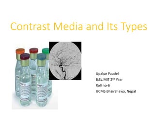
- 1. Contrast Media and Its Types Upakar Paudel B.Sc.MIT 2nd Year Roll no-6 UCMS Bhairahawa, Nepal
- 2. Introduction • Contrast agents are the substances which possess an atomic no or have an e- density which differs significantly from that of surrounding structures & permits the visualization of the details of internal organs that would not otherwise be demonstrable. 2
- 3. History • 1896, walter B. Cannon use radiopaque sustances. • 1904, colloidal silver was used in retrograde urography. • 1910, Krause use barium sulphate for visualisation of GIT. • 1923, Osborne reported the opacification of UT after the injection of 10% NaI soln. 3
- 4. History • 1928, compound with no of pyridine rings containing iodine . • 1952, first tri-iodinated compound (sodium acetrizoate) was introduced. • Until the early 1970s all CM were ionic compounds with osmolalities of 1200-2000 mosmal/kg. 4
- 5. History • Ionic monomer: 1954, diatrizoate (urografin) 1962, iothalamate (conray) 1962 – 1972, Metrizoate,Iodamide,Ioxithalamate, Ioglicate 5
- 6. History • Non-ionic monomers 1969 Metrizamide (Amipaque®) 1974 Iopamidol (Isovue®) 1976 Iohexol (Omnipaque®) 1979 Iopromide (Ultravist®) post 1980 Iopentol (Imagopaque®),Iomeprol,Ioxilan. 6
- 7. History • Ionic dimers 1978 Ioxaglate(Hexabrix®) 7 • Nonionic dimers Post 1980 Iodecol Iotrolan (Isovist®) Iodixanol (Visipaque®)
- 8. Function of contrast media • CM alters the attenuation of x-ray beam by altering the density of related structures. • The atomic no of the related structures also alter & helps in visualisation of similar density structures within the region of interest. 8
- 9. Classification of CM On the basis of physical properties: • Negative Contrast Media. • Positive Contrast Media 9
- 10. Negative CM • Gaseous substances which has low atomic number and specific weight, absorb X-rays to a lesser extent than the surrounding body structures. CO2 N2O Air O2 10
- 11. Positive CM • Substances which, because of their higher specific weight and atomic number, absorb X-rays to a greater extent than the body tissues. Barium sulphate Iodinated contrast media Contrast media for MRI and Ultrasound. 11
- 12. Barium sulphate • Ba Suspension is made up of pure Ba Sulphate. • Particles of Ba must be small (0.1-3μm),which makes more stable in suspension. • Barium, having atomic no 56, is a good contrast agent for GI examinations because it is very dense and won’t be absorbed by the GI tract. • In most situation it is diluted with water to give a lower density. 12
- 13. Ba sulphate • Solution has a pH of 5.3, which makes it stable in gastric acid. • Examinations of different parts of GI tract require Ba preparation with differing properties & different concentration. • A non ionic suspension medium is used, otherwise its particles would aggregate into clumps. 13
- 14. Advantage of Ba • Excellent coating which can be achieved. • Allowing the demonstration of normal and abnormal mucosal patterns. • Economic because of lower cost. 14
- 15. Iodinated CM • Most of IV CM contain Iodine which has 53 atomic no. & 127 atomic weight. • Total iodine content in the body is 50 mg. • It is Preferred because High contrast density due to high atomic no. Low toxicity. •Conc ranges from 140-370mgI/ml. 15
- 16. Iodinated CM • They are water soluble . • It is classified as: Ionic Non-ionic Ionic monomers CM High-osmolar Ionic dimer CM Low-osmolar Monoionic/diionic non-ionic monomer CM Low-osmolar Non-ionic dimer CM. Iso-osmolar 16
- 17. Ionic CM • In solution, ionic contrast media ionize into two particles, i.e. the cation and the anion. • The anion in an ionic contrast medium is the triiodinated benzoic acid part while the cation is usually sodium, calcium or meglumine. 17
- 18. Ionic monomers • Iodine to particle ratio is 3:2 • Sodium or meglumine act as cations. 18
- 19. Ionic monomers • Examples: Iothalamic Acid Has better neural tolerence but decrease cardiovascular tolerence. Eg. Conray Diatrizoic Acid Increase solubiliy Decrease plasma protein binding & increase glomerulus filtration. Improves patient tolerence. Eg. Urografin 19
- 20. Ionic dimer • Monoionic dimers ionize to give two particles but the number of iodine atoms present is 6. Therefore the iodine atom to particle ratio is 6:2, i.e.3 .Eg: Hexabrix. • Diionic dimers ionise to give three particles (two cations and one anion) with six iodineatoms present so that the iodine atom to particle ratio is 6:3, i.e. 2. 20
- 21. Non-ionic monomer • The molecule does not ionize & do not give two particles. • Formed by replacing the cation radical portion of benzene ring with a non-dissociating organic chain. • It remains as one single molecule when in solution because of an amide group on the acid part. 21
- 22. Non-ionic monomer • Less toxic effect becoz of removal of acid group & replacement of large no of OH group which make them more hydrophilic. 22
- 23. Non-ionic monomer • These molecules do not ionize in solution so that the iodine to particle ratio is 3:1. Therefore these are also ratio-3 molecules. • Eg: Iohexol (Omnipaque), Ioversol (Optiray) Iopromide (Ultravist). 23
- 24. Non-ionic dimer • These molecules will give six iodine atoms for every molecule so that their iodine atom to particle ratio is 6:1 which makes them ratio-6 molecules. • Disadvantage is high viscosity becoz of relatively large size of molecule. • Eg: Iodecol, Iotrolan (Isovist), Iodixanol (Visipaque®) 24
- 25. Osmolality • Osmolality is measure of no. of particles of solute in solution per kg of water. • Osmolality can be described as the property of a liquid to give up its water to or to take up water from another liquid separated by a semi- permeable membrane. • Hypo-osmolar liquid gives up water to the second liquid. • Hyper-osmolar liquid takes up water from the second liquid. • Iso-osmolar liquids have the same osmolality and do not exchange water. 25
- 26. Osmolality 26 Low and iso-osmolar compounds are tolerated better by the body and give rise to fewer side effects
- 27. Osmolality 27
- 28. Osmolality 28
- 29. Viscosity • Viscosity is a measure of the flow properties of solutions. • Iodine conc. determines the injection speed. 29
- 30. Characteristics of an ideal IV CM • provides maximum opacity to X-rays; • is biologically inert; • has high water solubility; • is chemically stable; • is selectively excreted; • has a low viscosity; • exerts minimal osmotic effects; • is safe; and • is not expensive 30
- 31. Application of CM •Contrast media may be introduced into the body via The gastro-intestinal tract (oral or rectal). Into the circulatory system (usually by the intravenous or intra-arterial routes) Into the cerebrospinal fluid (usually by the intrathecal route) Directly into a duct or tract (e.g. lymph vessels, lacrimal duct, salivary duct, etc). 31
- 32. Contd… • Barium Sulphate is used in GI tract: Ba Swallow, 200-250% w/v. Ba meal, 250% w/v. Ba follow-through, 60-100% w/v. Small bowel enema, 60% w/v. Barium enema, 115% w/v. CT of GI tract, 1-2% w/v. 32
- 33. Contd… • To achieve double contrast effect: for Oesophagus, Stomach & Duodenum- CO2 & less often, air is used. CO2 is administered orally in the form of gas producing granules/ powder. For large bowel, room air is administered per rectum via hand pump attached to the enema tube. 33
- 34. 34
- 35. • Ionic CM This type of CM can used in: Fistulography Sinography HSG MCU RGU CT GI tract (Oral CM) 35
- 36. • Non-ionic CM: This type of CM we can used widely. IVU, CT , Angiography Myelography- specially used Iohexol. Arthrography of joint with air. Non-ionic dimer are suitable for bronchography. 36
- 37. CM used in MRI • 1981, first CM enhanced, ferric chloride in GI tract. • 1984, gadolinium compound used as diagnostic IV MRI contrast agent. • Now a days, frequently MRI examinations are performed with CM. 37
- 38. Types of MRI CM • Ferromagnetic • Paramagnetic • Super-paramagnetic 38
- 39. Ferromagnetic • Retain magnetism even when the applied field is removed. • It may cause particle aggregation and cell function interference. • So unsafe for MR contrast agents. 39
- 40. Paramagnetic • Have magnetic moments which align to the applied field • Alignment return to normal after gradient field is turned off. • May be made soluble by chelation and hence can be used IV. • Maximum effect is on protons of water molecule, shortening the T1 relaxation time– increased signal intensity on T1 images. • Eg: Gadolinium. 40
- 41. Gadolinium • Gadolinium is a rare earth metal “heavy metal” • Gadolinium is chelated to diethylene triamine penta-acetic acid (DTPA) • Generic name is demeglumine gadopentate.(Magnevist) • By binding DTPA to the Gadolinium sites, only one “free” gadolinium site is available to attach to water molecules • Diffuse freely & excreated by kidneys • Gadolinium chelates are small mole. Wt. substance. 41
- 42. Super-paramagnetic • Are aggregation of paramagnetic ions in a crystalline lattice. • cause abrupt change in local magnetic field which results in rapid proton dephasing and reduction of T2 relaxation time – produce decreased signal intensity on T2 images. • Are less soluble than PM agents- so available only as colloidal suspensions. • Eg: Particals of iron oxide. 42
- 43. USG CM • Gas micro-bubbles are used • Should be less than 7 μm. • The gas molecules are encapsulated in palmitic acid, galactose or albumen. • US contrast media depend on interaction bet encapsulated micro- bubbles and US beam. • Allow imaging of vascular structures which cannot be evaluated even with sophisticated doppler techniques. 43
- 44. USG CM •Levovist Most widely used. Microbubbles of air enclosed by a thin layer of palmitic acid in a galactose sol. Stable in blood for 1- 4 min. •Echovist Precursor of levovist Bubbles in galactose but no palmitic acid. Can’t pass thro pulm beds Used for tubal patency. 44
- 45. USG CM • Albunex- Sonicated air micro-bubbles coated with human serum albumen. Used in echocardiography. Survives only a short time in left ventricle. Little enhancement of arterial tree. • EchoGen An emulsion of dodecafluoropentane which changes its phase converting into echogenic gas micro-bubbles by hypobaric activation prior to iv injection. 45
- 46. USG CM • SonoVue An aqueous suspension of stabilised sulphur hexafluoride micro-bubbles. After reconstitution of the lyophilisate with saline, the suspension is stable and can be used for upto 4 hrs. 46
- 47. Conclusion • Use of contrast media helps for easy diagnosis of diseases by tissue differentiation. • Choice of CM depends on the type of procedure being performed. • On going development of CM, more safe for patients. 47
- 48. References • A guide to radiological procedures: Stephen Chaman & Richard Nakielny, 5th edition. • Contrast media chemistry, pharmacology and pharmaceutical aspects, John Stephen Forte. • Clark’s special procedures. Latest edition. 48
- 49. ??? • What is contrast media? • Classify contrast media? • What is negative CM? • What is positive CM? • Applications of contrast media? • Differentiate ionic & non-ionic CM? • What are the uses of double contrast technique? • Characteristic of an ideal IV CM. • Types of MRI contrast media. 49
- 50. 50