Female fertility begins to decline many years prior to the onset of menopause despite continued regular ovulatory cycles. Although there is no strict definition of advanced reproductive age in women, infertility becomes more pronounced after the age of 35.
2. The infertility evaluation in women of advanced repro-
ductive age should include an assessment of ovarian reserve.
The term ovarian reserve describes a woman’s reproductive
potential with respect to ovarian follicle number and oocyte
quality. The measurement of serum basal FSH and estradiol
on day 3 of the menstrual cycle is often used to test for
ovarian reserve. FSH and estradiol should be measured in the
early follicular phase because accelerated follicular develop-
ment may be associated with reproductive aging. Elevated
FSH and estradiol levels are independent predictors of poor
prognosis in older women (14–16). Common criteria for
normal ovarian reserve are an early follicular phase FSH
level of Ͻ10 mIU/mL and an estradiol level of Ͻ80 pg/mL.
Higher cutoff values for FSH have been reported (as high as
20 to 25 mIU/mL for FSH) because of the use of different
FSH assay reference standards (13). The normal range for
FSH ideally should be determined in a normal fertile popu-
lation by each laboratory. In the event that such data are not
available, the laboratory’s stated upper limit for the follicular
phase can be used as an arbitrary and imperfect cutoff to
define abnormal.
The clomiphene citrate challenge test (CCCT), which is
another test of ovarian reserve, is performed by measuring a
day 3 FSH, administering clomiphene citrate 100 mg orally
on cycle days 5 to 9, and then measuring FSH on cycle day
10 (17–19). The test is considered to be abnormal if either
the day 3 or the day 10 FSH is above the threshold value for
the laboratory.
Women with abnormal basal FSH, estradiol, or CCCT
have lower live birth rates with ovulation induction and
intrauterine insemination (17, 19, 20). Women with dimin-
ished ovarian reserve also experience decreased responses to
ovulation induction, require higher doses of gonadotropin,
have higher IVF cycle cancellation rates, and experience
lower pregnancy rates through IVF (18, 21). In a general
infertility population, an abnormal CCCT predicts that a
successful pregnancy will be achieved about 5% of the time
(22). A single elevated day 3 FSH value connotes a poor
prognosis, even when values in subsequent cycles are normal
(23, 24).
Other tests of ovarian reserve under study include circu-
lating inhibin B levels, the gonadotropin-releasing hormone
agonist test, and small antral follicle count by ultrasound.
Women with decreased ovarian responsiveness to gonado-
tropin may have decreased serum inhibin B levels even when
FSH levels are normal (25). This finding suggests that in-
hibin B may be a more sensitive marker of ovarian reserve
than FSH. However, routine testing for serum inhibin B
levels is not recommended at this time due to limited avail-
ability of reliable assays and conflicting data regarding its
prognostic value (26). The gonadotropin-releasing hormone
agonist challenge test is not recommended for routine clin-
ical use because of limited data on its prognostic value. The
number of small antral follicles visible on transvaginal ul-
trasound appears to correlate directly with ovarian response
to gonadotropins (27, 28). In the future, transvaginal ultra-
sound may be an effective way of estimating ovarian reserve.
Currently it seems reasonable to test ovarian reserve by
using day 3 FSH and estradiol or the CCCT in all infertile
women age 35 and older who desire pregnancy. Ovarian
reserve testing may be considered in patients under age 35 with
a solitary ovary, history of ovarian surgery, poor response to
exogenous gonadotropins, exposure to chemotherapeutic agents
or ionizing radiation, and unexplained infertility.
COUNSELING
Preconception counseling should include a discussion of the
increased risks of aneuploidy, spontaneous abortion, and
obstetric complications such as delivery by cesarean section,
hypertension, and gestational diabetes (29) associated with
increasing maternal age. The rate of all clinically significant
cytogenetic abnormalities in live births increases from about
1/500 for women under 30 to 1/270 at age 30, 1/80 at age 35,
1/60 at age 40, and 1/20 at age 45 (30).
Counseling after ovarian reserve testing should include a
discussion of the results. While they may predict a lower
pregnancy rate, abnormal ovarian reserve test results do not
preclude the possibility of pregnancy and should not be pre-
sented to patients as absolute. Likewise, ovarian reserve testing
alone may yield falsely reassuring results, as advanced maternal
age and ovarian reserve test results are independent predictors
of infertility. Both should be used when counseling couples
regarding their chances for conception (22).
TREATMENT
Treatment options for age-related infertility include con-
trolled ovarian hyperstimulation with intrauterine insemina-
tion (COH/IUI), IVF, and oocyte donation. Except for oo-
cyte donation, these treatments are intended to accelerate the
time to conception rather than directly affect oocyte or
embryo quality. Expectant management, which should be
reserved for couples who do not desire medical intervention,
is also considered a treatment option but is less likely to
result in pregnancy in women of advanced reproductive age
(39). COH/IUI consists of gonadotropins administered to
initiate growth and ovulation of multiple follicles in con-
junction with the placement of washed sperm in the uterine
cavity.
COH/IUI has limited efficacy for women over 40 with
otherwise unexplained infertility, yielding a per cycle deliv-
ery rate of 5% or less (range 1.4% to 5.2%) (31–36). This
compares with a live birth rate per cycle of 17% to 22% for
women under 35 and 8% to 10% for women aged 35 to 40
(33, 37, 38). There have been no studies comparing COH/
IUI with IVF. Unfortunately, most reports have been retro-
spective case series or cohort studies that would be expected
to overestimate treatment effectiveness.
The presence of male factor, tubal disease, endometriosis,
or pelvic adhesions would argue for proceeding directly to
S249FERTILITY & STERILITYா
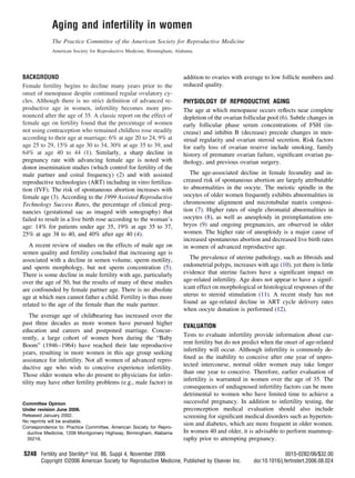
3. IVF in women of advanced reproductive age. Pregnancy
rates from IVF are generally higher than from COH/IUI but
also decline significantly with age. According to the 1999
Assisted Reproductive Technology Success Rates, live birth
rates per cycle were 32.2% in women under 35, 26.2% in
women aged 35 to 37, 18.5% in women aged 38 to 40, 9.7%
in women aged 41 to 42, and approximately 5% in women
43 and older (Fig. 1) (4).
In a recent multicenter review of 431 initiated IVF cycles
in women Ն41 years, there were no clinical pregnancies in
women Ն45 years and no deliveries in women Ն44 years of
age (40). This age-related decline in IVF success is related to
decreased ovarian responsiveness to gonadotropins and, more
importantly, to a marked decline in embryo implantation rates
(Table 1). Embryonic aneuploidy is likely the major reason for
implantation failure in older women (41).
The following alternative approaches have been described
for IVF treatment in women with decreased ovarian reserve:
1. Microdose GnRH agonist flare protocol or other flare (43,
44).
2. Use of a GnRH antagonist with gonadotropins.
3. Low-dose GnRH agonist suppression before gonadotro-
pin stimulation.
4. Assisted hatching of embryos.
5. Use of estrogen or oral contraceptives in the cycle prior to
gonadotropin stimulation.
Unfortunately, there are no randomized trials to compare
the relative efficacy of these approaches. Exclusion of ane-
uploid embryos with preimplantation genetic diagnosis
(PGD) may lower the spontaneous abortion rate in IVF
cycles (45). However, the technique is expensive and is not
yet widely available. Its role in the treatment of age-related
infertility has yet to be defined.
Nuclear (germinal vesicle) transfer is an experimental
technique in which the nucleus from the oocyte of an older
woman is transferred to the enucleated oocyte of a younger
woman. The safety and efficacy of this technique is currently
unknown (46).
No treatment other than oocyte donation has been shown
to be effective for women over 40 and for those with com-
promised ovarian reserve. Although the resulting child will
not be biologically related to the birth mother, oocyte dona-
tion yields the highest live birth rate of any ART treatment.
It is the treatment of choice for age-related infertility not
successfully addressed by other methods. Pregnancy rates
with oocyte donation are dependent on the age of the donor
rather than the recipient.
SUMMARY
● A relatively large group of women is experiencing age-
related infertility due to social trends that lead to deferred
childbearing and to the current age of the “Baby Boom”
generation.
● Age-related infertility is due to oocyte abnormalities and
decreased ovarian reserve.
● Clinical tests to estimate ovarian reserve include FSH and
estradiol levels in the early follicular phase (e.g., day 3) or
a clomiphene citrate challenge test.
FIGURE 1
Pregnancy and live birth rates for ART cycles
using fresh, nondonor eggs or embryos, by female
age, 1999. Data from reference 4. Gray line ϭ live
birth rate; solid line ϭ pregnancy rate. ASRM.
Aging and infertility in women. Fertil Steril 2002.
Source: Centers for Disease Control and
Prevention, American Society for Reproductive
Medicine, Society for Assisted Reproductive
Technology, RESOLVE. 1999 Assisted
reproductive technology success rates. Atlanta,
GA: Centers for Disease Control and Prevention,
2001.
ASRM Practice Committee. Aging and infertility in women. Fertil Steril 2006.
TABLE 1
Embryo implantation rates as a function of
female age (42).
Age Implantation rate
25–29 18.2%
30–34 16.1%
35–39 15.3%
40–44 6.1%
Reprinted with permission (Fertil Steril 1996;65:783–
790) (42).
Source: Centers for Disease Control and Prevention,
American Society for Reproductive Medicine, Society
for Assisted Reproductive Technology, RESOLVE.
1999 Assisted Reproductive Technology Success
Rates. Atlanta, GA. Centers for Disease Control and
Prevention; 2001 (4).
ASRM Practice Committee. Aging and infertility in women. Fertil Steril
2006.
S250 ASRM Practice Committee Aging and infertility in women Vol. 86, Suppl 4, November 2006
4. ● Evaluation and treatment of infertility should not be de-
layed in women 35 years and older.
● Treatment of infertility when the cause is limited to de-
creased ovarian reserve is empirical at present except for
oocyte donation.
Acknowledgments: This report was developed under the direction of the
Practice Committee of the American Society for Reproductive Medicine
as a service to its members and other practicing clinicians. While this
report reflects appropriate management of a problem encountered in the
practice of reproductive medicine, it is not intended to be the only
approved standard of practice or to dictate an exclusive course of
treatment. Other plans of management may be appropriate, taking into
account the needs of the individual patient, available resources and
institutional or clinical practice limitations. This report was approved by
the Practice Committee of the American Society for Reproductive Med-
icine in November 2001 and the Board of Directors of the American
Society for Reproductive Medicine in January 2002.
REFERENCES
1. Menken J, Trussell J, Larsen U. Age and infertility. Science 1986;233:
1389–94.
2. Schwartz D, Mayaux MJ. Female fecundity as a function of age: results
of artificial insemination in 2193 nulliparous women with azoospermic
husbands. Federation CECOS. N Engl J Med 1982;306:404–6.
3. Smith KE, Buyalos RP. The profound impact of patient age on preg-
nancy outcome after early detection of fetal cardiac activity. Fertil Steril
1996;65:35–40.
4. Centers for Disease Control and Prevention, American Society for
Reproductive Medicine, Society for Assisted Reproductive Technol-
ogy, RESOLVE. 1999 Assisted reproductive technology success rates.
Atlanta, GA: Centers for Disease Control and Prevention, 2001.
5. Kidd SA, Eskenazi B, Wyrobek AJ. Effects of male age on semen
quality and fertility: a review of the literature. Fertil Steril 2001;75:
237–48.
6. Faddy MJ, Gosden RG, Gougeon A, Richardson SJ, Nelson JF. Accel-
erated disappearance of ovarian follicles in mid-life: implications for
forecasting menopause. Hum Reprod 1992;7:1342–6.
7. Battaglia DE, Goodwin P, Klein NA, Soules MR. Influence of maternal
age on meiotic spindle assembly in oocytes from naturally cycling
women. Hum Reprod 1996;11:2217–22.
8. Angell RR. Aneuploidy in older women. Higher rates of aneuploidy in
oocytes from older women. Hum Reprod 1994;9:1199–200.
9. Benadiva CA, Kligman I, Munne S. Aneuploidy 16 in human embryos
increases significantly with maternal age. Fertil Steril 1996;66:248–55.
10. Nagele F, O’Connor H, Davies A, Badawy A, Mohamed H, Magos A.
2500 outpatient diagnostic hysteroscopies. Obstet Gynecol 1996;88:
900–1.
11. Noci I, Borri P, Chieffi O, Scarselli G, Biagiotti R, Moncini D, et al. Aging
of the human endometrium: a basic morphological and immunohistochem-
ical study. Eur J Obstet Gynecol Reprod Biol 1995;63:181–5.
12. Navot D, Drews MR, Bergh PA, et al. Age-related decline in female
fertility is not due to diminished capacity of the uterus to sustain
embryo implantation. Fertil Steril 1994;61:97–101.
13. Hershlag A, Lesser M, Montefusco D, Lavy G, Kaplan P, Liu HC, et al.
Interinstitutional variability of follicle-stimulating hormone and estra-
diol levels. Fertil Steril 1992;58:1123–6.
14. Smotrich DB, Widra EA, Gindoff PR, Levy MJ, Hall JL, Stillman RJ.
Prognostic value of day 3 estradiol on in vitro fertilization outcome.
Fertil Steril 1995;64:1136–40.
15. Frattarelli JL, Bergh PA, Drews MR, Sharara FI, Scott RT. Evaluation
of basal estradiol levels in assisted reproductive technology cycles.
Fertil Steril 2000;74:518–24.
16. Licciardi FL, Liu HC, Rosenwaks Z. Day 3 estradiol serum concentra-
tions as prognosticators of ovarian stimulation response and pregnancy
outcome in patients undergoing in vitro fertilization. Fertil Steril
1995;64:991–4.
17. Navot D, Rosenwaks Z, Margalioth EJ. Prognostic assessment of fe-
male fecundity. Lancet 1987;2:645–7.
18. Tanbo T, Dale PO, Lunde O, Norman N, Abyholm T. Prediction of
response to controlled ovarian hyperstimulation: a comparison of basal
and clomiphene citrate-stimulated follicle-stimulating hormone levels.
Fertil Steril 1992;57:819–24.
19. Scott RT, Leonardi MR, Hofmann GE, Illions EH, Neal GS, Navot D.
A prospective evaluation of clomiphene citrate challenge test screening
of the general infertility population. Obstet Gynecol 1993;82:539–44.
20. Buyalos RP, Daneshmand S, Brzechffa PR. Basal estradiol and follicle-
stimulating hormone predict fecundity in women of advanced repro-
ductive age undergoing ovulation induction therapy. Fertil Steril 1997;
68:272–7.
21. Toner JP, Philput CB, Jones GS, Muasher SJ. Basal follicle stimulating
hormone level is a better predictor of in vitro fertilization performance
than age. Fertil Steril 1991;55:784–91.
22. Scott RT, Opsahl MS, Leonardi MR, Neall GS, Illions EH, Navot D.
Life table analysis of pregnancy rates in a general infertility population
relative to ovarian reserve and patient age. Hum Reprod 1995;10:1706–10.
23. Scott RT Jr, Hofmann GE, Oehninger S, Muasher SJ. Intercycle vari-
ability of day 3 follicle-stimulating hormone level and its effect on
stimulation quality in in vitro fertilization. Fertil Steril 1990;54:297–302.
24. Martin JS, Nisker JA, Tummon IS, Daniel SA, Auckland JL, Feyles V.
Future in vitro fertilization pregnancy potential of women with variably
elevated day 3 follicle-stimulating hormone levels. Fertil Steril 1996;
65:1238–40.
25. Seifer DB, Scott RT Jr, Bergh PA, Abrogast LK, Friedman CI, Mack
CK, et al. Women with declining ovarian reserve may demonstrate a
decrease in day 3 serum inhibin B before a rise in day 3 follicle-
stimulating hormone. Fertil Steril 1999;72:63–5.
26. Corson SL, Gutmann J, Batzer FR, Wallace H, Klein N, Soules MR.
Inhibin-B as a test of ovarian reserve for infertile women. Hum Reprod
1999;14:2818–21.
27. Chang MY, Chiang CH, Hsieh TT, Soong YK, Hsu KH. Use of the
antral follicle count to predict the outcome of assisted reproductive
technologies. Fertil Steril 1998;69:505–10.
28. Frattarelli JL, Lauria-Costab DF, Miller BT, Bergh PA, Scott RT. Basal
antral follicle number and mean ovarian diameter predict cycle cancel-
lation and ovarian responsiveness in assisted reproductive technology
cycles. Fertil Steril 2000;74:512–7.
29. Gilbert WM, Nesbitt TS, Danielsen B. Childbearing beyond age 40:
pregnancy outcome in 24,032 cases. Obstet Gynecol 1999;93:9–14.
30. Hook EB. Rates of chromosome abnormalities at different maternal
ages. Obstet Gynecol 1981;58:282–5.
31. Agarwal SK, Buyalos RP. Clomiphene citrate with intrauterine insem-
ination: is it effective therapy in women above the age of 35 years?
Fertil Steril 1996;65:759–63.
32. Pearlstone AC, Fournet N, Gambone JC, Pang SC, Buyalos RP. Ovu-
lation induction in women age 40 and older: the importance of basal
follicle-stimulating hormone level and chronological age. Fertil Steril
1992;58:674–9.
33. Brzechffa PR, Buyalos RP. Female and male partner age and menotro-
phin requirements influence pregnancy rates with human menopausal
gonadotrophin therapy in combination with intrauterine insemination.
Hum Reprod 1997;12:29–33.
34. Corsan G, Trias A, Trout S, Kemmann E. Ovulation induction com-
bined with intrauterine insemination in women 40 years of age and
older: is it worthwhile? Hum Reprod 1996;11:1109–12.
35. Frederick JL, Denker MS, Rojas A, Horta I, Stone SC, Asch RH, et al.
Is there a role for ovarian stimulation and intra-uterine insemination
after age 40? Hum Reprod 1994;9:2284–6.
36. Campana A, Sakkas D, Stalberg A, Bianchi PG, Comte I, Pache T, et al.
Intrauterine insemination: evaluation of the results according to the
woman’s age, sperm quality, total sperm count per insemination and
life table analysis. Hum Reprod 1996;11:732–6.
S251FERTILITY & STERILITYா
5. 37. Brzechffa PR, Daneshmand S, Buyalos RP. Sequential clomiphene
citrate and human menopausal gondotrophin with intrauterine insemi-
nation: the effect of patient age on clinical outcome. Hum Reprod
1998;13:2110–4.
38. Guzick DS, Carson SA, Coutifaris C, Overstreet JW, Factor-Litvak P,
Steinkampf MP, et al. Efficacy of superovulation and intrauterine
insemination in the treatment of infertility. National Cooperative Re-
productive Medicine Network. N Engl J Med 1999;340:177–83.
39. Cohen MA, Chang PL, Uhler M, Legro R, Sauer MV, Lindheim SR.
Reproductive outcome after sterilization reversal in women of ad-
vanced reproductive age. J Assist Reprod Genet 1999;16:402–4.
40. Ron-El R, Raziel A, Strassburger D, Schachter M, Kasterstein E,
Friedler S. Outcome of assisted reproductive technology in women over
the age of 41. Fertil Steril 2000;74:471–5.
41. Munne S, Alikani M, Tomkin G, Grifo J, Cohen J. Embryo morphol-
ogy, developmental rates, and maternal age are correlated with chro-
mosome abnormalities. Fertil Steril 1995;64:382–91.
42. Hull MG, Fleming CF, Hughes AO, McDermott A. The age-related
decline in female fecundity: a quantitative controlled study of implant-
ing capacity and survival of individual embryos after in vitro fertiliza-
tion. Fertil Steril 1996;65:783–90.
43. Surrey ES, Bower J, Hill DM, Ramsey J, Surrey MW. Clinical and
endocrine effects of a microdose GnRH agonist flare regimen admin-
istered to poor responders who are undergoing in vitro fertilization.
Fertil Steril 1998;69:419–24.
44. Surrey ES, Schoolcraft WB. Evaluating strategies for improving ovar-
ian response of the poor responder undergoing assisted reproductive
techniques. Fertil Steril 2000;73:667–76.
45. Munne S, Magli C, Cohen J, Morton P, Sadowy S, Gianaroli L, et al.
Positive outcome after preimplantation diagnosis of aneuploidy in hu-
man embryos. Hum Reprod 1999;14:2191–9.
46. Zhang J, Wang CW, Krey L, Liu H, Meng L, Blaszczyk A, et al. In
vitro maturation of human preovulatory oocytes reconstructed by ger-
minal vesicle transfer. Fertil Steril 1999;71:726–31.
S252 ASRM Practice Committee Aging and infertility in women Vol. 86, Suppl 4, November 2006