1. 1
Tina Safavie
I. Literature Review
Trichomoniasis
Trichomoniasis is a commonly occurring, albeit infrequently reported sexually transmitted
infection. In contrast to most STIs, which are typically caused by viruses and bacterial etiologic agents,
trichomoniasis results from infection by the anaerobic protozoan Trichomonas vaginalis (T. vaginalis).
This flagellated parasite infiltrates the lower urogenital tract, and specifically resides in the female
urethra, vulva and vagina. Despite the variety of sexual activities, such as oral and anal sex, T. vaginalis
remains restricted to the urogenital regions for unknown reasons. Infection is spread through
unprotected sexual activity between uninfected and infected individuals. According to the Centers for
Disease Control, there are an estimated 3.7 million Americans currently afflicted with trichomoniasis. Of
this population, only about 30% develop a symptomatic form of the diseases. Given the lack of severe
disease complications and relative ease of treatment, public health officials tend to overlook the
importance of trichomoniasis infection. In fact, it has often been labeled as a “minor” STD (Poole et al
2013). Surprisingly, trichomoniasis cases do not need to be reported to the Centers for Disease Control.
Recently, however, clinicians recognize that “understanding the relation between T vaginalis and other
sexually transmitted infections would assist its use as a marker for the success of behavioral and
treatment interventions targeting other STDs including HIV,” (Bowden et al 2000). As such, the following
section of this paper is focused on elucidating the pathogenesis, clinical manifestations, and
epidemiology of trichomoniasis.
Pathogenesis
Trichomoniasis is caused by the protozoan parasite Trichomoniasis vaginalis (T. vaginalis).
Through light microscopic analysis, the protozoan is observed to have a pyriform and ameboid shape in
culture and in vivo respectively. In addition, T. vaginalis has a size of 9 by 7um, and contains four
2. 2
anterior flagella in a 9+2 arrangement. Along with possessing an undulating membrane, this nondividing
organism has unique energy producing organelles termed “hydrogenosomes.” These structures can be
visualized as paraxostylar and paracostal chromatic granules (Schwebke et al 2004). Another unique
feature of T. vaginalis is the presence of adherence factors, which allow it to colonize the cervicovaginal
epithelium within women. An axostyle, a rigid structure, extends from the posterior aspect of the
organism (Medscape 2013). Since the organism is non-dividing, it creates offspring to populate the
lower human urogenital tract through the process of binary fission (Schwebke et al 2004).
Epidemiology
One of the greatest challenges in addressing the current trichomoniasis epidemic is the
surprising lack of epidemiological data concerning its incidence and prevalence. With that in mind,
public health officials have recently embarked on a journey of documenting cases and risk factors in
relation to trichomoniasis. Recent investigations into the incidence of trichomoniasis suggest that the
disease annually affects about 3.7 million Americans. However, other sources indicate a much higher
incidence rate of 7.4 million new cases occurring annually (Schwebke et al 2004). Despite these high
numbers, epidemiologists estimate an even greater incidence rate due to the asymptomatic nature of
the disease in most women. In regards to the age distribution of the disease, statistical analysis
illustrates that trichomoniasis evenly affects women of all ages (Schwebke et al 2004). This is in stark
contrast with STIs such as C. trachomatis and Neisseria gonorrhoeae, which specifically affect younger
adults. Interestingly, however, CDC investigations indicate that of the majority of trichomoniasis cases
occur in young women between the ages of 15 - 24 years old. Moreover, trichomoniasis accounted for
about $34.2 million of health care costs among young women aged 15- 24 years in the year 2000 alone
(CDC 2013). Concerning trichomoniasis and socioeconomic status, anecdotal evidence indicates lower
prevalence of the disease amongst women in higher socioeconomic groups. Along the same lines, other
studies indicate that “prevalence maybe as high as 50% in women in the developing world and in
3. 3
minority groups in industrialized populations,” (Bowden et al 2000). For instance, the CDC found that
within the United States, white non-Hispanic women are least affected by trichomoniasis with a
prevalence of 1.8%. Mexican Americans have the next highest prevalence at a rate of 1.8%. Non-
Hispanic African Americans have the highest occurrence of 13.3% (CDC 2013). From this data, it is clear
that socioeconomic factors such as poverty, promiscuity and having only high school education or less
are significantly associated with trichomoniasis.
Clinical manifestations
Although the majority of trichomoniasis cases are asymptomatic, individuals affected by
symptoms have marked clinical manifestations (Schwebke et al 2004).. For instance, women faced with
the disease have yellowy green frothy vaginal discharge, pronounced unusual odor, and irritation. Other
symptoms include painful intercourse, dysuria, and lower abdominal pain. During infection, women
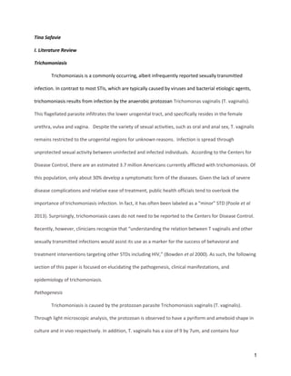
typically exhibit colpitis macularis or “strawberry cervix” as observed by colposcopy (figure 1). Colpitis
macularis results from microscopic, punctate hemorrhages of the cervix induced by the T. vaginalis
parasite and the host’s strong inflammatory response (Schwebke et al 2004). These symptoms appear
after an incubation period of 4-28 days. Often referred to as “trich,” if trichomoniasis is not treated,
infection with the parasite can persist for months or even years (Medicinenet 2013). It is also important
to note that researchers have yet to identify the reasons for the variation in immune responses to
trichomoniasis. However, it is hypothesized that clinical manifestations result from various biological
factors. These include fluctuations in hormonal levels, vaginal pH, and the strain and concentration of
existing commensal bacteria. Though individuals with trichomoniasis exhibit relatively minor symptoms,
the disease can cause complications such as pelvic inflammatory disease, low birth weight infants,
premature labor and increased risk for human immunodeficiency virus infection.
Trichomoniasis has been shown to increase susceptibility to HIV-1 infection. This is due a variety
of factors. For example, T. vaginalis causes an aggressive inflammatory and local cellular immune
4. 4
response within the cervix and vagina by the host immune system. Consequently, leukocytes and other
HIV target cells are quick to arrive to this vulnerable region. By increasing the number of CD4+ T Cells,
there is a greater likelihood that one could be infected by the human immunodeficiency virus. Another
mechanism by which T. vaginalis increases susceptibility to HIV-1 is inducing mucosal hemorrhaging
which degrades mechanical barriers to the virus. By disrupting the natural vaginal flora and producing
genital lesions, T. vaginalis enables direct viral access to the bloodstream. This strong biological
plausibility is supported by empirical studies from Africa documenting that Trichomonas may increase
HIV transmission by as much as threefold (Shafir et al 2009).
Although trichomoniasis has severe health complications, it is easily diagnosable and treatable
by medical professionals. For instance, clinicians utilize a visualization method to quickly detect the
presence of T. vaginalis. With this method, the motile organism is observed under a light microscope.
Here, the organism appears as the size of a white blood cell and contains a beating flagella that causes
jerky movements. Though this test is relatively inexpensive, the sensitivity is only about 60-70%. Other
indicators such as inflammation, elevated vaginal pH and olfactory detection of amines are also utilized
to diagnosis trichomoniasis (Medscape 2013). To treat trichomoniasis, patients are prescribed only a
single dose of 2 g the metronidazole antibiotic. If patients use this medication properly, then the
reported cure rate is 97%. Similar to other parasite and antibiotic interactions, the T. vaginalis parasite
has developed metronidazole resistance in approximately 2.5 - 5% of all cases. However, this antibiotic
resistance can be easily overcome by increasing the dose of metronidazole (Schwebke et al 2004).
Bacterial Vaginosis
Bacterial vaginosis (BV) is a condition responsible for causing most of the vaginal symptoms in
reproductive age women. When women are affected by BV, their natural commensal vaginal flora is
disrupted. Specifically, there is an increase in hydrogen peroxide-producing Lactobacillus spp (CDC
2013). There is also an increase in numbers of both facultative and anaerobic bacteria, including
5. 5
Gardnerella vaginalis. Similar to trichomoniasis and candidiasis, bacterial vaginosis is a relative mild and
easily treatable condition. However, left untreated, bacterial vaginosis can lead to severe health
implications. These include an increased risk for contracting human immunodeficiency virus and other
sexually transmitted diseases such as trichomoniasis and genital herpes. In pregnant women, BV
increases the risk of miscarriage,preterm labor, preterm delivery, and postpartum complications such as
endometritis and wound infections (Koumans et al 2007).
According to the CDC, 29.2% of the 14-49 age group or twenty million women are annually
affected by bacterial vaginosis. However, of this population, only 26% women ever reported symptoms.
With regards to race and ethnicity, BV occurs predominantly in non-Hispanic African American women.
For instance, the CDC found that there is a 51% prevalence rate or 2.75, 95% CI 2.2–3.5 amongst black
women with comparison to 23% prevalence in non-Hispanic white women (Koumans et al 2007).
Research also shows that 28% of non-Hispanic African American women that are not sexually active are
afflicted with bacterial vaginosis. This indicates that other factors rather than sexual activity may
influence contraction of bacterial vaginosis. These factors may include recent antibiotic use, hormonal
fluctuations (specifically decreased estrogen production), use of intrauterine devices and improper
douching (Medscape 2013). Despite the variation in these risk factors, all of these phenomena share the
similarity of disrupting the normal commensal vaginal flora.
When affected by BV, a small number of patients display symptoms such as gray, thin and
homogeneous vaginal discharge. This discharge may have small bubbles and adheres to the vaginal
epithelium. Along with painful urination and vaginal itching, affected women may also present with a
strong “fish-like” vaginal odor (CDC 2013). In contrast to candidiasis and trichomoniasis, bacterial
vaginosis does not cause abdominal pain or dyspareunia. Additionally, there is an absence of
vulvovaginal inflammation in the patient (Medscape 2013). The clinical diagnosis of bacterial vaginosis
relies on a combination of vaginal [symptoms highlighted above] and microscopic examination. For their
6. 6
microscopic examination, clinicians base their diagnosis of bacterial vaginosis on the analysis of the
patient’s vaginal discharge. In doing so, they assess the presence of three out of four criteria: (1) the
presence of clue cells on a saline smear [vaginal epithelium with “peppering” of coccobacilli] (2) an
elevated vaginal pH of 4.5 [3] a positive whiff test in which 10% KOH is mixed with vaginal fluid to
release volatile amines [4] gray, white, and thin discharge. To treat bacterial vaginosis, antibiotics are
used. These include antibiotics such as: metronidazole (Flagyl), clindamycin (Cleocin) oral or vaginal
suppositories, and metronidazole vaginal gel (MetroGel-Vaginal) (Medscape 2013).
Candidiasis
Commonly known as vaginal yeast infection, vaginal candidiasis occurs mostly when there is an
overgrowth of naturally occurring Candida within the Vagina. Human fungal diseases are predominantly
due to candida infections that are seldom life-threatening. In comparison to all other candida species,
Candida albicans specifically colonizes the skin, the gastrointestinal and reproductive tracts, and is the
leading cause of yeast infection. (Achkar et al 2010) Abnormal increases in this yeast population is
stimulated by sudden changes in the environment such as lowering of the vaginal acidity (pH). Though
candidiasis is a common fungal infection that can develop on the skin as rash, the throat/mouth as
thrush, and the blood as candidemia, this paper focuses on vulvo-viginal candidiasis (VVC) infections.
VCC is the second most significant source of vaginitis after bacterial vaginosis and is found in
approximately 40% of women who seek medical attention for vaginal infections (Kim et al 2011). The
transformation of C. albicans as a commensal fungal organism found into an opportunistic pathogen is
triggered by disturbances of the local microbiologic flora or environment (Kim et al 2011). Symptoms of
VCC vary from intense vaginal itching and burning to secretion of a white vaginal discharge. Diagnosis of
VCC is performed by using yeast culture, microscopy or vaginal pH measurements. Several risk factors
have been associated with candidal infection of the vagina. These risk factors include: the use of
7. 7
corticosteroids, reproductive hormone and oral contraceptives, antibiotics treatment,
immunodeficiency, diabetes and sexual behaviors. Azole-based antifungal drugs have been effective in
treating VCC and are accessible over-the-counter. Of these drugs, clotrimazole is the cheapest of the
anti-fungal treatments.
The clinical manifestations of VCC are similar to those of other vaginal conditions such as
chlamydia, trichomoniasis, gonorrhea, and bacterial vaginosis (Abbot 1995). Symptoms of VVC include
severe vaginal and labial itching, burning sensation, soreness, painful urination and vulvar inflammation.
Pain during intercourse, redness, irritation and swelling of the vulva are also reported. (El-Din et al.,
2001) In addition, a thick, odorless, flocculent, curdy and "cottage cheese-like" white vaginal discharge is
observed. Over 90% of VCC cases are caused by Candida Albicans. (Moreira & Paula, 2005)
Various methods are used to diagnose candidiasis. These include analysis of clinical symptoms,
vaginal culture, and measurement of vaginal pH. Yeast culture is most useful for the diagnosis when
patients have recurrent VCC infections. When diagnosing yeast through culture, clinicians rely on
Sabouraud’s medium, which allows them to distinguish between normal candidal colonization and true
infections (Tanaka, 1998). The vaginal pH of an individual affected with VCC is between 4.0 and 4.5.
Whereas a pH greater than 4.5 suggests other vaginal infections such as trichomoniasis and vaginosis. In
addition to yeast culture and vaginal pH analysis, simple microscopy can also be used to examine vaginal
discharge for VCC diagnosis. Microscopic analysis of a saline preparation or a wet mount has been
demonstrated to have a 40-60% accuracy rate. A 10% potassium hydroxide (KOH) preparation, however,
is said to be more sensitive (Sobel 2010). When observing the wet mount of sample positive for candida,
numerous filaments, hyphae, and buds are visible under the microscope. Since candida is a gram
positive organism, supplementing microscopy with gram staining will highly increase sensitivity of the
test (Anderson et al 2004). Another commercial diagnostic technique used to detect candida is the rapid
8. 8
agglutination test. This test provides results in as little as 2 minutes; however, precision of this test is
directly proportional to the number of yeast in the sample (Tanaka 1998).
Despite the widespread occurrence of VCC in healthy women, some behavioral and host-related
risk factors cause excessive growth of candida. Frequent sexual activity, receptive oral sex, usage of
high-estrogen oral contraceptives, spermicides use, and condom use were cited as some of the
behavioral aspects contributing to VCC incidences. Mismanaged diabetes, antibiotic treatment, and
genetic susceptibility and elevated reproductive hormone levels are other host-related risk factors. The
potential of hormones in causing VCC is evidenced by the fact that premenarchal girls and post-
menopausal women rarely contact this disease. Though the major source of VCC infections is the
patient’s own microflora, women can also acquire it sexually. Studies show that infected sexual partners
tend to have the same candida strains (Sobel 2010) Patients with uncontrolled type 2 diabetes mellitus
are particularly vulnerable to yeast infections due to the fact that hyperglycemia interferes with a
person’s immune response and facilitates Candida albicans’ attachment to the mucosal lining of the
vagina. Additionally, the diabetes provides the candida with carbon sources for germination, thereby
contributing to its proliferation (Nyirjesy et al 2013). With regards to HIV/AIDS infection, oral candidiasis
is so widespread in AIDS patients [as an opportunistic infection], that it is often used to indicate the
progression of HIV infection into AIDS. As such, the pathogen clearly takes advantage of the reduced
cell-mediated immunity of immonusuppresed individuals (Kim et al 2011). Moreover, antibiotics
indirectly select for C. albicans and stimulates its growth by 10 to 30% fold by eliminating the presence
of local gastrointestinal and vaginal microorganisms such as lactobacilli (Hawes et al 1996) VCC develops
in 28 to 30% of patients following antibiotic intake. Studies also suggest that VCC prevalence is higher in
non-Hispanic African American women compared to their white peers. Other findings indicate that
women with VCC are twice as likely to be co-infected with other STDs. Of the co-occurring STDs, most
patients are infected with HIV due to the damage to the vaginal mucosa. Consequently, women with
9. 9
VCC have increased amounts of HIV viral shedding and thus increase the potential for disease
transmission to partners.
VCC is not a reportable condition and asymptomatic VCC is pervasive. Since VCC is frequently
diagnosed and easily treatable with OTC medication without laboratory confirmation, it is difficult to
accurately assess the prevalence of this disease (Achkar et al 2010). Prior to FDA approval of OTC anti-
fungal drugs in the 1990s, up to 10 million women annually visited their gynecologist to treat their VCC.
As such, VCC accounted for roughly 1.8 billion dollars in health care costs in 1995. In addition, VCC
treatment accounted for nearly 60% of drugstore feminine health care revenues, generating about 290
million dollars in sales of vaginal anti-fungal treatment that same year. (Achkar et al 2010). Currently,
estimates indicate that 13 million cases of VCC are recorded annually and that up to 75% of women of
childbearing age experience at least one episode of VCC during their lifetime (Sobel 2007). The highest
VCC incidence is seen in women between the ages of 16 to 30 years. (Tanaka 1998) At least half of
women have recurrent VVC infections (RVCC) due to continuous exposure through their sexual
partner(s). As such, these individuals have a reservoir of the pathogens in their gut or incomplete
clearance of a previous infection (El-Din et al. 2001) Though VCC rarely causes death, the disease affects
the social life and work of infected women by incapacitating them from functioning normally to
complete their daily tasks. Acute cases of VCC and RVCC can cause significant morbidity by affecting a
woman’s self-esteem as well as causing stress, pain and substantial productivity loss. Overall, VCC has a
negative impacts on women’s quality of life. (Achkar et al 2010)
VCC is generally treated successfully and rapidly using a single dose regimen or a short course
topical and oral anti-fungal treatments for 1 to 7 days. (Nyirjesy et al 2003) The CDC and the Infectious
Disease Society of America (IDA) categorized VCC infection into two types: uncomplicated VCC (90% of
all cases) and complicated VCC (remaining 10% of patients). The treatment recommendations are
adjusted according to these classification (CDC 2013). The IDSA and the CDC suggest administering
10. 10
either an oral or topical antimycotic or flucanazole to treat uncomplicated VCC. Their advice for
treatment of complicated VCC was to intravaginally apply an azol cream every day for at least a week.
Another option is the oral intake of 150 mg flucanazole every 72 hours, twice. For RVCC, the IDSA
recommends using the latter treatment for an extended period, 6 months usually. Alternatively, RVCC is
also treated with oral or topical probiotics therapy (Sobel 1992). Patients should feel relief from VCC
symptoms four to seven days post treatment initiations. Nevertheless, the IDSA cautions patients not to
rely on self-diagnosis of VCC since anti-fungal drugs overuse can leads to development of resistant
strains and further irritate the vagina (Pappas et al., 2004).
Our basic understanding of the reasons leading to C. albican infections remains poor. Thus, this
is an area of significant ongoing scientific investigations. Since VCC is not a reportable disease, estimates
regarding its prevalence are probably inaccurate. In addition, over-the-counter access to effective anti-
fungal treatments encourages self-diagnosis and further impedes future documentation and
epidemiological studies of this condition. Chances are, incidences of VCC are grossly underestimated.
Prospective viable prevention strategies could involve the development of countermeasures permitting
to overcome the host’s genetic susceptibilities to candidiasis and the yeast’s genetic factors facilitating
its growth and germination in the vagina.
II. Materials and methods
In order to conduct a thorough analysis on the incidence of trichomoniasis, bacterial vaginosis
and candidiasis, data was collected from approximately 143 data collection sheets. Information of
interest was collected from the charts of specifically female patients who visited the HAHSTA clinic in
January and early February 2013. In this study, we collected information regarding the race,
socioeconomic status [determined by the form of payment - i.e. medicaid/medicare, private, out of
pocket], ethnicity and age of patients. These particular factors are important for elucidating patient
demographics of these three diseases of interest. We abstracted information on symptoms since the
11. 11
literature indicates that vaginal symptoms are one of the most common reasons for gynecological visits.
Furthermore, we recorded additional information about the type of vaginal discharge if they were
provide so as to determine whether trichomoniasis, candidiasis and vaginosis each correlates with
specific kinds of discharge. In order to identify potential disease risk factors, we also documented
whether or not the patient consumed illicit substances in their recent history [>90 days]. However, it is
worth noting that Schedule I drugs such as heroin and cocaine were listed alongside non-drugs such as
alcohol and tobacco. The clinic’s geographical location and its patient population’s epidemiological
profile gives significance to this information since the District of Columbia has the nation’s highest rate
of HIV/AIDS infection. In addition, patient responses to questions regarding prior STD infections was
also marked. The rationale being to examine if past infections or current co-infections are indicative or
play role in the patient’s present diagnosis. Given the fact that multiple partners increase susceptibility
to infection, we also documented the gender of the patients’ sex partners [male or female], the number
of partners in the past 90 days, and the degree of condom use [always, frequently, occasionally, never].
An epidemiologist from the CDC assisted in developing the assessment tools and collecting the
information. To observe the data collection form that we generated, please reference the following two
pages.
14. 14
III. Results
Table 1. Socio-economic background of Patients
Number of Patients
Age 15-25 87
25-35 34
35-45 13
>45 16
Race African-Am. 116
white 17
other 10
Payment Method Private insurance 17
Public insurance 65
Out-of-pocket 24
Other 37
The 143 women whose information we collected ranged from ages 16 to 74 years; the majority
of patients (83%) were women of childbearing age [ 15 to 35 years]. 116 of these patients were
black(81%), 17 (12%) were white, and 10 (7) were of other races (listed as Hispanic, multiracial or
unknown). 17 patients (12%) had private insurance while 65 (45%) had public insurance. 24 women paid
out-of-pocket while 37 (26%) others had not specified their payment method.
Table 2. STDs Prevalence: Number of those who tested positive
Trich Candi Chla HSV
Hep
B
Hep
A HPV Syph Gon BV HIV Total
Prior 11 ---- (1) 38 3 0 0 5 5 14 --- 3 80
Current 4 17 18 ---- ---- ---- ---- 6 10 16 0 71
Total 15 18 56 3 0 0 5 5 24 16 3 151
--- means that prior/current diagnosis was not listed in the chart
15. 15
The total number of reported sexually transmitted diseases. These included both prior and current STD
diagnosis, totaling to 151 overall 15 (10%) trichomoniasis, 18 (12%) candidiasis and 16 (11%) vaginosis
infections. There is little to say about the association between trichomoniasis, candidiasis and vaginosis
infections and other conditions. However, it is clear that there are higher causes of both chlamydia and
gonorrhea.
Table 3. Trichomoniasis, Bacterial Vaginosis & Candidiasis Symptoms
Current Diagnoses
Trich Candid BV
Discharge
Type
creamy 2 6 12
watery 0 1 1
bloody 0 1 0
cheesy 0 6 0
mucoid 0 1 1
creamy
&
cheesy
1 2
0
Foamy 0 0 1
Missing 0 0 1
Other
Symptoms
Dysuria 1 3 1
Itching 1 2 3
Odor 0 2 6
Other 0 2
From our analysis, we observed that 20 women tested positive for Trichomoniasis, Bacterial Vaginosis or
Candidiasis. Many of those who tested positive for candidiasis (30%) sustained creamy, cheesy vaginal
discharges; while others(10%) had vaginal odor, (10%) vaginal itching, and dysuria (15) Of the four
individuals who were presently affected with trichomoniasis, only 10% sustained creamy discharge while
5% had either vaginal dysuria, vaginal itching or vaginal odor. For those testing positive for bacterial
vaginosis, the majority (12) had creamy discharge accompanied with odor (6).
16. 16
Table 4. Trichomoniasis & Candidiasis Age as a Risk Factors
There seem to be some correlation between Candidiasis and age. The data appears to be in agreement
with the reported literature. For instance, most of the patients diagnosed with candidiasis were of
childbearing age [15 to 35]. Similarly, Trichomoniasis (11) and Bacterial Vaginosis (8) also affected
women of childbearing age.
Table 5. Trichomoniasis & Candidiasis Sexual Behavior as a Risk Factors
Prior Diagnosis Current Diagnoses
Trich Candid BV Trich Candid BV
Sex
Partners
Gender
Female 1 0 0 0 0 0
Male 10 1 0 4 17 16
Number
0 0 0 0 0 2 0
1 7 0 0 2 11 14
2 3 0 0 1 3 1
3 1 0 0 0 1 1
Unknown 1 0 0 0 0 0
New
Patner
Yes 2 0 0 0 4 2
No 5 0 0 2 9 10
Unknown 4 0 0 1 5 4
In terms of sexual behavior, most of the women were heterosexual; only one patient reported having a
female as a partner. Since the populations of homosexual and heterosexual patients are not of
comparable sizes, we cannot infer much about the effect of sexual preference on the transmissions of
candidiasis, bacterial vaginosis and trichomoniasis. The same can be said about assessing the risk of the
Prior Diagnosis Current Diagnoses
Age Trich Cand BV Trich Cand BV
15-25 2 2 11 8
25-35 6 1 3 5
35-45 2 0 1 1
>45 1 1 1 2 2
total 11 1 0 4 17 16
17. 17
number of sexual partners and condom use. Most of the women indicated that they had only one
partner and few stated that they always used condom during intercourse.
Discussion
According to our literature review, socioeconomic status, sexual activity, age and other factors
play a huge role in bacterial vaginosis, trichomoniasis, and candidiasis infection. However, when we
collected and subsequently analyzed relevant data from 143 female patients at the HAHSTA clinic in SE
DC, we were unable to find significant connections between the disease burden and these factors. We
can confidently infer that this phenomenon resulted from the absence of a substantial pool of data.
Upon reviewing literature for similar population studies done by the CDC regarding candidiasis,
trichomoniasis, and bacterial vaginosis, we observed that vaginal swabs and other relevant information
were collected from up to 4646 women (Sternberg 2007). As such, we can further conclude the reasons
for our discrepancies in data and our inability for comprehensive analysis was the small population size.
Perhaps if we had collected data from 300-500 patients, we could’ve acquired more information
regarding the risk factors for bacterial vaginosis, trichomoniasis, and candidiasis infection.
Although we were unable to provide strong conclusions regarding our data, our endeavors at
the HAHSTA clinic were not futile. This is because we were able to create and optimize an assessment
tool for effectively collecting large amounts of data regarding patients. Not only are we able to further
collect and analyze information regarding the three diseases of interest quickly and efficiently, but we
can also make connections between prior and current STDs, race/ethnicity, age, socioeconomic status,
HIV infections, and drug use. The mechanism by which we organized and developed our assessment
eliminates the limitations for analyzing various types of data. Moreover, we believe that this will be an
excellent tool for projects and studies carried out by future students interning at the HAHSTA clinic.
Since we spent so much time perfecting this tool, future students can start the internship by delving
quickly into data collection as opposed to taking time to build a strong foundation.
20. 20
References:
1. Abbott, J. (1995). Clinical and microscopic diagnosis of vaginal yeast infection: a prospective
analysis. Annals of emergency medicine, 25(5), 587–591.
2. Achkar, J. M., & Fries, B. C. (2010). Candida Infections of the Genitourinary Tract. Clinical
Microbiology Reviews, 23(2), 253–273. doi:10.1128/CMR.00076-09
3. Anderson MR, Klink K, & Cohrssen A. (2004). Evaluation of vaginal complaints. JAMA, 291(11),
1368–1379. doi:10.1001/jama.291.11.1368
4. http://emedicine.medscape.com/article/254342-overview
5. CDC. (2013). Genital / Vulvovaginal Candidiasis (VVC). Retrieved from
http://www.cdc.gov/fungal/candidiasis/genital/
6. El-Din, S. S., Reynolds, M. T., Ashbee, H. R., Barton, R. C., & Evans, E. G. V. (2001). An
investigation into the pathogenesis of vulvo-vaginal candidosis. Sexually Transmitted Infections,
77(3), 179–83.
7. Hawes, S. E., Hillier, S. L., Benedetti, J., Stevens, C. E., Koutsky, L. A., Wølner-Hanssen, P., &
Holmes, K. K. (1996). Hydrogen peroxide—producing lactobacilli and acquisition of vaginal
infections. Journal of Infectious Diseases, 174(5), 1058–1063.
8. Moreira, D., & Paula, C. R. (2006). Vulvovaginal candidiasis. International journal of gynaecology
and obstetrics: the official organ of the International Federation of Gynaecology and Obstetrics,
92(3), 266–267. doi:10.1016/j.ijgo.2005.12.007
9. Nyirjesy, P., & Sobel, J. D. (2003). Vulvovaginal candidiasis. Obstetrics and Gynecology Clinics of
North America, 30(4), 671–684. doi:10.1016/S0889-8545(03)00083-4
10. Nyirjesy, P., & Sobel, J. D. (2013). Genital mycotic infections in patients with diabetes.
Postgraduate medicine, 125(3), 33–46. doi:10.3810/pgm.2013.05.2650
21. 21
11. Pappas, P. G., Rex, J. H., Sobel, J. D., Filler, S. G., Dismukes, W. E., Walsh, T. J., & Edwards, J. E.
(2004). Guidelines for treatment of candidiasis. Clinical Infectious Diseases, 38(2), 161–189.
12. http://emedicine.medscape.com/article/230617-overview
13. Sutton, M., M. Sternberg, EH Koumans, and G. McQuillan. "The Prevalence of Trichomonas
Vaginalis Infection among Reproductive-age Women in the United States, 2001-2004." Clinical
Infectious Diseases (2007): n. pag. Print.
14. http://www.cdc.gov/std/trichomonas/stdfact-trichomoniasis.htm
15. Sobel, J. D. (1992). Pathogenesis and treatment of recurrent vulvovaginal candidiasis. Clinical
infectious diseases, 14(Supplement 1), S148–S153.
16. Tanaka, D. (1998). MANAGEMENT OF VAGINAL CANDIDIASIS: A REVIEW. Journal August 1998,
17(8), 746–747.
17. Poole, Danielle, and Scott McClelland. "Global Epidemiology of Trichomonas Vaginalis." BMJ.
(2013): 418-22. Web.
18. Jane R. Schwebke and Donald Burgess “Trichomoniasis.” 1128/CMR.17.4.794-803.2004. Clin.
Microbiol. Rev. 2004, 17(4):794.
19. Bowden, Francis, and Geoffrey Garnett. "Trichomonas Vaginalis Epidemiology: Parameterising
and Analysing a Model of Treatment Interventions." Sexually Transmitted Infectiosn (2000): 248-
56. Online.
20. http://www.mayoclinic.com/health/bacterial-vaginosis/DS01193
21. Shafir, Shira C., Frank J. Sorvillo, and Lisa Smith. "Current Issues and Considerations Regarding
Trichomoniasis and Human Immunodeficiency Virus in African-Americans." Clinical Microbiology
Reviews22.1 (2009): 37-45. American Society for Microbiology. Web. 21 Feb. 2010.
22. 22
22. Sutton M, Sternberg M, Koumans EH, McQuillan G, Berman S, Markowitz L. The prevalence of
Trichomonas vaginalis infection among reproductive-age women in the United States, 2001-
2004. Clin Infect Dis. 2007 Nov 15;45(10):1319-26.
23. Koumans, Emilia. "The Prevalence of Bacterial Vaginosis in the United States, 2001–2004;
Associations With Symptoms, Sexual Behaviors, and Reproductive Health." Sexually Transmitted
Diseases: 34.11 (2007): 864-69. Web. 30 Dec. 2013.
<http://journals.lww.com/stdjournal/Fulltext/2007/11000/The_Prevalence_of_Bacterial_Vagino
sis_in_the.6.aspx>.