Funaria ( bryophytes)
•Als PPTX, PDF herunterladen•
38 gefällt mir•34,261 views

funaria, description, structure , reproduction, plant body

Empfohlen
Weitere ähnliche Inhalte
Was ist angesagt?
Was ist angesagt? (20)
Ähnlich wie Funaria ( bryophytes)
Ähnlich wie Funaria ( bryophytes) (20)
Mehr von SyedaFari2
Kürzlich hochgeladen
Kürzlich hochgeladen (20)
Funaria ( bryophytes)
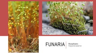
- 1. FUNARIA Bryophytes Prepared by Syeda Fari
- 2. SYSTEMATIC POSITION Division: Bryophta Class: Bryopsida Order: Funariales Family: Funariaceae Genus: Funaria
- 3. INTRODUCTION Common terrestrial moss Have dark green, velvety patches and grows in moist shady places Funaria hygrometrica is most common and worldwide species It colonize and grows best in presence of calcium, potassium , nitrogen, and phospohorus
- 4. VEGETATIVE MORPHOLOGY Plant body is gametophytic Differentiated into prostrate, green, filamentous structure Erect shoot arise from protonema called as gametophores( juvenile gametophyte) Protonema is short lived Each gametophore consists of stem and leaves. Sex organs are present on apices Rhizoids are present at the base of gametophore , absorbs nutrients
- 5. LEAVES Leaves are small, ovate, sessile and green Born on prostrate branches Leaves on lower branches are colorless and on upper branches are green and large in size These are called as foliage leaves and arranged in spiral fashion Leaves also surround sex organs, these are larger in size
- 6. REPRODUCTION Gametophyte reproduces by vegetative multiplication and sexual process Sporophyte reproduces by production of spore
- 7. VEGETATIVE REPRODUCTION By death of prostrate branches Development of gammae cups Protonemal growth. In which fragments are formed and each fragment grows into new protonema, buds and give rise to gametophores. It is done after the wounding of sporophyte Apospory ( buds develop on protonmea, and it is converted into diploid gametophore .
- 8. SEXUAL REPRODUCTION Funaria is monecious , Antheridia and archegonia are present on the apices of separate erect branches called gametophores. These organs are intermingled with the sterile hairs called paraphyses. These paraphyses contains chloroplast and apical cell is globose , meet with antheridia to cover it. Paraphyses also hold water by capillary , and helps in preventing desiccation .
- 9. MALE BRANCH Antheridia develop in group and present on the convex shaped apex of leafy branch gametophore called as male branch. The antheridia are intermingled with paraphyses Structure It is club shaped born on stalk. The main body contains a mass of spermatogenesis cells surrounded by layer of jacket cells. The free distal end of the antheridium is differentiated into cap like structure , the operculum . It helps in dehiscence .
- 10. FEMALE BRANCH The gametophore bearing archegonia is called as female branch.it arises from the base of male branch. The apex of the branch flattens into a receptacle on which archegonia develop in clusters in intermingled with paraphyses. Structure A mature archegonium is a flask shaped which is borne on short stalk. it has basal swollen part, the venter, and elongated neck. The venter is surrounded by a two layered jacket whereas the around the neck is single layer.
- 13. FERTILIZATION AND POST FERTILIZATION CHANGESDehiscence of antheridium is done by the rain or dew drops from the apical end of male branch. The jacket cells imbibe water and split up from the operculum forming a small pore. The male gametes move in a mass. At the same time the neck canal cell and venter canal cell disintegrate to form mucilage . It absorbs moisture , swell up and forces the disintegration of apical cell of neck. The male sperms are attracted chemotactically , and after this fertilization takes place. The zygote enlarges in size to fill up venter and secret a thick wall around it. After fertilization, the venter cells divide and form protective sheath called calyptra around the sporogonium
- 15. SPOROGONIUM The sporophyte in funaria is commonly called as sporogonium Spores are produced by meiosis
- 16. STRUCTURE OF SPOROGONIUM It is borne at the end of female branch It has following parts 1. Massive foot 2. Long seta 3. Pear shaped capsule
- 17. FOOT Foot is embedded in the apical tissue of female branch . It absorbs water and nutrients from the gametophyte
- 18. SETA The seta is long and it carries capsule at the apex. it consist of central conducting strand composed of thin walled cells surrounded by cortex and epidermis , covered by cuticle
- 19. CAPSULE Capsule is pear shaped, and highly organized spore producing structure At younger stage it is green, but as it matures it turns dark brown The apical part of capsule is covered by remains of ruptured calyptra The capsule has considerable tissue differentiation
- 22. INTERNAL STRUCTURE OF SPORGONIUM Foot : it is bulbus mass of tissues embedded in the apical tissue of female branch Seta : it consist of central conducting strand composed of thin walled cells surrounded by cortex and epidermis , covered by cuticle Capsule : capsule has three distinct regions. 1. Apophysis 2. theca 3. Operculum
- 23. APOPHYSIS Basal swollen, sterile region of capsule, The wall is made up of epidermis that contains stomata Below this, a photosynthetic spongy layer is present that has intercellular spaces. In the center a strand of thin walled, vertically, elongated cells are present that are conducting.
- 24. THECA Central part of capsule Sterile central part called as collumela is present. Collumela is surrounded by barrel shaped spore sac that contain spore mother cells Outer to these sac, there is a wide air space called as trabeculae that connects capsule wall to the wall of spore sac The wall of theca consist of 1. Epidermis 2. Hypodermis 3. Two celled thick photosynthetic spongy layer
- 25. OPERCULUM The conical, cap like terminal region of capsule It consist of 3-4 layered thin walled cells covered with epidermis Below the operculum is peristome ( ring of tooth like segments) it consist of 16 long doubled, incurved teeth and 16 thin walled inner segments . The peristome is attached to the ring of thin walled cells that form rim of capsule The peristome teeth are hygroscopic and respond to the slight change in moisture Annulus : the lower 2 layer of cells are thin walled and constitute annulus . The degeneration of annulus results in loosening and dropping off of operculum
- 26. PERISTOME