Weitere ähnliche Inhalte
Andere mochten auch
정품레비트라 구입방법 카톡:DDF11 & DDF11.KR 칵스타 판매,칵스타 구입,칵스타 파는곳,칵스타 팝니다,칵스타 가격,칵스타 종류,칵...정품레비트라 구입방법 카톡:DDF11 & DDF11.KR 칵스타 판매,칵스타 구입,칵스타 파는곳,칵스타 팝니다,칵스타 가격,칵스타 종류,칵...kim ming
Ähnlich wie NMT_Nature (20)
Mehr von Stephen Brand (6)
NMT_Nature
- 1. ARTICLES
N-myristoyltransferase inhibitors as new
leads to treat sleeping sickness
Julie A. Frearson1
, Stephen Brand1
, Stuart P. McElroy1
, Laura A. T. Cleghorn1
, Ondrej Smid1
, Laste Stojanovski1
,
Helen P. Price4
, M. Lucia S. Guther1
, Leah S. Torrie1
, David A. Robinson1
, Irene Hallyburton1
,
Chidochangu P. Mpamhanga1
, James A. Brannigan3
, Anthony J. Wilkinson3
, Michael Hodgkinson4
, Raymond Hui5
,
Wei Qiu5
, Olawale G. Raimi2
, Daan M. F. van Aalten2
, Ruth Brenk1
, Ian H. Gilbert1
, Kevin D. Read1
, Alan H. Fairlamb1
,
Michael A. J. Ferguson1
, Deborah F. Smith4
& Paul G. Wyatt1
African sleeping sickness or human African trypanosomiasis, caused by Trypanosoma brucei spp., is responsible for ,30,000
deaths each year. Available treatments for this disease are poor, with unacceptable efficacy and safety profiles, particularly
in the late stage of the disease when the parasite has infected the central nervous system. Here we report the validation of a
molecular target and the discovery of associated lead compounds with the potential to address this lack of suitable
treatments. Inhibition of this target—T. brucei N-myristoyltransferase—leads to rapid killing of trypanosomes both in vitro
and in vivo and cures trypanosomiasis in mice. These high-affinity inhibitors bind into the peptide substrate pocket of the
enzyme and inhibit protein N-myristoylation in trypanosomes. The compounds identified have promising pharmaceutical
properties and represent an opportunity to develop oral drugs to treat this devastating disease. Our studies validate T. brucei
N-myristoyltransferase as a promising therapeutic target for human African trypanosomiasis.
Protein N-myristoylation is a ubiquitous eukaryotic co- and post-
translational modification and is required for the membrane target-
ing and biological activity of many important proteins1,2
. The
N-myristoylation reaction—the transfer of C14:0 myristic acid from
myristoyl-coenzyme A (CoA) to the amino group of amino-terminal
glycine residues within specific sequence contexts3
—is catalysed by
the enzyme myristoyl-CoA-protein N-myristoyltransferase (NMT;
InternationalUnion ofBiochemistryand MolecularBiologyaccession
EC 2.3.1.97)4
. In T. brucei, NMT activity is encoded by a single gene,
which has been shown to be essential for parasite growth using
RNA interference5
. The effects of NMT knockdown on T. brucei are
probably complex as more than 60 proteins are predicted to be
N-myristoylated in this organism6
. Experimentally validated targets
for NMT include ADP ribosylation factors (ARF)7
, ADP ribosylation-
like factors (ARL)8
, a calpain-type protease (CAP5.5, systematic gene
nameTb927.4.3950)9
and,intherelatedLeishmaniamajorandT.cruzi
parasites, hydrophilic acylated surface proteins10
and flagellar
calcium-binding protein11
, respectively. The predicted pleiotropic
effects of NMT inhibition on trypanosome physiology make it an
attractive target for therapeutic intervention. NMT has also been con-
sidered as an anticancer12
, antifungal13
and antiviral14
target. Fungal
NMT orthologues have been shown to be druggable, although broad-
spectrum activity has not been achieved.Nevertheless,becausethere is
good evidence from these programmes that selectivity over human
NMT is possible, NMT has been proposed as a target for the treatment
of human African trypanosomiasis and other parasitic diseases15,16
.
There are two human isozymes sharing 77% identity (HsNMT1 and
HsNMT2)17
of which HsNMT2 is the closest human homologue to
T. brucei NMT (TbNMT), with overall 55% identity and 69% simi-
larity. On the basis of 31 residues that are within 5 A˚ of DDD85646 in
the active site, this rises to 83% identity and 90% similarity.
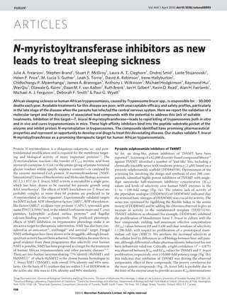
Pyrazole sulphonamide inhibitors of TbNMT
So far, no drug-like, potent inhibitors of TbNMT have been
reported18
. Screening of a 62,000 diversity-based compound library19
against TbNMT identified a number of ‘lead-like’ hits, including a
chemically tractable series with moderate potency (2 mM) based on a
pyrazole sulphonamide scaffold (DDD64558). Optimization of the
screening hit, involving the design and synthesis of over 200 com-
pounds, identified highly potent inhibitors of TbNMT with single-
digit nanomolar half-maximum inhibitory concentration (IC50)
values and levels of selectivity over human NMT enzymes in the
1- to .100-fold range (Fig. 1a). The relative lack of activity of
the piperidine analogue (DDD85635) of DDD85602 indicated that
the terminal basic nitrogen of DDD85602 was crucial for activity. The
series was optimized by rigidifying the flexible linker to the amine
moiety of DDD85602 and by adding the chlorines observed to give an
increase in activity in the unelaborated template (DDD73234).
TbNMT inhibitors so obtained (for example, DDD85646) inhibited
the proliferation of bloodstream form T. brucei in culture with the
best compounds yielding half-maximum effective concentration
(EC50) values between 0.8 and 3 nM and clear windows of selectivity
(.200-fold) with respect to proliferation of a prototypical mam-
malian cell type (MRC5). We attribute the increased selectivity at
the cellular level to differences in cell biology between host and para-
site, although differential cellular pharmacokinetic behaviour has not
been definitively ruled out. Critically, a tight correlation (r2
5 0.875)
was observed between IC50 and EC50 values for TbNMT and T. brucei
proliferation, respectively, over a 10,000-fold potency range (Fig. 1b);
this indicates that inhibition of TbNMT was driving the observed
antiparasitic effect of these compounds. The poorer correlation for
the most potent compounds (Fig. 1b, lower left) is probably due to
the limit of the enzyme assay to provide accurate IC50 determinations
1
Drug Discovery Unit, Division of Biological Chemistry and Drug Discovery, 2
Division of Molecular Microbiology, College of Life Sciences, University of Dundee, Dundee DD1 5EH, UK.
3
Structural Biology Laboratory, Department of Chemistry, 4
Centre for Immunology and Infection, Department of Biology and Hull York Medical School, University of York, Heslington,
York YO10 5YW, UK. 5
Structural Genomics Consortium, University of Toronto, MaRS South Tower, 7th Floor, 101 College Street, Toronto, Ontario M5G 1L7, Canada.
Vol 464|1 April 2010|doi:10.1038/nature08893
728
Macmillan Publishers Limited. All rights reserved©2010
- 2. for such highly active, tight-binding inhibitors (see Supplemen-
tary Fig. 1). As a result of its impressive potency in inhibiting
both TbNMT and T. brucei proliferation in vitro, together with its
promising physicochemical properties, DDD85646 was assessed for
efficacy in animal models of trypanosomiasis.
TbNMT inhibitor cures acute trypanosomiasis in vivo
DDD85646 is moderately bioavailable (approximately 20%) and
demonstrates good exposure after oral dosing at 10 and 50mgkg21
to female NMRI mice, with the free drug level above the effective
concentration calculated to achieve 99% inhibition of parasite growth
(EC99) for T. brucei brucei (T. b. brucei) proliferation for over 6 and
10 h, respectively (Fig. 2a). Furthermore, this compound cured all
animals in the T. b. brucei acute mouse model of human African tryp-
anosomiasis at a minimal oral dose of 12.5 mg kg21
(twice a day for 4
days) (Fig. 2b). Cure of all animals was also obtained with shorter oral
dosing schedules: 100 mgkg21
twice a day for 1 day and 25mgkg21
twice a day for 2 days. Notably, DDD85646 also cured all animals at
50 mgkg21
(twice a day for 2 days) in the more refractory but clinically
relevantT.b. rhodesiensemodelof human Africantrypanosomiasis (see
Supplementary Fig. 2). This reduced sensitivity in vivo is not due to
reduced sensitivity to the compound in vitro (T. b. rhodesiense, EC50
0.6nM), but may be a result of the known precedent for this species to
occupy privileged sites in vivo. Notably, the efficacy observed for
DDD85646 was comparable to the responses observed for the clinically
used drugs pentamidine and melarsoprol in the T. b. brucei model
(minimal full cure doses were 1 mgkg21
and 0.5 mgkg21
(intraperi-
toneal), respectively). Furthermore, despite the minimal window of in
vitro enzyme selectivity between TbNMT and mammalian (human)
NMT (Fig. 1a and see later), this compound was well tolerated at
efficacious doses.
TbNMT inhibitors are trypanocidal
Addition of DDD85646 resulted in rapid killing of trypanosomes
both in vivo and in vitro (Fig. 3a, b). Parasite counts dropped to below
r2
= 0.875
a b
NH
NN
O2
S
OMe
NH
NN
O2
S
Cl Cl
Br
NH
NN
O2
S
ClCl
NNH
NN
O2
S O2
S
N
NH
NH
NN
N
N
NH
N
N
NH
N
DDD64558
TbNMT
HsNMT
T. brucei EC50
MRC5 EC50
TbNMT
HsNMT
T. brucei EC50
MRC5 EC50
0.34 µM
(ND)
16 µM
>50 µM
TbNMT
HsNMT
T. brucei EC50
MRC5 EC50
2 nM
4 nM
2 nM
400 nM
TbNMT
HsNMT
T. brucei EC50
MRC5 EC50
0.14 µM
8 µM
1.6 µM
>50 µM
TbNMT
HsNMT
T. brucei EC50
MRC5 EC50
11 µM
87 µM
50 µM
>50 µM
1.9 µM
22 µM
21 µM
55 µM
DDD85602 DDD85635
DDD85646
DDD73234
In-house library
synthesis
(120 compounds)
Constrain
amine
30 follow-up
compounds
purchased 1,000
100
10
1
0.1
0.01
0.001
0.0001
T.bruceiEC50(µM)
0.001 0.01 0.1 1 10 100
TbNMT IC50 (µM)
Figure 1 | IdentificationofNMTleadseries inhibitors. a, Chemicalevolution
of DDD85646 from the initial high-throughput screening hit DDD64588.
Combining the structure–activity relationships from the two strategies led to
the development of the potent compound DDD85646. Potencies were
determined for all compounds synthesized against recombinant TbNMT and
HsNMT, as well as against bloodstream form T. brucei and MRC5
proliferation in vitro. ND, not determined. b, Correlation between the
inhibition of recombinant TbNMT and bloodstream form T. brucei
proliferation for 175 members of the pyrazole sulphonamide series. Data
shown are replicates of between 2 and 22 independent potency
determinations using 10-point curves. Robustness of TbNMT and
trypanosome proliferation assays are exemplified through routinely reported
parameters of Z9 (0.703 6 0.050, n 5 169 and 0.695 6 0.095, n . 1,000 for
TbNMT andtrypanosomeassays, respectively) andreproducible potenciesof
standards (DDD73498 (TbNMT assay) pIC50 5 6.52 6 0.14, n 5 276 and
pentamidine (trypanosome assay) pEC50 5 8.37 6 0.41, n 5 497), where p is
thenegativelogarithmoftheIC50 andEC50 values.r2
isthesquareofthelinear
regression correlation coefficient.
a
Total 50 mg kg–1
Free 50 mg kg–1
Total 10 mg kg–1
Free 10 mg kg–1
Time (min)
0 200 400 600
Bloodconcentration(ngml–1
)
1
10
100
1,000
10,000
EC99
EC50
Days after infection
10 20 30
Survival(%)
0
20
40
60
80
100 Control
50.0 mg kg–1
25.0 mg kg–1
12.5 mg kg–1
6.25 mg kg–1
3.13 mg kg–1
1.56 mg kg–1
b
Figure 2 | TbNMT inhibitor cures acute trypanosomiasis in vivo. a, Mean
total and free blood concentration time profiles after singleoral administration
of DDD85646 at 10 and 50 mg kg21
free-base to female NMRI mice (n 53 per
dosegroup). EC99 is calculatedfrom the averageEC50 of 2.4661.8 nMandHill
slope of 4.8460.6 (n 5 5). Solid lines are total plasma concentrations and
dashed lines are the predicted free plasma concentrations (fractionunbound in
plasma50.063).Dataaremean6s.d.b,Kaplan–Meiersurvivalplotforfemale
NMRI mice (n 55 per dose group) after infection with T. b. brucei s427 (BS
221) (inoculum 1 3 104
parasites). Oral treatment with DDD85646 started 3
days after infection at the indicated doses (all twice a day for 4 days).
NATURE|Vol 464|1 April 2010 ARTICLES
729
Macmillan Publishers Limited. All rights reserved©2010
- 3. detectable levels within 12 h of dosing mice at 50 mg kg21
twice a day.
Addition of DDD85646 (50 nM) to bloodstream form T. brucei
cultures in vitro also resulted in rapid killing with numbers of motile
cells reduced to below detectable levels between 24 and 48 h. The
apparent differences in the kinetics of death between the in vivo and
in vitro systems are probably a combination of the harsher in vivo
environment for drug-damaged trypanosomes and the fact that com-
pound exposure reached higher concentrations in vivo (up to
,1 mM), compared with 50 nM in vitro.
The trypanocidal mechanism of compound action was confirmed
by subjecting T. brucei treated in vitro with 50 nM DDD85646 to live/
dead fluorescence-activated cell sorting (FACS) analysis, which
showed .95% cell death within 24 h of treatment (see Supplemen-
tary Fig. 3). Furthermore, wash-out experiments showed that death
was irreversible after 48 h of exposure to 50 nM compound (data not
shown). Microscopic examination of the trypanosomes treated with
DDD85646 in vivo and in vitro showed the same abnormal morpho-
logy, that is, the development of a large vesicular structure (Fig. 3c). A
scanning electron micrograph of a treated trypanosome clearly shows
the rounded and swollen features of this phenotype compared to
control (Fig. 3d). Notably, rapid cell killing with a similar morpho-
logical phenotype has been previously observed after treatment with
myristate analogues, such as 10-(propoxy)decanoic acid20
. This mor-
phology closely resembles the ‘BigEye’ phenotype observed in blood-
stream form T. brucei when endocytosis is disrupted by the
knockdown of clathrin heavy chain, TbRAB521,22
or TbARF17
,
leading to expansion of the flagellar pocket. Parasites with enlarged
flagellar pockets are clearly visible in the DDD85646-treated tryp-
anosome population (Fig. 3e). Additional studies are required to
understand fully the cellular effects of NMT inhibition.
Inhibitor acts ‘on target’
Incubation of bloodstream form T. brucei with [3
H]myristic acid
results in the biosynthetic radiolabelling of myristoylated substrates,
particularly thehighlyabundant variant surface glycoprotein(VSG)23
.
Using this method, but including a detergent lysis step that activates
an endogenous phospholipase C (PLC) to release the [3
H]myristate
label from the glycosylphosphatidylinositol (GPI) anchor of VSG24
,
a number of putative N-[3
H]myristoylated proteins were visualized
by SDS–polyacrylamide gel electrophoresis (PAGE) and fluoro-
graphy (Fig. 4a, lane 2). The labelling of most of these proteins was
eliminated by prior treatment with DDD85646 (Fig. 4a, lane 1). To
confirm that most of the proteins labelled with [3
H]myristic acid
were indeed N-myristoylated, a duplicate gel was treated with 0.2 M
NaOH in methanol before fluorography to remove base-labile
hydroxy- or thioester linked [3
H]myristate (Fig. 4a, lanes 3 and 4).
ThreefaintDDD85646-insensitive bandswereremoved(Fig.4a,com-
pare lanes 1 and 2 with lanes 3 and 4); these probably include traces of
residual GPI-anchored VSG at 55 kilodalton (kDa)24
and thioester-
myristoylated GPI-PLC at 42 kDa25
. To assess whether DDD85646
specifically inhibited N-[3
H]myristoylation, the same cells were
labelled in parallel with [35
S]methionine. Pre-treatment of parasites
with DD85646 had no effect on [35
S]methionine incorporation into
proteins, showing that the compound has no effect on general protein
synthesis (Fig. 4a, lanes 5 and 6).
Further evidence that DDD85646 was acting on target in the tryp-
anosome was obtained by overexpressing TbNMT (fivefold) in a
tetracycline-inducible manner; this resulted in an eightfold reduction
in DDD85646 potency against these cells (Fig. 4b).
Another independent approach was taken using one of the few
known substrates of TbNMT in T. brucei, TbARF1. This protein
has a central role in endocytosis and Golgi–lysosome trafficking,
where tetracycline-induced expression of a constitutively active
GTP-locked mutant (Q71L) causes rapid cell death in blood-
stream form T. brucei in an N-myristoylation-dependent manner7
.
Short-term treatment (5 h) with 10 nM DDD85646 rescued these
TbARF1(Q71L)-expressing cells from death (Fig. 4c), presumably
by preventing N-myristoylation of newly produced TbARF1(Q71L)
protein. Although the Q71L mutant protein could not be detected by
western blotting, as previously reported 7
, the inducible expression of
the related, but myristoylation-blocked, G2A/Q71L mutant in the
presence of DDD85646 does show that its effect on cell survival
was not due to interference with tetracycline induction of these
mutants.
Taken together, these data provide strong evidence that DDD85646
acts to inhibit TbNMT in bloodstream form trypanosomes and that
this is directly linked to inhibition of proliferation. These data also
provide a potential link between inhibition of TbNMT and disruption
of the function of the TbNMT substrate TbARF1, known to operate in
protein trafficking and endocytic processes.
Inhibitor binds in TbNMT peptide pocket
The target product profile for a new drug to combat human African
trypanosomiasis, as defined by the Drugs for Neglected Diseases ini-
tiative, requires compounds that are safe and efficacious against both
the stage 1 disease, when parasites are present in the blood, lymph and
interstitial fluids, and the stage 2 disease, when parasites are also
present in the central nervous system (CNS). The DDD85646 com-
pound does not yet meet these criteria and will have to be optimized
for selectivity over human NMT and for diffusion into the CNS, while
retaining excellent potency against TbNMT and bloodstream form
T. brucei cells. To achieve these objectives, a detailed understanding
of the interaction between DDD85646 and TbNMT is required.
Characterization of the mode of inhibition of the early hits revealed
competition with the peptide substrate as a probable mode of inhibi-
tion for the series. Thus, a shift in IC50 from 1 to 4.3 mM for an early hit
was seen when the peptide substrate concentration in the assay was
increased from 0.5 to 16 mM (Fig. 5a). Surface plasmon resonance
(SPR) studies confirmed a 1:1 binding stoichiometry of DDD85646
with TbNMT (Fig. 5b) and showed that the binding affinity of the
compound was increased in the presence of myristoyl-CoA from
33 nM to 1 nM (data not shown). Accurate determination of binding
a b
c
In vivo
In vitro
– DDD85646 + DDD85646
d e
Time (h)
0 12 24 36
Trypanosomes(ml–1
)
Trypanosomes(ml–1
)
105
106
107
108
109
1010
109
108
107
106
105
103
102
104
Time (h)
0 24 48 72
Figure 3 | TbNMT inhibitors have rapid trypanocidal effects in vitro and in
vivo. a, Parasitaemia in mice (n 5 3 per group) with (red) or without (black)
DDD85646 treatment (50 mg kg–1
, oral, twice a day); for method see Fig. 2b.
Arrows represent dose administration times. Data are mean 6 s.d. b, T. b.
brucei proliferation in culture determined by counting motile parasites in
the presence (red) or absence (black) of 50 nM DDD85646. Data are
mean 6 s.d. for 3 determinations. c, Blood smears of infected mice and
culture samples were stained by Giemsa and observed by light microscopy.
Treated cells showed typical BigEye phenotype. d, Scanning electron
micrograph of T. b. brucei treated with 10 nM DDD85646 for 24 h. Inset
shows an untreated control cell. e, Transmission electron micrograph of
sagittal section of flagellar pocket of T. b. brucei treated with 5 nM
DDD85646 for 72 h. Inset shows a section of flagellar pocket of an untreated
control cell. Asterisks mark flagellar pockets. Dashed lines, cell detection
limits. Scale bars, 500 nm.
ARTICLES NATURE|Vol 464|1 April 2010
730
Macmillan Publishers Limited. All rights reserved©2010
- 4. affinity using SPR also showed a potential small window of selectivity
between TbNMT and human NMT with binding constants of 1 and
14 nM, respectively. Owing to tight binding of this compound (see
Supplementary Fig. 1), this selectivity was less clear in the biochemical
assay, which recorded 2 and 4 nM (IC50), respectively. Furthermore,
this emerging selectivity is clearly driven by differing off-rates, and
optimization around this parameter will not only be important in
improving selectivity, but also in sustaining a high residency time of
binding to TbNMT. The structure of TbNMT has yet to be solved.
However, using L. major NMT (LmNMT) as a model system, the
binding of DDD85646 in the peptide substrate binding site has been
confirmed by X-ray crystallography (Fig. 5c, for stereo view see
Supplementary Fig. 4). LmNMT has 74% overall sequence identity
with TbNMT and 94% identity within the peptide-binding site (see
Supplementary Fig. 5). The binding mode shows the piperazine inter-
acting with the carboxy-terminal carboxylate of NMT by means of a
tightly coordinated water molecule as opposed to a direct hydrogen
bond. The sulphonamide makes water-bridged interactions with the
highly conserved residue His 219 and with the backbone NH group of
Gly 397. The geometry of the sulphonamide creates a significant bend
in the structure, allowing the pyrazole to fit into a lipophilic pocket
where it acts as a hydrogen-bond acceptor from Ser 330. The binding
mode is of particular interest because all but the latter interactions are
through water-mediated hydrogen bonds; a mode not readily pre-
dicted using computational techniques. Overlaying the structure of
Saccharomyces cerevisiae NMT in complex with substrate peptide
(Protein Data Bank accession 1IID)26
with the LmNMT complex
shows that DDD85646 occupies thepeptide bindingsite withthebasic
piperazine moiety mimicking the N terminus of the substrate (see
Supplementary Fig. 6).
Conclusions
We have presented evidence that our model TbNMT inhibitor,
DDD85646, kills bloodstream form T. brucei by acting on TbNMT
in situ. There are probably several downstream consequences of
TbNMT inhibition in the parasite, as the enzyme has over 60 putative
Kd
(nM)
14.05
1.043.20 × 10–4
5.05 × 10⁻³
kon
(M–1 s–1
) koff
(s–1
)
3.09 × 10⁵
3.59 × 10⁵
100
75
25
0
–25
0.1 1 10 100
[DDD64558] (µM)
50
Inhibition(%)
a
c
b 70
50
40
30
20
10
0
–10
60
Response
–100 0 100 300 400200 500 600 700
HsNMT
TbNMT
TbNMT
Time (s)
IC50
1.0 µM
IC50
4.3 µM
Figure 5 | Characterization of pyrazole sulphonamide interactions with
NMT. a, DDD64558 potency against TbNMT (IC50) determined in the
presence of 0.5 mM (filled circles) and 16 mM (open circles) CAP5.5 peptide
substrate. Each data point represents mean 6 s.d. (n 5 4). b, Kinetics of
binding of DDD85646 to TbNMT and HsNMT1 determined by SPR. kon or
ka is the second-order rate constant for association and koff or kd is the first-
order rate constant for dissociation, where the dissociation constant Kd 5
koff/kon. c, X-ray crystal structure of DDD85646 bound to LmNMT. The left
panel shows the LmNMT binding site with protein backbone (pink ribbon),
solvent accessible surface (grey), DDD85646 (stick representation, carbon
atoms gold, nitrogen blue, chlorine green, oxygen red and sulphur yellow),
myristoyl CoA (carbon atoms cyan) and an omit map (Fo 2 Fc, 3.0 sigma)
around DDD85646 (blue). The right panel shows a stick representation of
DDD85646 and residues forming the active site (carbon atoms grey). Key
residues are highlighted (carbon atoms yellow) as are water molecules (red
spheres) and hydrogen bonds (dashed lines).
[3
H]Myristic acid [35
S]Methionine
+ NaOH– NaOH – NaOH
DDD85646 + + + –––
-
-
-
-
-
-
-
kDa
191
97
64
51
39
28
19
-Front
1 2 3 4 5 6
b
Myc
NMT
BiP
c
– DDD85646 + DDD85646
– Tet – Tet+ Tet + Tet
0
20
40
60
80
100
SM(wildtype)
SMARF1(Q71L)::Myc
SMARF1(G2A/Q71L)::Myc
SMARF1(G2A/Q71L)::Myc
Myc
α-tubulin
SM(wildtype)
SMNMT::Myc
SMNMT::Myc
Tet – +–
Tet + +– +
DDD85646 + –– +
[DDD85646] (nM)
1 10 100Inhibition(%)
Livecells(%)
0
20
40
60
80
100
EC50
2.1 nM
EC50
16.6 nM
a
Figure 4 | Pyrazole sulphonamide series acts ‘on target’ in the
trypanosome. a, Fluorographs of SDS–PAGE gels loaded with lysates of
bloodstream form T. b. brucei cells labelled with either [3
H]myristic acid
(lanes 1–4) or [35
S]methionine (lanes 5 and 6) after pre-incubation with (1)
or without (2) 0.5 mM DDD85646 for 6 h. Gels were incubated with or
without 0.2 M NaOH in MeOH, as indicated, before fluorography. b, Wild-
type (‘single marker’, SM) parasites and T. b. brucei overexpressing Myc-
tagged NMT were incubated with 0–100 nM DDD85646 for 64 h; motile cells
were counted using a haemocytometer. Filled circles, T. brucei
overexpressing NMT (n 5 3); open circles, wild-type cells (n 5 3). Levels of
Myc-tagged NMT expression were confirmed by western blotting. c, T. b.
brucei expressing ARF1(Q71L) (GTP-locked form of ARF1) under
tetracycline control were treated with 10 nM DDD85646 for 6 h. Cells were
then subjected to live/dead FACS analysis. Data shown represent
mean 6 s.d. from two independent experiments. Levels of Myc-tagged ARF1
mutant expression were analysed by western blotting. Tet, tetracycline. BiP,
binding protein, an endoplasmic reticulum chaperone.
NATURE|Vol 464|1 April 2010 ARTICLES
731
Macmillan Publishers Limited. All rights reserved©2010
- 5. substrates6
, and this no doubt explains the speed of killing and the
marked morphological changes observed on treatment with this com-
pound. The emergence of the BigEye phenotype, and the possible link
to TbARF1 activity, suggests that disturbance of endocytosis is one
mechanism by which TbNMT inhibitors act in bloodstream form
T. brucei. Interestingly, knockdown of TbNMT levels in bloodstream
formT.bruceiby RNAinterference to 16% of wild-type levels,although
fatal in vitro and in vivo, does not lead to the BigEye phenotype but
rather to the accumulation of vesicles close to the flagellar pocket27
.
It is notable that, despite the small window of selectivity between
human NMT and TbNMT, DDD85646 shows promising selectivity at
the cellular level. One might speculate that T. brucei cells are hyper-
sensitive to NMT inhibition because of unique or unusual aspects of
their biochemistry and/or cell biology. In this context, the extremely
high endocytic rate of bloodstream form T. brucei (some 9 times faster
than fibroblasts and 2.6 times faster than macrophages), combined
with the entire endocytic/exocytic process occurring in the flagellar
pocket,isnoteworthy28
. Theparasite’srequirementforthishighendo-
cytic rate relates to its need to remove antibody from the cell surface
and to recycle the protective VSG coat29
.
Finally, we may conclude that TbNMT is one of the few T. brucei
proteins that has been comprehensively validated as a drug target for
human African trypanosomiasis. The TbNMT inhibitors described
meet many requirements for a greatly needed new therapeutic agent
for human African trypanosomiasis. Further optimization of this
series towards improved CNS penetration and selectivity is currently
underway. In the meantime, DDD85646 will serve as an excellent
chemical tool for investigation of the biology of protein
N-myristoylation across a range of organisms.
METHODS SUMMARY
Enzyme activity assays were performed in scintillation proximity format18
, with
minor modifications. Proliferation assays were performed using resazurin as an
indicator of metabolic activity30
. TbNMT was immobilized onto NTA sensor
chips using standard immobilization protocols and kinetics of binding of com-
pound analysed on Biacore T-100. Details of all standard methods including
compound exposure studies and efficacy studies, metabolic labelling, micro-
scopy and crystallography can be found in Methods. Chemical synthesis details
can be found in Supplementary Materials.
Full Methods and any associated references are available in the online version of
the paper at www.nature.com/nature.
Received 30 September 2009; accepted 10 February 2010.
1. Farazi, T. A., Waksman, G. & Gordon, J. I. The biology and enzymology of protein
N-myristoylation. J. Biol. Chem. 276, 39501–39504 (2001).
2. Resh, M. D. Trafficking and signaling by fatty-acylated and prenylated proteins.
Nature Chem. Biol. 2, 584–590 (2006).
3. Maurer-Stroh, S., Eisenhaber, B. & Eisenhaber, F. N-terminal N-myristoylation of
proteins: prediction of substrate proteins from amino acid sequence. J. Mol. Biol.
317, 541–557 (2002).
4. Bhatnagar,R.S.,Futterer,K.,Waksman,G.&Gordon,J.I.Thestructureofmyristoyl-
CoA: protein N-myristoyltransferase. Biochim. Biophys. Acta 1441, 162–172 (1999).
5. Price, H. P. et al. Myristoyl-CoA: protein N-myristoyltransferase, an essential
enzyme and potential drug target in kinetoplastid parasites. J. Biol. Chem. 278,
7206–7214 (2003).
6. Mills, E., Price, H. P., Johner, A., Emerson, J. E. & Smith, D. F. Kinetoplastid PPEF
phosphatases: dual acylated proteins expressed in the endomembrane system of
Leishmania. Mol. Biochem. Parasitol. 152, 22–34 (2007).
7. Price, H. P., Stark, M. & Smith, D. F. Trypanosoma brucei ARF1 plays a central role in
endocytosis and Golgi-lysosome trafficking. Mol. Biol. Cell 18, 864–873 (2007).
8. Price, H. P., Panethymitaki, C., Goulding, D. & Smith, D. F. Functional analysis of
TbARL1, an N-myristoylated Golgi protein essential for viability in bloodstream
trypanosomes. J. Cell Sci. 118, 831–841 (2005).
9. Hertz-Fowler, C., Ersfeld, K. & Gull, K. CAP5.5, a life-cycle-regulated,
cytoskeleton-associated protein is a member of a novel family of calpain-related
proteins in Trypanosoma brucei. Mol. Biochem. Parasitol. 116, 25–34 (2001).
10. Denny, P. W., Gokool, S., Russell, D. G., Field, M. C. & Smith, D. F. Acylation-
dependent protein export in Leishmania. J. Biol. Chem. 275, 11017–11025 (2000).
11. Wingard, J. N. et al. Structural insights into membrane targeting by the flagellar
calcium-binding protein (FCaBP), a myristoylated and palmitoylated calcium
sensor in Trypanosoma cruzi. J. Biol. Chem. 283, 23388–23396 (2008).
12. Selvakumar, P. et al. Potential role of N-myristoyltransferase in cancer. Prog. Lipid
Res. 46, 1–36 (2007).
13. Georgopapadakou, N. H. Antifungals targeted to protein modification: focus on
protein N-myristoyltransferase. Expert Opin. Investig. Drugs 11, 1117–1125 (2002).
14. Hill, B. T. & Skowronski, J. Human N-myristoyltransferases form stable complexes
with lentiviral Nef and other viral and cellular substrate proteins. J. Virol. 79,
1133–1141 (2005).
15. Bowyer, P. W. et al. N-myristoyltransferase: a prospective drug target for
protozoan parasites. ChemMedChem 3, 402–408 (2008).
16. Sheng, C. et al. Homology modeling and molecular dynamics simulation of
N-myristoyltransferase from protozoan parasites: active site characterization and
insightsintorationalinhibitordesign.J.Comput.AidedMol.Des.23,375–389(2009).
17. Giang, D. G. & Cravatt, B. F. A second mammalian N-myristoyltransferase. J. Biol.
Chem. 273, 6595–6598 (1998).
18. Panethymitaki, C. et al. Characterization and selective inhibition of myristoyl-
CoA: protein N-myristoyltransferase from Trypanosoma brucei and Leishmania
major. Biochem. J. 396, 277–285 (2006).
19. Brenk, R. et al. Lessons learnt from assembling screening libraries for drug
discovery for neglected diseases. ChemMedChem 3, 435–444 (2008).
20. Doering, T. L. et al. An analog of myristic acid with selective toxicity for African
trypanosomes. Science 252, 1851–1854 (1991).
21. Allen, C. L., Goulding, D. & Field, M. C. Clathrin-mediated endocytosis is essential
in Trypanosoma brucei. EMBO J. 22, 4991–5002 (2003).
22. Hall, B., Allen, C. L., Goulding, D. & Field, M. C. Both of the Rab5 subfamily small
GTPases of Trypanosoma brucei are essential and required for endocytosis. Mol.
Biochem. Parasitol. 138, 67–77 (2004).
23. Ferguson, M. A. J. & Cross, G. A. M. Myristylation of the membrane form of a
Trypanosomabruceivariantsurfaceglycoprotein.J.Biol.Chem.259,3011–3015(1984).
24. Ferguson, M. A. J., Low, M. G. & Cross, G. A. M. Glycosyl-sn-1,2-
dimyristylphosphatidylinositol is covalently linked to Trypanosoma brucei variant
surface glycoprotein. J. Biol. Chem. 260, 4547–4555 (1985).
25. Armah, D. A. & Mensa-Wilmot, K. S-myristoylation of a
glycosylphosphatidylinositol-specific phospholipase C in Trypanosoma brucei. J.
Biol. Chem. 274, 5931–5938 (1999).
26. Farazi, T. A., Waksman, G. & Gordon, J. I. Structures of Saccharomyces cerevisiae
N-myristoyltransferase with bound myristoylCoA and peptide provide insights
about substrate recognition and catalysis. Biochemistry 40, 6335–6343 (2001).
27. Price, H. P., Guther, M. L., Ferguson, M. A. & Smith, D. F. Myristoyl-CoA:protein
N-myristoyltransferase depletion in trypanosomes causes avirulence and
endocytic defects. Mol. Biochem. Parasitol. 169, 55–58 (2010).
28. Overath, P. & Engstler, M. Endocytosis, membrane recycling and sorting of GPI-
anchored proteins: Trypanosoma brucei as a model system. Mol. Microbiol. 53,
735–744 (2004).
29. Engstler, M. et al. Hydrodynamic flow-mediated protein sorting on the cell surface
of trypanosomes. Cell 131, 505–515 (2007).
30. Patterson, S. et al. Synthesis and evaluation of 1-(1-(benzo[b]thiophen-2-
yl)cyclohexyl)piperidine (BTCP) analogues as inhibitors of trypanothione
reductase. ChemMedChem 4, 1341–1353 (2009).
Supplementary Information is linked to the online version of the paper at
www.nature.com/nature.
Acknowledgements This work was supported by grants from the Wellcome Trust
(WT077705, WT083481, WT077503 and WT085622), Scottish Funding Council
(HR04013) and by the Translational Biology Theme of SULSA. We thank the
European Regional Development Fund and the Wolfson Foundation for grants that
provided relevant infrastructure for this work. The Structural Genomics
Consortium is a registered charity (number 1097737) that receives funds from the
Canadian Institutes for Health Research, the Canadian Foundation for Innovation
and Genome Canada through the Ontario Genomics Institute, GlaxoSmithKline,
Karolinska Institutet, the Knut and Alice Wallenberg Foundation, the Ontario
Innovation Trust, the Ontario Ministry for Research and Innovation, Merck, the
Novartis Research Foundation, the Swedish Agency for Innovation Systems, the
Swedish Foundation for Strategic Research and the Wellcome Trust. We would like
to thank all members of the Drug Discovery Unit for their technical assistance in
this study, particularly B. Rao, I. Collie and D. James.
Author Contributions The project management team responsible for experimental
design and coordination of research activities comprised S.B., R.B., A.H.F., M.A.J.F.,
J.A.F., I.H.G., K.D.R., D.M.F.vA., P.G.W. and D.F.S. J.A.B., M.H. and A.J.W. optimized
expressionandproducedtheactiveTbNMTusedforscreeningintheDrugDiscovery
Unit. Biological studies were carried out by S.P.M., O.S., L.S.T., M.L.S.G., I.H. and
H.P.P.; chemical syntheses by L.A.T.C. and S.B.; structural biology and modelling by
D.A.R., O.G.R., C.P.M., R.H. and W.Q.; and pharmacological studies by L.S.
Author Information Atomic coordinates and structure factors for the crystal
structures have been deposited with the Protein Data Bank under accession codes
3H5Z and 2WSA for LmNMT with bound myristoyl CoA and with bound
DDD85646, respectively. Reprints and permissions information is available at
www.nature.com/reprints. The authors declare competing financial interests:
details accompany the full-text HTML version of the paper at www.nature.com/
nature. Correspondence and requests for materials should be addressed to P.G.W.
(p.g.wyatt@dundee.ac.uk).
ARTICLES NATURE|Vol 464|1 April 2010
732
Macmillan Publishers Limited. All rights reserved©2010
- 6. METHODS
Determination of exposure in rat after acute oral dosing. Female NMRI mice
(25–35 g; Harlan Laboratories; n 5 3 per dose group) were administered
DDD85646 at 10 and 50 mg kg21
orally. The dose solutions were prepared on
the day of dosing and the vehicle was 5% dimethylsulphoxide (DMSO) in deio-
nized water, or 5% DMSO and 40% PEG400 in deionized water, respectively.
Blood samples (10 ml) were collected from the tail vein of each animal into
micronic tubes (Micronic BV) containing deionized water (20 ml) at 0.25, 0.5,
1, 2, 4, 6 and 8 h post-dose and stored at 280 uC to await analysis.
Sample extraction of blood was performed by a method based on protein
precipitation using acetonitrile and a structural analogue of the analyte of interest
as internal standard. Blood extracts were analysed by UPLC/MS/MS using a
Quattro Premier XE mass spectrometer (Waters). Calibration curves were con-
structed in blood to cover at least three orders of magnitude (that is,
1–1,000 ngml21
).
Efficacy studies. DDD85646 was tested in vivo against T. b. brucei using a modi-
fication of theapproach described31
. In brief, female NMRI mice (25–35 g; Harlan
Laboratories; 3–5 per group) were injected intraperitoneally with 1 3 104
blood-
stream forms of T. b. brucei strain s427 (also known as BS 221 or MITaT 1.2).
These bloodstream forms come from a stock of cryopreserved stabilates contain-
ing 10% glycerol. The stabilate was suspended in phosphate-saline-glucose to
obtain a trypanosome concentration of 5 3 104
ml21
. Each mouse was injected
with 0.2 ml. DDD85646 was administered orally from day 3 to day 6 of the
experiment and parasitaemia levels monitored up to day 30. Animals with para-
sitaemia .108
ml21
were humanely killed as previous studies had shown that
animals do not survive a further 24 h32
. The day on which mice were killed was
recorded.
NMT enzyme assay. NMT assays18,33
were carried out at room temperature
(22–23 uC) in 384-well white optiplates (Perkin Elmer). Each assay was per-
formed in a 40 ml reaction volume containing 30 mM Tris buffer, pH 7.4,
0.5 mM EDTA, 0.5 mM EGTA, 1.25 mM dithiothreitol (DTT), 0.1% (v/v)
Triton X-100, 0.125 mM [3
H]myristoyl-coA (8 Curie (Ci) mmol21
), 0.5 mM bio-
tinylated CAP5.5, 5 nM NMT and various concentrations of the test compound.
The IC50 values for HsNMT1 and HsNMT2 were essentially identical against 80
compounds tested and, for logistical reasons, only HsNMT1 was used in later
studies.
Test compound (0.4 ml in DMSO) was transferred to all assay plates using a
Cartesian Hummingbird (Genomics Solution) before 20 ml of enzyme was added
to assay plates. The reaction was initiated with 20 ml of a substrate mix and
stopped after 15 min (HsNMT1 or HsNMT2) or 50 min (TbNMT) with 40 ml
of a stop solution containing 0.2 M phosphoric acid, pH 4.0 and 1.5 M MgCl2
and 1 mg ml21
PVT SPA beads (GE Healthcare). All reaction mix additions were
carried out using a Thermo Scientific WellMate (Matrix). Plates were sealed and
read on a TopCount NXT Microplate Scintillation and Luminescence Counter
(Perkin Elmer).
ActivityBase from IDBS was used for data processing and analysis. All IC50
curve fitting was undertaken using XLFit version 4.2 from IDBS. A four-
parameter logistic dose response curve was used using XLFit 4.2 Model 205.
All test compound curves had floating top and bottom and pre-fit was used
for all four parameters.
Compound efficacy and trypanocidal activity in cultured T. brucei parasites.
Bloodstream T. b. brucei s427 were cultured at 37 uC in modified HMI9 medium
(56 mM 1-thioglycerol was substituted for 200 mM 2-mercaptoethanol) and
quantified using a haemocytometer. For the live/dead assay, cells were analysed
using a two-colour cell viability assay (Invitrogen) as described previously7
.
Routine screening of test compounds against parasites was performed in 96-well
plates using a modification30
of the cell viability assay previously described34
. Cell
culture plates were stamped with 1 ml of an appropriate concentration of test
compound in DMSO followed by the addition of 200 ml trypanosome culture
(104
cells ml21
) to each well, except for one column which received media only.
MRC5 cells were cultured in DMEM, seeded at 2,000 cells per well and allowed to
adhere overnight. One-microlitre of test compound (10 point dilutions from
50 mM to 2 nM) was added to each well at the start of the assay. Culture plates of
T. brucei and MRC5 cells were incubated at 37 uC in an atmosphere of 5% CO2
for 69 h, before the addition of 20 ml resazurin (final concentration, 50 mM). After
a further 4 h incubation, fluorescence was measured (excitation 528 nm; emis-
sion 590 nm) using a BioTek flx800 plate reader.
SPR analysis. The interaction between NMT and DDD85646 was assessed using a
Biacore T100 instrument. Histidine-tagged TbNMT and HsNMT1 were immo-
bilized onto an NTA sensor chip (GE Healthcare) to levels of ,6,000 response
units (RU) using standard immobilization protocols (GE Healthcare).
Experiments were carried out at 25 uC using HBS-P1 running buffer (GE
Healthcare) containing 100nM myristoyl-CoA. DDD85646 binding was tested
by injecting varying concentrations of inhibitor (0.05 nM to 1,000nM) at a flow
rate of 30 ml min21
. Different concentrations of DDD85646 were prepared so that
the final DMSO concentration was 0.1%. Kinetic data analysis was performed
using Biacore T100 Evaluation Software 2.0.1 and fitted to the standard 1:1
interaction model to give results.
Overexpression of NMT and mutant ARF1 studies. The plasmid vector pM2cN
was a gift from D. Horn and S. Alsford. The T. brucei NMT open reading frame
was amplified from genomic DNA using suitable primers and cloned into the
plasmid vector pM2cN. The resulting construct encodes the target protein with
an N-terminal Myc epitope tag under the control of a tetracycline-inducible T7
promoter. Mid-log phase T. brucei BSF ‘single marker’ (T7RPOL TETR NEO)
s427 (subsequently termed SM) were electroporated with 10 mg of NotI-digested
pM2cN-TbNMT using methods described8
. Expressionof Myc-tagged NMT was
induced in stable cell lines by incubating parasites in 1 mg ml21
tetracycline for
24 h. Immunoblotting8
of parasite lysates was performed using the following
primary antibodies: mouse anti-Myc (Invitrogen, 1:1,000 dilution), rabbit
anti-TbNMT (1:500 dilution) and rabbit anti-BiP (gift from J. Bangs, used at
1:10,000 dilution), and mouse anti-a-tubulin, TAT1 (gift from K. Gull). Parental
and NMT overexpressing cell lines (induced with tetracycline for 24 h) were
seeded in 24-well plates at 5 3 105
ml21
in triplicate, containing 0–100 nM of
DDD85646. Motile cells were counted using a haemocytometer.
Production and transfection of the constructs pM2-TbARF1(Q71L) and
pM2-TbARF1(G2A/Q71L) have been described previously7
. Parental and
mutant cell lines were seeded in 10 ml cultures at 5 3 105
ml21
. DDD85646
(10 nM) with or without tetracycline (1 mg ml21
) was added to cultures and
flasks incubated at 37 uC with 5% CO2 for 6 h. A set of cultures was set up in
which tetracycline was added for 1 h before addition of DDD85646 and incu-
bated for a further 5 h. Treated cells were then analysed using a Live/Dead two
colour cell viability assay as described previously7
and immunoblotted using
anti-Myc antibody8
.
Transmission electron microscopy. T. brucei cells were collected by centrifu-
gation at 1,000g for 10 min and fixed in 1 pellet-volume of 4% paraformaldehyde
in 0.1 M piperazine-N,N9-bis(2-ethanesulphonic acid) for 30 min at room tem-
perature (22–23 uC). Fixed cells were spun at 13,000g for 10 min, the supernatant
was removed, and the pellet was resuspended in 1% aqueous osmium tetroxide
(to fix and stain lipids), dehydrated, and set in Durcupan epoxy resin (Sigma).
Sections were cut using a Leica Ultracut UCT system and analysed using a Philips
Tecnai 12 transmission electron microscopy instrument.
Metabolic labelling. Live bloodstream form T. brucei (107
) were incubated with
and without 0.5 mM DDU85646 compound in 10 ml HMI9 for 6 h at 37 uC in a
5% CO2 incubator and subsequently labelled with 50 mCi ml21
[3
H]myristic acid
or 20 mCi ml21
[35
S]methionine for 1 h at 37 uC. The cells were washed in tryp-
anosome dilution buffer and lysed in 1% Triton X-100 in 10 mM sodium phos-
phate buffer, pH 8 and incubated for 10 min at 37 uC. Duplicate aliquots of
lysates were run in 4–12% Nupage gels, stained with Coomassie blue and sub-
sequently treated, or not, with 0.2 M NaOH in MeOH for 1 h at room temper-
ature (22–23 uC). The gels were washed and soaked in En3
Hance, dried and
exposed to film.
Crystallography. LmNMT protein was obtained from the Structural Genomics
Consortium parasitic disease project (courtesy of R. Hui). The protein construct
consisted of 6xHis, TEV cleavage site and LmNMT 5–421, which was prepared in
10 mM HEPES, pH 7.5, 500 mM NaCl at 9 mg ml21
for crystallization. A
200 mM stock solution of DDD85646 was prepared in DMSO and added to
the protein solution to a final concentration of 10 mM. Myristoyl-CoA was also
added to the sample to a final concentration of 1 mM before crystallization.
Crystallization was carried out using vapour diffusion methods with 2 ml of
protein–ligand complex mixed with 2 ml of reservoir solution. The reservoir
solution contained 26% PEG 1500, 0.2 M NaCl, 0.1 M Na-cacodylate, pH 5.6.
X-ray diffraction data for LmNMT 1 DDD85646 were measured at the
European Synchrotron Facility at beamline ID14-1. The data were integrated
and scaled using the HKL suite35
. The structure was solved by molecular replace-
ment as implemented in MOLREP36
using the native LmNMT coordinates (PDB
accession 3H5Z) as a search model. The structure was refined using REFMAC537
and manual alteration of the model carried out using COOT38
. Ligand coordi-
nates and topology files were created using the PRODRG server39
. Data collec-
tion and refinement statistics are presented in Supplementary Table 1.
31. Thuita, J. K. et al. Efficacy of the diamidine DB75 and its prodrug DB289, against
murine models of human African trypanosomiasis. Acta Trop. 108, 6–10 (2008).
32. Sienkiewicz, N., Jaroslawski, S., Wyllie, S. & Fairlamb, A. H. Chemical and genetic
validation of dihydrofolate reductase-thymidylate synthase as a drug target in
African trypanosomes. Mol. Microbiol. 69, 520–533 (2008).
33. Bowyer, P. W. et al. Molecules incorporating a benzothiazole core scaffold inhibit
the N-myristoyltransferase of Plasmodium falciparum. Biochem. J. 408, 173–180
(2007).
doi:10.1038/nature08893
Macmillan Publishers Limited. All rights reserved©2010
- 7. 34. Ra¨z, B., Iten, M., Grether-Bu¨hler, M., Kaminsky, R. & Brun, R. The Alamar BlueHassay to determine drug sensitivity of African trypanosomes (T.b.rhodesiense and
T.b.gambiense) in vitro. Acta Trop. 68, 139–147 (1997).
35. Otwinowski, Z. & Minor, W. Processing of X-ray diffraction data collected in
oscillation mode. Methods Enzymol. 276, 307–326 (1997).
36. Vagin, A. & Teplyakov, A. MOLREP: an automated program for molecular
replacement. J. Appl. Cryst. 30, 1022–1025 (1997).
37. Murshudov, G. N., Vagin, A. A. & Dodson, E. J. Refinement of macromolecular
structures by the maximum-likelihood method. Acta Crystallogr. D 53, 240–255
(1997).
38. Emsley, P. & Cowtan, K. Coot: model-building tools for molecular graphics. Acta
Crystallogr. D 60, 2126–2132 (2004).
39. Schu¨ttelkopf,A.W.& van Aalten, D.M.F.PRODRG:atoolforhigh-throughputcrys-
tallography ofprotein-ligand complexes. Acta Crystallogr. D 60, 1355–1363(2004).
doi:10.1038/nature08893
Macmillan Publishers Limited. All rights reserved©2010