
Empfohlen
Weitere ähnliche Inhalte
Was ist angesagt?
Was ist angesagt? (20)
Ähnlich wie Hand-spaces and arterial arches.pptx
Ähnlich wie Hand-spaces and arterial arches.pptx (20)
Kürzlich hochgeladen
Kürzlich hochgeladen (20)
Hand-spaces and arterial arches.pptx
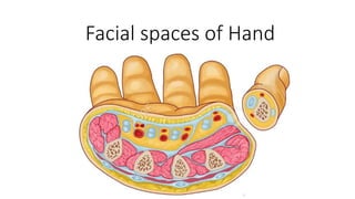
- 1. Facial spaces of Hand
- 2. Spaces Of Hand • Palmar spaces • Thenar space • Midpalmar space • Web space • Pulp space of fingers
- 6. Thenar space • Boundaries Anterior 1. Palmar aponeurosis 2. Flexor tendon of index finger 3. Short muscles of thumb 4. Lumbricals (1 & 2) Posterior 1. Adductor pollicis (trans) Radial Lateral palmar septum Medial Intermediate palmar septum
- 7. Thenar space • Proximally • Distal margin of flexor retinaculum • Distally • Distal transverse palmar crease • Communication • With I web space • Through lumbrical canal
- 8. Midpalmar space • Boundaries Anterior 1. Palmar aponeurosis 2. Flexor tendons of medial 3 fingers 3. Lumbricals (3 & 4) Posterior 1. Fascia over the interossei of 3rd & 4th space Radial Intermediate palmar septum Medial Medial palmar septum
- 9. Midpalmar space • Proximally • Distal margin of flexor retinaculum • Distally • Distal palmar crease • Communication • From distal end • With 2nd, 3rd & 4th lumbrical canals • Lumbrical canal • Space surrounds the tendon of lumbrical
- 10. Web space • 4 subcutaneous spaces • Extent • From free margin of web • To the metacarpophalangeal joint • Content • Fat • Superficial transverse palmar ligament • Tendons of • Interossei & lumbicals • Digital nerves & vessels
- 11. Fibrous flexor sheath • Fibrous sheath covering the digits • On anterior surface of the fingers and attached to the sides of the phalanges • Forms an Osteofibrous blind Tunnel • To retain the tendons in position • Covering the long flexor tendons • Thumb • Flexor pollicis longus • Medial 4 fingers • Flexor digitorum profundus • Flexor digitorum superficialis
- 12. Fibrous flexor sheath - Extent • From • Head of Metacarpal • Proximal end is opened • To • Base of Distal phalanx • Distal end is closed
- 13. Fibrous flexor sheath • 2 types of arrangement • Annular (5) • Joints (3) • Middle of proximal & middle phalanges (2) • Cruciate (4) • Between the annular arrangement • Thick • Opposite to phalanges • Thin • Opposite to joints • To allow free movement
- 14. Pulp space • Subcutaneous space • Between • Distal phalanx • & • Skin • Proximal • Fusion of fibrous flexor sheath • Space is divided into many compartments by fibrous septa
- 15. Applied anatomy • Infection of this region • Known as whitlow or felon • Distension of any compartment by pus • Presses on nerve endings • Necrosis of distal 4/5th of distal phalanx may happen • If whitlow is not treated • Why • Blood supply is from the branches arising within this space • Proximal 1/5th part of distal phalanx is getting supply from the branch which does not traverse the pulp space
- 16. Dorsal subcutaneous space • Lies immediately below the skin in the dorsum of the hand
- 17. Arterial supply to hand • Hand has rich arterial supply • Many anastomoses • Allows the hand get blood supply • Grasping or applying pressure • Supplied by • Ulnar and radial arteries
- 18. Palmar arches • Superficial palmar arch • Above long flexor tendons • Deep palmar arch • Below long flexor tendons
- 19. Superficial palmar arch - location • Lies beneath • Palmar aponeurosis • Superficial to • Long flexor tendons • Lumbrical • Palmar digital branches of median nerve
- 20. Superficial palmar arch - formation • Ulnar artery enters palm • Above the flexor retinaculum • Lateral to pisiform • Beneath palmaris brevis • Here it divides into • Superficial • Deep • Superficial branch turns laterally
- 21. • Ulnar artery is main contribution • Superficial branch of ulnar artery • Continues as superficial palmar arch • Mode of completion • By one of the following branches of radial artery • Superficial palmar branch • Arteria princeps pollicis • Arteria radialis indicis Superficial palmar arch - formation
- 22. Superficial palmar arch - branches • 1 proper palmar digital branch • Supplies ulnar side of little finger • 3 common digital branches • Reaches the web between medial 4 fingers • Each artery divides into • 2 proper palmar digital arteries • To supply adjacent fingers • Superficial palmar arch supplies • Medial 31/2 fingers
- 23. • At web • Common palmar digital • Branch from superficial palmar arch • Palmar metacarpal • Branch from deep palmar arch
- 24. Relations • Superficially • Palmaris brevis & palmar aponeurosis • Deep • Flexor digitorum tendons • Flexor digiti minimi • Lumbricals • & • Median nerve branches
- 25. Radial artery • Proximal to flexor retinaculum • Superficial palmar branch • Passes into palm by • Over the thenar muscles • or • Piercing through thenar muscles • Usually superficial palmar branch will anastomose • To form superficial palmar arch
- 26. Radial artery • At wrist • Lies beneath tendons of • Abductor pollicis longus • Extensor pollicis brevis • Crosses anatomical snuff box • Enters the palm • In between the 2 heads of dorsal interossei • In the palm • Passes in between oblique and transverse heads of adductor pollicis
- 27. Radial artery • In the palm • Between the 2 heads of adductor longus it gives • Arteria princeps pollicis • Passes beneath oblique head of adducor longus along I metacarpal • At proximal phalanx divides into 2 branches • Supplies both sides of thumb • Arteria radialis indicis • Passes in between • Transverse head of adductor longus and first dorsal interosseous • Supplies • Radial side of index finger
- 28. Deep palmar arch • Continuation of the radial artery • Passes medially • Lies deep to • Oblique head of adductor pollicis • Long flexor tendons • Lumbricals • Passes transversely on • Bases of metacarpals • Interossei • Completed on the medial side by • Deep branch of ulnar artery • Surface marking • At the level of proximal border of outstretched thumb • 1 cm proximal to superficial palmar arch
- 29. Deep palmar arch • Formed by continuation of radial artery and completed by deep branch of ulnar artery • Branches • 3 palmar metacarpal arteries • 3 perforating • With dorsal metacarpal arteries • Recurrent branches • Anastomose with anterior carpal arch
- 30. Dorsal metacarpal arteries • 1st dorsal metacarpal • From radial • 2nd , 3rd, & 4th dorsal metacarpal • From dorsal carpal arch
- 31. Allen's test • To test for adequate anastomoses between the radial and ulnar arteries • Compress both the radial and ulnar arteries at the wrist • Then release pressure from one or the other • and • Determine the filling pattern of the hand
- 32. How to control bleeding in hand • Compression of brachial artery • Against humerus • Why not at wrist by arresting ulnar & radial arteries • There is communication between palmar & dorsal carpal arches with arterial arches of palm