2. 32 Chapter 4. How to Conduct Urodynamic Studies
do help to dissect out the relative severity of the different
components in patients with mixed incontinence and thus
guide you as to the main thrust of treatment.This is described
in the case history at the end of this chapter.
In general, urodynamics are very worthwhile in the follow-
ing cases (in descending order):
Patients with failed continence surgery need detailed uro-
dynamic studies.
Patients with symptoms or a past history of voiding difficulty
(previous prolonged catheter or self-catheterization post-
op or postpartum) need voiding cystometry.
Patients with mixed symptoms and cystocele who are con-
sidering surgery should have detailed urodynamics,
possibly with ring pessary in situ (see “Occult” Stress
Incontinence).
Patients with mixed stress and urge leak need cystometry
at least, to determine the relative severity of the two
problems.
Patients with pure stress incontinence symptoms who have
failed physiotherapy should have cystometry with some
form of imaging, to check whether there is undiagnosed
detrusor overactivity or incomplete emptying.
Patients with pure urge symptoms who have failed bladder
training and anticholinergic therapy should also have
cystometry with imaging, to look for an undiagnosed
stress incontinence component or incomplete emptying
(the latter may be worsened by the anticholinergic
drugs).
Different Forms of Urodynamic Studies
The term “urodynamics” is a general phrase, used to describe
a group of tests that assess the filling and voiding phase of the
micturition reflex, to determine specific abnormalities.
Some of these tests are not “physiological.” For example,
inserting catheters into the urethra and a pressure balloon
into the rectum, then expecting the patient to fill and empty
3. 33Different Forms of Urodynamic Studies
as she normally does, may not give a “true” picture of that
woman’s micturition cycle. Nevertheless, the tests have been
standardized over the last 40 years, in accordance with the
Standardization Committee of the International Conti-
nence Society (ICS), and are performed in a similar fashion
across the world. Therefore, abnormalities are interpreted
in a standard way and have a common meaning in clinical
practice.
The tests that are generally used include the following:
Uroflowmetry: Measuring the patient’s flow rate when
voiding in private, onto a commode that is connected to a
collecting device that measures the rate of fall of urine upon
the device.
Simple cystometry: Inserting a single catheter into the
bladder that measures pressure, with no correction for
abdominal pressure, during a filling cycle, not widely used in
the Western world.
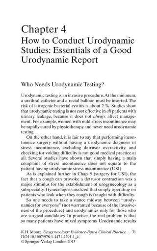
Twin channel subtracted cystometry: Inserting a pressure
recording line into the bladder as well as a filling catheter,
along with an abdominal pressure recording line (rectal bal-
loon), that records a filling cycle. The abdominal pressure is
subtracted from the bladder pressure to give the detrusor
pressure (see Fig. 4.1 and later figures).
Voiding cystometry: The same as twin channel cystometry
above, but the patient is asked to void into a uroflow com-
mode while the pressure lines are in situ, so that the contrac-
tility of the detrusor muscle during the voiding phase is
measured.
Videourodynamics: The same as voiding cystometry above,
but radiopaque X-ray contrast dye is used to fill the bladder.
The test is done in the X-ray department, and the bladder/
urethra is filmed during cough and other provocation. In
males, filming is continued during the voiding phase, but
60 % of women are not able to void in these public condi-
tions. Post-void films are taken to check residual.
Voiding cystometry with ultrasound: The same as voiding
cystometry, but ultrasound imaging is undertaken during
cough and other provocation, and post-void image is taken.
4. 34 Chapter 4. How to Conduct Urodynamic Studies
Urethral pressure profile: Tests the function of the external
urethral sphincter, performed in selected cases. Similar infor-
mation is available from leak point pressure testing.
The frequency volume chart and the pad test are also part
of urodynamic assessment, but these are discussed in Chap. 5
(Outcome Measures).
Practical Advice About How to Perform
Urodynamic Studies
This section gives practical advice for a registrar or resident/
house officer who is newly attached to a urogynecology
department. For information about the medical physics of the
tests, books by Abrams [1] or Cardozo and Staskin [4] are
recommended.
FILLING
warm
sterile
water
Subtraction
Water Inflow Catheter
Bladder
Pressure Catheter
Transducer
Transducer
Pves
Pabdo
Rectal Pressure Catheter
Figure 4.1 Schematic diagram of twin channel cystometry
5. 35PracticalAdviceAbout How to Perform Urodynamic Studies
Calibration of the Equipment
In essence, one must check that the equipment is correctly
functioning and measures what it is supposed to measure.
Calibration of the urine flow machine involves pouring a
known quantity of fluid into the uroflow equipment at a rea-
sonably slow rate and then checking that the volume poured
in equals the volume measured and that the computer calcu-
lated the flow rate correctly.
Calibration of the cystometry equipment involves check-
ing that a column of fluid 100 cm high yields a pressure read-
ing of 100 cmH2
O water pressure,then zeroing the transducers
to atmospheric pressure (room air) so that zero pressure
gives a zero reading. For detailed discussion, see suggested
further reading.
General Clinical Guidelines
When a patient presents for urodynamics studies, you need to
“troubleshoot” to make sure that the test can be correctly
performed on the day.
If she has symptoms of acute urinary tract infection (dysu-
ria, foul-smelling urine, excessive frequency, strangury, or
hematuria), then the test should be postponed, a midstream
urine culture taken,and antibiotics prescribed.This is because
instrumentation of the lower urinary tract in the presence of
infection can cause septicemia.
In many units, there is a substantial delay between the first
visit date and the date of the urodynamic test. In these cases,
you should review the patient’s status quickly before starting
the test.
If the patient was given a therapeutic trial of anticholin-
ergic therapy at the first visit but was not given clear instruc-
tions to stop them 1–3 weeks before the test (and is still taking
them), then cystometry may not diagnose detrusor overactiv-
ity, so the test may need to be postponed so anticholinergic
tablets can be stopped.
6. 36 Chapter 4. How to Conduct Urodynamic Studies
If the patient had mild symptoms and has been attending
a physiotherapist or nurse continence advisor in the mean-
time, she may be cured of her incontinence and no longer
need the test.
Explaining the Test to the Patient
This is best done by the urodynamics nurse, who must form a
trusting relationship with the patient. In our unit, that same
nurse may have been involved in taking her initial history or
will often be involved in following up the patient’s response
to treatment subsequently.
Urodynamic testing does involve some minor discomfort
with passage of urethral and rectal catheters, but if performed
in a dignified and sympathetic manner, most patients say that
it was just slightly uncomfortable. In a teaching unit, only one
medical student should “watch” the procedure. Actually, we
ask the student to position the lamp, type in data on the com-
puter, and help the patient off the couch, so they do not
“watch” the patient but are actively involved. Patients do not
like to feel like a goldfish in a bowl, especially when they are
being asked to leak.
Before starting to fill, the nurse or doctor also explains the
concepts of first desire to void, strong desire to void, and
maximum cystometric capacity (see below). It is important
for patients to know we will stop filling if they have too much
discomfort.
Uroflowmetry
Ideally, the patient should come to the urodynamics test with
a comfortably full bladder, then pass urine in a private
uroflowmetry cubicle. Because many patients empty their
bladder just before seeing a doctor, this is not always possible
(no matter what letter you send beforehand).
A normal urine flow rate (shown in Fig. 4.2) looks like a
bell-shaped tracing.The maximum flow rate should be at least
7. 37Uroflowmetry
15 ml/s, but this cannot be judged unless the voided volume is
at least 150–200 ml. This is because flow rate depends on the
volume in the bladder. For example, if you drink several pints
of beer, you will pass urine rapidly. If you only drink the occa-
sional small cup of tea, your flow rate will trickle out.
Other parameters that are measured include the total
duration of flow time to empty the bladder and the average
flow rate (i.e., the volume voided divided by the flow time).
Typical abnormalities of flow rate in women include inter-
mittent prolonged flow rate with evidence of abdominal
straining, suggestive of outflow obstruction. This most com-
monly occurs after surgery for stress incontinence that has
overcompensated the urethral support. It is also seen in
women with a cystourethrocele, in which the urethra may be
kinked during voiding.
Normal values for flow rate in relation to volume voided
have been derived from a study of several hundred normal
women (Haylen et al. [7]; see Fig. 4.3). These “nomograms”
Figure 4.2 Normal uroflow curve. Maximum flow rate 23 ml/s, aver-
age 14 ml/s, voided volume 410 ml/s, flow time 31 s
8. 38 Chapter 4. How to Conduct Urodynamic Studies
allow you to determine what centile of the population a
patient’s flow rate represents. Flow rates below the tenth
centile are considered abnormal.
The other common abnormality in elderly women is an
underactive detrusor; see Fig. 4.4c.The peak flow rate is poor;
15
Urineflow(ml/sec)
Time (sec)
a b c
Figure 4.4 (a) Normal. (b) Abdominal straining. (c) Underactive
detrusor (Reprinted with permission from Prolapse and urinary
incontinence. Leader [12]; Reproduced by permissions of Edward
Arnold)
70 95th
90th
75th
50th
25th
10th
5th
60
50
40
30
20
10
0
VOIDED VOLUME (ml)
MAXIMUMURINEFLOWRATE(ml/s)
0 100 200 300 400 500 600
Figure 4.3 Liverpool nomogram for maximum urine flow rate in
women
9. 39Uroflowmetry
the average flow rate is poor, but there is no evidence of
abdominal straining. The detrusor contraction is intrinsically
weak, but this needs to be proven by voiding cystometry.
Less common voiding abnormalities are described in the
section on voiding cystometry (detrusor hyperactivity with
impaired contractility, DHIC, seen in the elderly with mild
neurological dysfunction and detrusor sphincter dyssynergia,
seen only in neuropathic disease such as multiple sclerosis).
After uroflowmetry, residual urine volume is measured
either by catheterization, if the patient is about to undergo
cystometry, or by ultrasound.A simple “bladder scan” (Bard)
may be used, which automatically calculates the residual vol-
ume. Alternatively, standard transabdominal or trans-vaginal
ultrasound is used to measure the residual volume, and for-
mulae that calculate the volume of a sphere are then used by
the clinician to calculate the residual amount (e.g.,
width×depth×height×0.7).
Performance of Cystometry
To pass the bladder catheters, the urethra is cleansed with
sterile saline; a sterile drape is placed around the urethra;
Lignocaine gel is applied to the urethra, then the filling line
and the pressure recording line (similar to a central venous
pressure manometry line) are inserted into the urethra.
Usually, the manometry line is inserted into the distal cathe-
ter hole, so the patient only feels one line going into the ure-
thra, then the manometry line is disconnected from the filling
line by pulling it backward slightly once it is in the bladder.
The vesical pressure line is then attached to the domed trans-
ducer unit, which feeds into the software of the urodynamic
equipment. See Fig. 4.5.
Some units employ a catheter that has a micro-tip pressure
transducer embedded into the distal end, so that an external
transducer is not needed and the slight artifactual delay
encountered in the fluid-filled system is avoided. Such micro-
tip transducer catheters are quite costly (1,500–1,800 Euros
per catheter) and are quite delicate, so they may last roughly
10. 40 Chapter 4. How to Conduct Urodynamic Studies
6 months to 2 years of normal use. The fluid-filled pressure
recording lines are single-use items, costing a few Euros per
set. Each unit makes its own decision about which catheter
type to use, generally on the basis of cost.
Passing the Rectal Catheter
The very small rectal balloon/transducer catheter is attached to
the abdominal pressure recording line (usually prepackaged by
the manufacturer).The balloon is coated in sterile lubricant,then
placed into the rectum. One should not push the finger into the
patient’s rectum; this is unpleasant and unnecessary. Just gently
insert the balloon about 3 cm into the rectal ampulla.As an alter-
native, a vaginal balloon may also be used to record intravaginal
Figure 4.5 Bladder filling line, vesical pressure line, and rectal bal-
loon
11. 41Twin Channel Cystometry
pressure which is equivalent, but this is usually not successful in
parous women as the balloon slips out in the erect position.
Twin Channel Cystometry
After connecting the bladder pressure recording line and the
abdominal pressure recording line to the transducer domes,
insert fluid into the line to exclude air bubbles, then zero the
recording pressure using the software of the urodynamic pro-
gram. The software program will subtract the abdominal
pressure (Pabdo) from the vesical pressure (Pves) to yield
the true detrusor pressure (Pdet).
The bladder is then filled with warm sterile water. Medium
filling rate (10–100 ml) is advised in nonneuropathic patients.
Generally a rate of 50–75 ml is used, via a peristaltic pump to
prevent backflow into the bladder during a rise in detrusor
pressure. The following parameters are important in a full
urodynamic report:
Results of free uroflowmetry if available.
Initial residual urine volume (after the patient has per-
formed free uroflowmetry)—normal residual=less than
50 ml.
Whether pain or resistance to catheterization is noted
(may suggest urethral stenosis).
The first desire to void, when patient first notes that she
would look for a toilet—normal FDV=150–200 ml.
Normal desire, when patient would normally stop work
and go to toilet—normal desire usually=350–400 ml.
Maximum cystometric capacity, when patient would not
tolerate any more fluid. Although the patient should
not be pushed to the point of bladder pain, we use the
example that if she were driving in the country, she
would get out of her car and go behind the bushes to
void—normal MCC=450–500 ml.
The filling line is then removed (because it has a diameter
sufficient to obstruct the outflow of urine during the
next steps).
12. 42 Chapter 4. How to Conduct Urodynamic Studies
A supine cough is performed, while the urethra is visually
inspected to look for a stress leak. Reassure the patient
that there is only sterile water in the bladder and that
all linen is discarded after each test, so her leaking will
not spoil the linen. At this point, a cough-provoked
detrusor contraction may be seen.
Supine tap water provocation is performed, while asking if
urgency is increased by the sound of running water (and
rise in detrusor pressure is checked for).
The patient then stands erect.
The transducer levels are readjusted so that they remain at
the level of the symphysis pubis (e.g., raise them for a
tall patient).
Erect tap water stimulus is performed (as for supine).
Erect cough is performed, with the legs widely apart.
Reassure the patient again that if any fluid escapes, it is
only sterile water; there is no urine in the bladder, and
this is an important part of the test.
The patient then sits down on the uroflow commode; the
transducers are lowered so they remain at the symphy-
sis pubis, and voiding cystometry commences.
Urodynamic Diagnoses Available
from the Filling Phase
The diagnoses that may be made during the filling phase
Abrams et al. [2] are as follows:
Urodynamic stress incontinence (USI) is the involuntary
leakage of fluid during increased abdominal pressure, in the
absence of a detrusor contraction (Fig. 4.6).
Detrusor overactivity is a urodynamics observation charac-
terized by involuntary detrusor contractions during the filling
phase which may be spontaneous or provoked.The most com-
mon picture is that of systolic detrusor pressure waves, seen
during the filling phase (Fig. 4.7). The same picture is seen
when the sound of running tap water provokes a detrusor
contraction.
13. 43Urodynamic Diagnoses Available from the Filling Phase
A less well-understood phenomenon is detrusor overactiv-
ity seen as a gradual linear rise in bladder pressure (Fig. 4.8)
that persists after filling stops, in association with urgency.
This is often termed “low compliance DO.”
Finally, two less common but important variants of systolic
overactivity are cough-provoked DO and erect-provoked
DO. Cough-provoked DO is usually quite clearly seen on the
tracing (Fig. 4.9).
But erect-provoked DO often needs careful scrutiny to
exclude artifact. A common problem is that the abdominal
pressure transducer is not readjusted when the patient stands
up (it is not repositioned to the level of the pubic symphysis).
If a short patient stands up from the table, her pubic bone
may drop to well below its original site when she was lying on
FDV
SDV
MAX CAP
150
300
leak
450
Filling
Volume
mls
mls/sec
cmH2O
cmH2O
cmH2O
Pves
Pabdo
Pdet
Vol
Voided
500
250
0
60
40
20
0
60
40
20
0
60
40
20
0
60
40
20
0
Figure 4.6 Urodynamic stress incontinence, with a normal FDV,
SDV, and MCC, no detrusor contractions (Pves and Pdet remain
flat) but obvious leak of fluid with cough
14. 44 Chapter 4. How to Conduct Urodynamic Studies
the couch; Pabdo then becomes negative. Because Pves
minus Pabdo equals Pdet, if you subtract a falsely negative
Pabdo, you will get a falsely positive Pdet when the patient
stands (see Fig. 4.18 given as part of the case history at end of
this chapter).
What Is Sensory Urgency, Now Termed Bladder
Oversensitivity?
For many years, patients who suffered from frequency,
urgency, and nocturia, in whom urodynamic testing revealed
a stable bladder but a very early first desire to void (less than
100–150 ml) and a small maximum cystometric capacity (less
than 400 ml), were diagnosed as having sensory urgency
Jarvis [10]. These patients often found bladder filling unduly
FDV
MAX CAP
leak
Cough
Filling
Volume
mls
mls/sec
cmH2O
cmH2O
cmH2O
Pves
Pabdo
Pdet
Vol
Voided
500
250
0
60
40
20
0
60
40
20
0
60
40
20
0
60
40
20
0
Figure 4.7 Detrusor overactivity with systolic waves of detrusor
contractions, seen at FDV and at MCC. Stress leak does not occur
15. 45Urodynamic Diagnoses Available from the Filling Phase
uncomfortable. More recently, the International Continence
Society has termed such patients as being on the mild end of
the spectrum of “bladder pain syndrome.” The severe end of
the spectrum is frank interstitial cystitis (see Chap. 12, these
patients mainly complain of suprapubic pain).The milder end
of the spectrum is now called bladder oversensitivity.
A problem arises in that repeat twin channel cystometry
(and ambulatory cystometry, a research tool) will reveal
detrusor overactivity in at least one third of cases of “sensory
urgency.”
The management of patients with a small capacity stable
bladder is therefore usually empirical. One starts out treating
as for detrusor overactivity, because they do meet the clinical
mls
mls/sec
leak
FDV
MAX CAP
120
280
cmH2O
cmH2O
cmH2O
500
250
0
60
40
20
0
60
40
20
0
60
40
20
0
60
40
20
0
Filling
Pves
Pabdo
Pdet
Vol.
Voided
Volume
Figure 4.8 Low compliance detrusor overactivity
16. 46 Chapter 4. How to Conduct Urodynamic Studies
criteria for the symptom complex of overactive bladder. If
the patient does not respond, then cystoscopy to look for
features of interstitial cystitis is reasonable. This area is
controversial.
Features of the Atonic Bladder
During the Filling Phase
Patients with a very late FDV (more than 400–500 ml) and a
very large MCC (more than 650–750 ml) have characteristics
of an atonic bladder, but this condition should not really be
diagnosed until voiding cystometry has been performed, to
prove that the detrusor is underactive.
Before going on to describe voiding cystometry, a sum-
mary of videourodynamic testing and twin channel cystometry
with ultrasound imaging is given.
500
250Filling
Pves
Pabdo
Pdet
mls/sec
Flow Rate
Volume
mls
FDV
Max cap
leak
0
60
40
20
0
60
40
20
0
60
40
20
0
60
40
20
0
Figure 4.9 Cough-provoked detrusor overactivity
17. 47Videourodynamics
Videourodynamics
Videourodynamic Testing
This involves installation of a radiopaque dye (e.g., Hypaque)
dissolved in warm water, while screening intermittently using
a fluoroscopy unit with image intensifier in the radiology
department. A fluoroscopy table that rises to the erect posi-
tion is needed, with a platform on the bottom of the table, so
that the erect patient can turn to the side for filming of the
lateral view of the bladder neck and urethra (see Fig. 4.10).
This study is termed videocystourethrography (VCU) where a
videotape can be made of the screening images that most
software packages can superimpose upon the cystometry
tracing and store for later review.
Because VCU involves exposure to X-ray and installation
of iodine-containing medium which patients may be allergic
to, not to mention the costs of using the fluoroscopy unit, it is
only needed in selected cases.
Figure 4.10 Patient in erect position during screening on videocys-
tourethrography
18. 48 Chapter 4. How to Conduct Urodynamic Studies
VCU was the initial “gold-standard” urodynamics test and
is still important for male patients in whom prostatic outflow
obstruction needs to be delineated from simple detrusor
overactivity. In men, the voiding phase is always screened.
Also, in men with neurogenic incontinence, VCU allows
clearer definition of any contribution from prostatic outflow
obstruction. Finally, VCU allows detection of vesicoureteric
reflux which may threaten the upper urinary tract.
In the female, studies have shown that about 60 % of
women cannot void in the upright position on a screening
table with a collecting funnel between their legs.
During a cough, the bladder neck may be slightly open,
forming the shape of a bird’s beak, with fluid entering the
proximal urethra (called “beaking”; see Fig. 4.11). In more
severe cases, the urethra may open widely in the shape of a
funnel during cough (called “funneling”). In the worst-case
scenario, as soon as the patient stands, the bladder funnels
open widely, and fluid pours out onto the floor. These
Figure 4.11 “Beaking” on VCU
19. 49“Occult” Stress Incontinence
findings have been classified using various grading systems
Herschorn [8].
VCU is very helpful in women with failed previous conti-
nence surgery. In the anteroposterior view, typical features of
previous colposuspension or sling can be seen, with slightly
“dog-ear”-shaped indentation just lateral to the bladder
neck. Sometimes although these lateral indentations are
partly evident, the urethrovesical junction may still be hyper-
mobile on the lateral view, suggesting that the sutures are no
longer effective.
The patient in Fig. 4.11 had undergone Macroplastique
injections to the midurethra, which explains the slightly
asymmetrical picture of the “beak.”
In other cases, the sutures are very evident; the bladder
neck does not open appreciably, but fluid still leaks out. This
is typically suggestive of intrinsic sphincteric deficiency; that
is, the urethral musculature is intrinsically weak. Many clini-
cians would seek to quantify this by performing an abdomi-
nal leak point pressure or a urethral pressure profile (see
below).
Value of VCU in Cystocele
In patients symptomatic of cystocele (often worse at the end
of the day, not when you examine them in the morning
clinic), a cystocele may be very evident in the erect position
with a full bladder that was not clearly seen when examined
in the supine position. At the end of the voiding phase, you
may also see urine trapping in the cystocele (when screening
in the erect position to check post-void residual; see
Fig. 4.12).
“Occult” Stress Incontinence
One problem in urogynecology is that a patient with cysto-
cele but no appreciable incontinence may begin leaking after
an anterior repair. This is because the cystocele may involve
20. 50 Chapter 4. How to Conduct Urodynamic Studies
the upper portion of the urethra, so when the cystocele
descends during cough, the urethra is kinked off, masking the
incipient incontinence. It is very disturbing when the patient
comes to the postoperative visit complaining of stress incon-
tinence for the first time. This is known as “occult” stress
incontinence. The likelihood of this occurring ranges from 7
to 28 %, depending upon the publication (for review, see
Haessler et al. [6]).
Such patients may have to replace their cystocele manu-
ally before they can have a good stream of urine. If they do
not digitate the cystocele, they can have initial hesitancy, need
to strain to start, and have terminal dribble. In such cases, it is
Figure 4.12 Urine trapping in a dependant cystocele after voiding
21. 51Ultrasound
worthwhile to conduct VCU (or twin channel cystometry)
with a ring pessary in situ, as this is likely to unmask the
occult incontinence. This allows one to incorporate a specific
procedure for incontinence into the repair operation (for
example see Schierlitz et al. [15]).
Ultrasound
Because of the costs and X-ray exposure involved with VCU,
ultrasound imaging has become popular as part of urody-
namic testing.
Initially, ultrasound imaging of the pelvis used transab-
dominal scanning which gave poor definition of the bladder
neck. The next step was to use trans-vaginal scanning, which
allowed better definition of the bladder neck but could not be
performed during a stress provocation test (because the vagi-
nal probe interfered with urethral leakage). In the last
decade, trans-perineal scanning has allowed good visualiza-
tion of the bladder neck. See Fig. 4.13. Using this technique,
one can assess the following:
Hypermobility of the bladder neck region
Fluid in the proximal urethra (Fig. 4.13)
Beaking and funneling of the urethra
The main difficulties are that:
Ultrasound scanning is not easy to perform in the erect
position.
Trans-perineal scanning does not easily yield a lateral
view that is helpful in previous failed continence
surgery.
Therefore, trans-perineal scanning occupies an intermedi-
ate position in terms of accurate anatomical assessment of
complex incontinence (somewhere between simple “eyeball-
ing” of leakage on twin channel cystometry and full radio-
logical imaging with VCU).
22. 52 Chapter 4. How to Conduct Urodynamic Studies
a
b
Figure 4.13 Determination of bladder neck descent and retrovesi-
cal angle: ultrasound images show the midsagittal plane at rest (a)
and on Valsalva (b). S symphysis pubis, U urethra, B bladder, Ut
uterus, V vagina, A anal canal, R rectal ampulla, L levator ani (From:
Dietz [5], with permission)
23. 53Voiding Cystometry
Voiding Cystometry
During voiding cystometry, the patient sits on the uroflow
commode with the pressure transducers in situ.All staff leave
the room while she voids in private (Fig. 4.14).The maximum
and average flow rates (Q Max and Q Ave) are measured, as
in a free uroflow, but the maximum detrusor pressure at the
point of maximum flow (Pdet at Q Max) is also measured.
The findings may be as follows.
Figure 4.14 Voiding cystometry
24. 54 Chapter 4. How to Conduct Urodynamic Studies
In outflow obstruction, Q Max and Q Ave are low, but the
detrusor pressure is high (the detrusor is trying to overcome
the obstruction, so Pdet at Q Max is high, called “high pres-
sure, low flow”).
Also in outflow obstruction, abdominal straining may be
seen on Pabdo channel.
In an underactive detrusor, the Q Max and Q Ave are low,
but the detrusor pressure at Q Max is also low (called “low
pressure, low flow”), which is a feature of the atonic bladder.
Diagnoses Made After Voiding Cystometry
Outflow Obstruction
In women, the most common cause of obstruction is previous
continence surgery or prolapse kinking the urethra (see
Fig. 4.15).The high detrusor pressure with the low flow rate is
typical. If sufficient voiding efficiency can be generated (often
with abdominal straining, giving an intermittent pattern),
then the residual may be minimal.
Atonic Bladder
As mentioned, some features of bladder atony (large volume
at FDV and MCC) are seen during filling, but during voiding,
the most important feature emerges, of low detrusor pressure
with low flow rate. Generally, there is a substantial residual.
In women, this may be seen with diabetic autonomic neu-
ropathy, or it may be a marker of a neurological lesion at the
level of the sacral cord.
Detrusor Hyperactivity with Impaired
Contractility (DHIC)
This is another cause of an underactive detrusor in elderly
women. During the filling phase, there may be mild detrusor
overactivity (see Fig. 4.16). During voiding, there is an initial
26. 56 Chapter 4. How to Conduct Urodynamic Studies
burst of detrusor activity at the start of flow (detrusor hyper-
activity), but it is not sustained through the whole flow
(impaired contractility). This condition is thought to be due
to atherosclerotic changes of the blood vessels supplying the
spinal cord, so that there is relative impairment of the coor-
dination of the micturition reflex Resnick and Yalla [14].
Detrusor Sphincter Dyssynergia (DSD)
In women with multiple sclerosis or spinal cord injury, you may
see severe detrusor overactivity during the filling phase, then
during voiding,very high detrusor pressures and an intermittent
flow rate without abdominal straining,due to intermittent spasm
of the urethra. It is due to poor coordination of the spinal relays
mls
mls/sec
leak
FDV
MAX CAP
300
480
cmH2O
cmH2O
cmH2O
500
250
0
60
40
20
0
60
40
20
0
60
40
20
0
60
40
20
0
Filling
Pves
Pabdo
Pdet
Vol.
Voided
Volume
Figure 4.16 Detrusor hyperactivity with impaired contractility. Note
detrusor overactivity during filling phase, but poorly sustained con-
tractility during voiding. Q Max 8 ml/s, Q Ave 3.5 ml/s, and residual
volume was 120 ml
27. 57Special Urodynamic Tests
of the impulses that signal the command to void. These should
evoke synchronous relaxation of the urethra with contraction of
the detrusor,but in DSD the synchrony is impaired due to spinal
cord pathology (for review, see Jung and Chancellor [11]).
Special Urodynamic Tests
Urethral Pressure Profilometry
With about 200 ml fluid in the bladder, a double lumen
fluid-filled manometry catheter or a flexible micro-tipped
pressure recording catheter with one transducer mounted
at the end and one 6 cm along is withdrawn from the blad-
der into the urethra. A mechanical puller device is used so
that withdrawal occurs at about 5–10 cm/min. First, a resting
urethral pressure profile (UPP) is made, to record the rise in
pressure as the catheter at the 6 cm position passes through
the urethral sphincter area. See Fig. 4.17. The urethral clo-
sure pressure equals urethral pressure (Pura) minus the
bladder pressure (Pves). In a continent woman, Pura
exceeds Pves. In most continent women, the urethral clo-
sure pressure is greater than 60 cmH2
O pressure (although
the UPP has been criticized because there is no absolute
cutoff between continence and incontinence for this test).A
resting closure pressure of less than 20 cmH2
O is consid-
ered very low and is one indicator of intrinsic sphincteric
deficiency (ISD).
Next, the catheter is reinserted into the bladder and with-
drawn through the urethra while the patient gives a series of
short hard coughs (a stress UPP). Even while coughing, Pura
should exceed Pves. In the incontinent woman, the Pves
repeatedly exceeds the Pura during the cough, yielding a
“negative stress profile.”
Abdominal or Valsalva Leak Point Pressure Test
At a volume of 200–250 ml, with a simple manometry line
in the bladder (as for cystometry setup), the patient is
28. 58 Chapter 4. How to Conduct Urodynamic Studies
asked to give a series of progressively harder coughs or
Valsalva maneuvers. The intravesical pressure required to
produce leakage from the external meatus (in the absence
of a detrusor contraction) is called the leak point pressure
(LPP). An LPP of less than 60 cm is thought to indicate
intrinsic sphincteric deficiency: 60–100 cmH2
O is equivocal,
and a pressure of more than 100 cm is often taken to indi-
cate that the leak is due to urethral hypermobility. The test
D-E:PucD-E:PuraE:Pves
12.7.11UPP
12/7/11 9:31.
cmH2O
1:05.5MUCP23CM1*
cmH2OcmH2O
0
20
40
60
80
-20
-20
0 1:15 1:40
1
0
20
40
60
80
-20
0
20
40
60
80
Figure 4.17 Urethral pressure profile test in stress incontinence
29. 59Special Urodynamic Tests
is controversial because test–retest reliability has been
difficult to document and correlation with other measures
of incontinence severity is not high.
Triple Lumen (Trantner) Catheter Test for Urethral
Diverticulum, Now Replaced by MRI
The triple lumen catheter test, with radiological screening,
was previously the standard test for diagnosis of urethral
diverticulum. The catheter had two balloons; the smaller bal-
loon was filled with 8 ml water and compressed gently against
the internal urethral meatus. The larger balloon was filled
with 20 ml of water and compressed against the external ure-
thral meatus, so that fluid could not escape the urethra.
Radiopaque dye injected into the urethra would be forced
into the urethral diverticulum, thus delineating it on X-ray
screening.
In the last 5 years, urogynecological MRI and ultrasound
imaging have improved, so that these are the preferred diag-
nostic test for detection of urethral diverticulum.See Fig.4.18,
for ultrasound image of diverticulum.
Although excluding the diagnosis of urethral diverticu-
lum is an important part of urogynecology investigation, the
condition is not commonly encountered (about 3 % of
women with recurrent UTI and post-micturition dribbling).
Therefore, it is not further discussed in this “practical” text
(but see Nichols and Randall [13] or Cardozo [3] for full
review).
Note Regarding Diagnostic Tests
for Vesicovaginal Fistulae
Because vesicovaginal fistulae are not common in theWestern
world, details of diagnosis and management are outside the
scope of this text. For full review, see Hilton [9].
30. 60 Chapter 4. How to Conduct Urodynamic Studies
Example of Report
Case History, with Example of a Full Urodynamic
Report, Illustrating Contribution of Urodynamic
Studies to Management
Mrs. Brown is a 47-year-old para 2+0 lady. Twelve years ago,
after her second delivery (Kielland’s forceps), she noted
leakage with standing up from the sitting position, with mixed
Figure 4.18 MRI of urethral diverticulum (arrow)
31. 61Example of Report
stress and urge incontinence.She had twin channel cystometry
elsewhere; results are lost. Afterward, she was given 6 weeks
of Ditropan 5 mg TDS, which she did not tolerate because of
dry mouth. Pelvic floor physiotherapy was not performed. She
told the doctor she did not want any more tablets but would
like an operation. She underwent a colposuspension and went
home with a suprapubic catheter for 10 days.
She was dry for about 2 years but did notice persistent
daytime urge with nocturia. Since then, she has had gradually
increasing leakage when arising from a sitting position. She
often has to go back to the toilet to revoid.
On examination, with bladder partly full, stress leak is not
seen.The anterior vaginal wall is not hypermobile.The retro-
pubic area is rather fixed to the back of the pubic bone, more
so on the left than the right. She had a weak 2-s pelvic floor
contraction.
Summary, provisional diagnosis: This patient may have
failed continence surgery with recurrent stress leak, or she
may have an overactive bladder, or she may have both.
Obstruction is also a possibility to explain her need to revoid.
Clearly, careful urodynamics are essential.
Urodynamic Result
Initial Residual: 90 ml. First desire to void=190 ml. Strong
desire to void=230 ml. Maximum capacity=380 ml.
During filling phase, systolic detrusor contractions were
seen, Max P det of 21 cm. Supine tap water=increase in Pdet to
28 cmH2
O. Supine cough=no stress leak. Erect provoca-
tion=increased detrusor pressure to Pdet 35 cmH2
O with leak.
During multiple erect coughs, the patient leaked a small
amount of fluid; on screening, asymmetrical beaking of the
bladder neck was seen, with fluid leak.
In lateral view, the bladder neck did not descend.
Voiding cystometry—Q Max 25 ml/s; Q Ave 9 ml/s. Flow
rate was intermittent and prolonged, with abdominal strain-
ing. Pdet at Q Max was 45 cmH2
O; final residual was 110 ml.
See Fig. 4.19
32. 62 Chapter 4. How to Conduct Urodynamic Studies
Comments
Mrs. Brown has a reduced bladder capacity (380 ml), with
detrusor contractions provoked by filling, supine tap water,
and erect provocation to a maximum of 38. She does have
some stress incontinence with an asymmetrical appearance of
the urethra, in keeping with findings on examining the retro-
pubic vagina. Her maximum flow rate is fine, but her average
flow rate is poor, with abdominal straining suggesting relative
outflow obstruction, in keeping with initial and final residuals
of 90 ml/110 ml.
mls
mls/sec
FDV
MAX CAP
190
380
cmH2O
Supine
tapwater
Supine
Cough
Erect
position
commences
Erect
provoked
D.O.
Erect
cough
stress
leak
intermittent
prolonged
flow with
Abdominal
stratining
cmH2O
cmH2O
500
250
0
60
40
20
0
60
40
20
0
60
40
20
0
60
40
20
0
Filling
Pves
Pabdo
Pdet
Flow
Voided
Volume
Figure 4.19 Urodynamic study of Mrs. Brown
33. 63References
Diagnosis: Marked Detrusor Overactivity (DO)
with Mild Degree of Obstruction; Mild Stress
Incontinence Management
Treat the DO with bladder training, including pelvic floor
muscle physiotherapy.Teach double emptying techniques.At
6 weeks, start anticholinergics, for example, tolterodine (less
dry mouth), but recheck post-void residual 6 weeks later.
If increased, you may need to consider clean intermittent
self-catheterization. After this therapy, if stress incontinence
persists, consider collagen/Macroplastique.
Note: If this patient had undergone pelvic floor training
initially, with alternative anticholinergic therapy, the current
situation may not have arisen.
Conclusions
Urodynamic testing requires careful attention to detail, both
in the selection and counseling of the patient during the test,
in performance of the provocation maneuvers, and in analysis
of the results, to obtain precise diagnoses of the components
of the continence disorder. Unlike an ECG that can be per-
formed by a technician, this test requires a trained clinician in
order to yield the maximum information.
References
1. Abrams P. Urodynamics. 3rd ed. London: Springer; 2006.
2. Abrams P, Cardozo L, Fall M, Griffiths D, Rosier P, Ulmsten U, et al.
The standardisation of terminology of lower urinary tract function:
report from the standardisation Sub-committee of the International
Continence Society. Neurourol Urodyn. 2002;21:167–78.
3. Cardozo L. Urethral problems. In: Urogynaecology. New York:
Churchill Livingstone; 1997. p. 377–86. Chapter 24.
4. Cardozo L, Staskin D, editors. Textbook of female urology and
urogynaecology. Thirdth ed. London: Martin Dunitz; 2010. p. 257–
304. Chapters 29–32.
5. Dietz HP. Pelvic floor imaging in incontinence: what’s in it for the
surgeon? Int Urogynecol J. 2011. doi:10.1007/s00192-011-1402-7.
34. 64 Chapter 4. How to Conduct Urodynamic Studies
6. Haessler AL, Lin LL, Ho MH, Betson LH, Bhatia NN. Reevaluating
occult incontinence. Curr Opin Obstet Gynecol. 2005;17:535–40.
7. Haylen BT,Ashby D, Sutherst JR, et al. Maximum and average urine
flow rates in normal male and female populations – the Liverpool
nomograms. Br J Urol. 1989;64:30–8.
8. Herschorn S. Videourodynamics. In: Cardozo L, Staskin D, editors.
Textbook of female urology and urogynaecology. London: Martin
Dunitz; 2001. p. 264–74. Chapter 24.
9. Hilton P. Surgical fistulae and obstetric fistulae. In: Cardozo L,
Staskin D, editors. Textbook of female urology and urogynaecology.
London: Martin Dunitz; 2001. p. 691–720. Chapters 55, 56.
10. Jarvis GJ. The management of urinary incontinence due to primary
vesical sensory urgency by bladder drill. Br J Urol. 1982;54:374–6.
11. Jung SY, Chancellor MB. Neurological disorders. In: Cardozo L,
Staskin D, editors. Textbook of female urology and urogynaecology.
London: Martin Dunitz; 2001. p. 837–53. Chapter 65.
12. Leader LR, et al. Handbook of obstetrics and gynaecology. 4th ed.
London: Chapman & Hall; 1996. p. 406.
13. Nichols DH, Randall CL. Urethral diverticulum and fistulae. In:
Vaginal surgery. 4th ed. Baltimore: Williams and Wilkins; 1996.
p. 422–5. Chapter 18.
14. Resnick NM, Yalla SV. Detrusor hyperactivity with impaired
contractile function: an unrecognized but common cause of inconti-
nence in elderly patients. JAMA. 1987;257:3076–81.
15. Schierlitz L, Dwyer P, Rosamilia A, Murray C, Thomas E, Fitzgerald
E, Hiscock R, De Souza A. A prospective randomised controlled
trial comparing vaginal prolapse repair with and without tension
free vaginal tape (TVT) in women with severe genital prolapse and
occult stress incontinence: long term follow up. Int Urogynecol
J. 2010;21(Suppl):S2–3.