
Empfohlen
Weitere ähnliche Inhalte
Was ist angesagt?
Was ist angesagt? (20)
Ähnlich wie REPRODUCTIVE SYSTEM.pptx
Ähnlich wie REPRODUCTIVE SYSTEM.pptx (20)
Kürzlich hochgeladen
Kürzlich hochgeladen (20)
REPRODUCTIVE SYSTEM.pptx
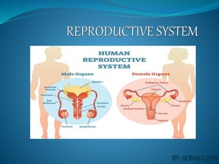
- 2. STRUCTURAL OUTLINE Female reproductive system External genitalia (vulva) Internal genitalia Vagina Uterus Uterine tubes Ovaries The reproductive cycle Menopause Breasts
- 3. Male reproductive system Scrotum Testes Seminal vesicles Ejaculatory ducts Prostate gland Urethra and penis Ejaculation
- 5. INTRODUCTION The ability to reproduce is one of the properties distinguishing living from non-living matter. The more primitive the animal, the simpler the process of reproduction. In mammals, including humans, the process is one of sexual reproduction, in which the male and female organs differ anatomically and physiologically, and the new individual develops from the fusion of two different sex cells (gametes). The male gametes are called spermatozoa and The female gametes are called ova
- 6. The functions of the female reproductive system are: 1. Formation of ova 2. Reception of spermatozoa 3. Provision of suitable environments for fertilisation and fetal development 4. Parturition (childbirth) 5. Lactation, the production of breast milk, which provides complete nourishment for the baby in its early life.
- 7. External genitalia (vulva) The external genitalia are known collectively as the vulva, and consist of the : 1. Labia majora and labia minora, 2. The clitoris, 3. The vaginal orifice, 4. The vestibule, the hymen and the vestibular glands (Bartholin’s glands).
- 9. Labia majora These are the two large folds forming the boundary of the vulva. They are composed of skin, fibrous tissue and fat and contain large numbers of sebaceous and eccrine sweat glands. Labia majora or "greater lips" are the part around the vagina containing two glands (Bartholin’s glands) which helps lubrication during intercourse. At puberty, hair grows on the mons pubis and on the lateral surfaces of the labia majora.
- 10. Labia minora These are two smaller folds of skin between the labia majora, containing numerous sebaceous and eccrine sweat glands. "lesser lips" are the thin hairless ridges at the entrance of the vagina, which joins behind and in front. In front they split to enclose the clitoris
- 11. Clitoris The clitoris corresponds to the penis in the male and contains sensory nerve endings and erectile tissue. The clitoris is a small pea-shaped structure. It plays an important part in sexual excitement in females.
- 12. Vestibular glands The vestibular glands (Bartholin’s glands) are situated one on each side near the vaginal opening. They are about the size of a small pea and their ducts open into the vestibule immediately lateral to the attachment of the hymen. They secrete mucus that keeps the vulva moist.
- 13. Blood supply, lymph drainage and nerve supply Arterial supply . This is by branches from the internal pudendal arteries that branch from the internal iliac arteries and by external pudendal arteries that branch from the femoral arteries. Venous drainage .This forms a large plexus which eventually drains into the internal iliac veins.
- 14. Lymph drainage. This is through the superficial inguinal nodes. Nerve supply. This is by branches from pudendal nerves. Perineum • The perineum is a roughly triangular area extending from the base of the labia minora to the anal canal. • It consists of connective tissue, muscle and fat. It gives attachment to the muscles of the pelvic floor
- 15. Internal genitalia Vagina, Uterus, Two uterine tubes Two ovaries.
- 16. Vagina The vagina is a fibromuscular tube lined with stratified squamous epithelium It runs obliquely upwards and backwards at an angle of about 45° between the bladder in front and rectum and anus behind. In the adult, the anterior wall is about 7.5 cm long and the posterior wall about 9 cm long. The difference is due to the angle of insertion of the cervix through the anterior wall.
- 17. Hymen. The hymen is a thin layer of mucous membrane that partially occludes the opening of the vagina. It is normally incomplete to allow for passage of menstrual flow and is stretched or completely torn away by sexual intercourse, insertion of a tampon or childbirth.
- 18. Structure of the vagina The vaginal wall has three layers: an outer covering of areolar tissue, a middle layer of smooth muscle and an inner lining of stratified squamous epithelium that forms ridges or rugae. It has no secretory glands but the surface is kept moist by cervical secretions. Between puberty and the menopause, Lactobacillus acidophilus, bacteria that secrete lactic acid, are normally present maintaining the pH between 4.9 and 3.5. The acidity inhibits the growth of most other micro-organisms that may enter the vagina from the perineum or during sexual intercourse
- 19. Blood supply, lymph drainage and nerve supply Arterial supply. An arterial plexus is formed round the vagina, derived from the uterine and vaginal arteries, which are branches of the internal iliac arteries. Venous drainage. A venous plexus, situated in the muscular wall, drains into the internal iliac veins. Lymph drainage. This is through the deep and superficial iliac glands.
- 20. Functions of the vagina • The vagina acts as the receptacle for the penis during sexual intercourse (coitus), and provides an elastic passageway through which the baby passes during childbirth.
- 21. Uterus The uterus is a hollow muscular pear-shaped organ, that is located anteroposteriorly in the pelvic cavity. It lies in the pelvic cavity between the urinary bladder and the rectum . It is about 7.5 cm long, 5 cm wide and its walls are about 2.5 cm thick. It weighs between 30 and 40 grams
- 22. Parts of the uterus For the descriptive purpose uterus can be divided into three distinct parts: 1. Fundus 2. Body 3. Cervix
- 24. Fundus. This is the dome-shaped part of the uterus above the openings of the uterine tubes. Body. This is the main part. It is narrowest inferiorly at the internal os where it is continuous with the cervix. Cervix (‘neck’ of the uterus). This protrudes through the anterior wall of the vagina, opening into it at the external os.
- 25. Layers of the uterus Perimetrium Myometrium Endometrium
- 26. Perimetrium This is peritoneum, which is distributed differently on the various surfaces of the uterus. Anteriorly it lies over the fundus and the body where it is folded on to the upper surface of the urinary bladder. This fold of peritoneum forms the vesicouterine pouch. Posteriorly the peritoneum covers the fundus, the body and the cervix, then it folds back on to the rectum to form the rectouterine pouch (of Douglas).
- 28. Myometrium This is the thickest layer of tissue in the uterine wall. It is a mass of smooth muscle fibres interlaced with areolar tissue, blood vessels and nerves. Endometrium Made up of the Columnar epithelial cells . Consist mucous secreting tubular glands
- 29. Blood supply, lymph drainage and nerve supply Arterial supply. This is by the uterine arteries, branches of the internal iliac arteries. Venous drainage internal illiac vein. Lymph drainage. Deep and superficial lymph vessels drain lymph from the uterus and the uterine tubes to the aortic lymph nodes and groups of nodes associated with the iliac blood vessels.
- 30. Supporting structures to the uterus The uterus is supported in the pelvic cavity by surrounding organs, muscles of the pelvic floor and ligaments that suspend it from the walls of the pelvis Broad ligaments. These are formed by a double fold of peritoneum, one on each side of the uterus. Round ligaments. These are bands of fibrous tissue between the two layers of broad ligament.
- 32. Uterosacral ligaments. These originate from the posterior walls of the cervix and vagina and extend backwards, one on each side of the rectum, to the sacrum. Transverse cervical (cardinal) ligaments. These extend one from each side of the cervix and vagina to the side walls of the pelvis.
- 33. Uterine tubes The uterine (Fallopian) tubes are about 10 cm long and extend from the sides of the uterus between the body and the fundus. They lie in the upper free border of the broad ligament and their trumpet-shaped lateral ends, penetrate the posterior wall, opening into the peritoneal cavity close to the ovaries. The end of each tube has fingerlike, projections called fimbriae. The longest of these is the ovarian fimbria, which is in close association with the ovary.
- 34. Structure The uterine tubes are covered with peritoneum (broad ligament), have a middle layer of smooth muscle and are lined with ciliated epithelium. Blood and nerve supply and lymphatic drainage are as for the uterus.
- 35. Functions The uterine tubes propel the ovum from the ovary to the uterus by peristalsis and ciliary movement. The secretions of the uterine tube nourish both ovum and spermatozoa. Fertilisation of the ovum usually takes place in the uterine tube, and the zygote is propelled into the uterus for implantation.
- 36. Ovaries The ovaries are the female gonads (glands producing sex hormones and the ova), and they lie in a shallow fossa on the lateral walls of the pelvis. They are 2.5–3.5 cm long, 2 cm wide and 1 cm thick. Each is attached to the upper part of the uterus by the ovarian ligament and to the back of the broad ligament by a broad band of tissue, the mesovarium. Blood vessels and nerves pass to the ovary through the mesovarium.
- 37. Structure • The ovaries have two layers of tissue. Medulla. • This lies in the centre and consists of fibrous tissue, blood vessels and nerves. Cortex. This surrounds the medulla. It has a framework of connective tissue, or stroma, covered by germinal epithelium. • It contains ovarian follicles in various stages of maturity, each of which contains an ovum.
- 38. Functions The ovary is the organ in which the female gametes are stored and develop prior to ovulation. Their maturation is controlled by the hypothalamus and the anterior pituitary gland,
- 39. Breasts The breasts or mammary glands are accessory glands of the female reproductive system. They exist also in the male, but in only a rudimentary form. Structure The mammary glands or breasts consist of varying amounts of glandular tissue, responsible for milk
- 41. Each breast contains about 20 lobes, each of which contains a number of glandular structures called lobules, where milk is produced. Lobules open into lactiferous ducts, which drain milk towards the nipple. breast itself is covered in subcutaneous fat. In the lactating breast, glandular tissue proliferates (hyperplasia) to support milk production, and recedes again after lactation stops.
- 42. The nipple. This is a small conical eminence at the centre of the breast surrounded by a pigmented area, the areola. On the surface of the areola are numerous sebaceous glands (Montgomery’s tubercles), which lubricate the nipple during lactation.
- 44. Blood supply, lymph drainage and nerve supply Arterial supply. The breasts are supplied with blood from the thoracic branches of the axillary arteries and from the internal mammary and intercostal arteries. Venous drainage. This is formed by an anastomotic circle round the base of the nipple from which branches carry the venous blood to the circumference, and end in the axillary and mammary veins.
- 45. Lymph drainage. This is mainly into the superficial axillary lymph vessels and nodes. Nerve supply. The breasts are supplied by branches from the 4th, 5th and 6th thoracic nerves,
- 47. The functions of the male reproductive organs are: production, maturation and storage of spermatozoa delivery of spermatozoa in semen into the female reproductive tract.
- 48. Scrotum The scrotum is a pouch of pigmented skin, fibrous and connective tissue and smooth muscle. It is divided into two compartments, each of which contains one testis, one epididymis and the testicular end of a spermatic cord.
- 49. Testes The testes are the male reproductive glands and are the equivalent of the ovaries in the female. They are about 4.5 cm long, 2.5 cm wide and 3 cm thick and are suspended in the scrotum by the spermatic cords. They are surrounded by three layers of tissue. 1) Tunica vaginalis. 2) Tunica albuginea. 3) Tunica vasculosa.
- 51. Structure In each testis are 200–300 lobules, and within each lobule are 1–4 convoluted loops of germinal epithelial cells, called seminiferous tubules. Between the tubules are groups of interstitial cells (of Leydig) that secrete the hormone testosterone after puberty. Functions Spermatozoa (sperm) are produced in the seminiferous tubules of the testes
- 52. Spermatic cords The spermatic cords suspend the testes in the scrotum. Each cord contains a testicular artery, testicular veins, lymphatics. Seminal vesicles is a 5 cm long tube that joins with the deferent duct to forms the common ejaculatory duct. Functions The seminal vesicles contract and expel their stored contents, seminal fluid, during ejaculation. Seminal fluid, which forms 60% of the volume of semen,
- 53. Ejaculatory ducts The ejaculatory ducts are two tubes about 2 cm long, each formed by the union of the duct from a seminal vesicle and a deferent duct. They pass through the prostate gland and join the prostatic urethra, carrying seminal fluid and spermatozoa to the urethra
- 55. Prostate gland: The gland weighs about 8 g in youth, but progressively enlarges (hypertrophies) with age and is likely to weigh about 40 g by the age of 50. Functions The prostate gland secretes a thin, milky fluid that makes up about 30% of the volume of semen, and gives it its milky appearance. It contains a clotting enzyme, which thickens the semen in the vagina, increasing the likelihood of semen being retained close to the cervix.
- 56. Urethra 19-20 cm long Prostatic urethra Membranous urethra Penile urethra Two sphincter.
- 57. Penis
- 60. 100 million spermatozoa per mL. If not ejaculated, sperm gradually lose their fertility after several months and are reabsorbed by the epididymis.
- 61. THANK YOU